Original Article
Assessment of Radiation Dose from Radioactive Wedge Filters during High-Energy X-Ray Therapy
Geum-mun Back*, Sung Ho Park†, Tae-Hyung Kim‡
*Department of Radiation Oncology, Asan Medical Center, Seoul, †Department of Neurosurgery, Ulsan University Hospital, Ulsan,
‡Department of Radiological Science, Kangwon National University, Samcheok, Korea
Copyright © 2017 Korean Society of Medical Physics
CCThis is an Open-Access article distributed under the terms of the Creative Commons Attribution Non-Commercial License (http://creativecommons.org/licenses/by- nc/4.0) which permits unrestricted non-commercial use, distribution, and reproduction in any medium, provided the original work is properly cited.
Introduction
In radiation therapy, 4-, 6-, 10-, and 15-MV photon beams generated by a linear accelerator or 6-, 9-, 12-, 15-, 18-, and 21-MeV electron beams are typically used. There is dose contamination owing to photo neutrons in high- energy photon therapy. The method for evaluating the neutrons generated from linear accelerators is covered in NCRP and AAPM reports.1,2) These neutrons are generated from the nuclear reaction of high-energy photons and are referred to as Giant Resonance Neutrons (GRN). For each material that reacts with photons, there is a threshold energy, which for tungsten and lead are 6.19 and 6.74 MeV, respectively. While there is no generation of neutrons from
a 6-MV photon beam, nuclear reactions occur between a 10-MV photon beam and the heavy nuclei. The NCRP and AAPM reports suggest the phosphorus activation method using P-31, the moderated foil method using In-115 and Au-197, and the activated rem meter method using BF3 as the methods for measuring neutrons. In radiation therapy, the effects of neutron doses depend on the depth.
According to studies by C. Ma et al., an 18-MV photon beam is 1/10,000 times less effective on a patient’s skin, and the difference is 100 times greater in the direction of depth.3) According to studies by A. Brahme et al., the respective differences in the proton, neutron, and alpha doses are 1/1,000, 1/10,000, and 1/10,000 times greater at a depth of 5.5 cm.4) The neutrons generated in the
Progress in Medical Physics 28(2), June 2017 https://doi.org/10.14316/pmp.2017.28.2.45 pISSN 2508-4445, eISSN 2508-4453
This paper evaluated the amount of radiation generated by wedge filters during radiation therapy using a high-energy linear accelerator, and the dose to the worker during wedge replacement.
After 10-MV photon beam was irradiated with wedge filter, the wedge was removed from the linear accelerator, and the dose rate and energy spectrum were measured. The initial measurement was approximately 1 uSv/h, and the radiation level was reduced to 0.3 uSv/h after 6 min. The effective half-life derived from the dose rate measurement was approximately 3.5 min, and the influence of AI-28 was about 53%. From the energy spectrum measurements, a peak of 1,799 keV was measured for AI-28, while the peak for Co-58 was not measured in the control room. The peaks for Au-106 and Cd-105 were found only measurement was done without wedge removement from the linear accelerator. The additional doses received by the radiation worker during wedge replacement were estimated to be 0.08−0.4 mSv per year.
Keywords: Radiation dose, Wedge filter, Linear accelerator Received 17 March 2017
Revised 4 June 2017 Accepted 1 July 2017
Corresponding author Sung Ho Park ([email protected]) Tel: 82-52-250-7137 Fax: 82-52-250-7138
PMP
Geum-mun Back, et al:Radiation Dose from Radioactivated Wedge
46
www.ksmp.or.kr
components of linear accelerators lead to the irradiation of materials, causing radionuclides with long and short half-lives to contribute to the exposure of maintenance personnel and workers, respectively. Petrovic analyzed radionuclides using HPGe after irradiating with 500 MU of an 18-MV photon beam in a 20×20 irradiation field, and reported the dose rate and effective dose by measuring the dose for 10 min.7) Based on this data, an additional annual dose of 0.25~1.7 mSv was estimated, and it was recommended that workers enter the treatment room 2~4 min after irradiation. Fischer conducted radionuclide measurements after irradiating with 15-MV 400 MU in 15×15 and 0×0 irradiation fields.8) Afterward, Fischer measured the radionuclides after irradiating with 400 MU in four types of linear accelerators with an average workload of 9,000~15,000 MU/d and a maximum dose rate of 200~400 MU/min, and reported the dose and effective dose evaluated for each radionuclide.9) Israngkul performed radionuclide analysis and dose rate measurements after irradiating with 1,000 MU in 2×2, 5×5, 10×10, 15×15, and 30×30 irradiation fields.10)
The purpose of this study was to evaluate the amount of radiation generated by the wedge filter during treatment with a linear accelerator of 10 MV or larger capacity, and to assess the dose received by the worker during wedge replacement.
Materials and Methods
A 10-MV photon beam was emitted with the wedge
attached in 0×0, small 5×5, and large 25×25 irradiation fields that were selected to evaluate the IMRT treatment, and each field was irradiated with 5 Gy. Five Gy is a nominal value for 500 MU, which corresponds to 5 Gy under standard measurement conditions (SSD 100 cm, 10×10 irradiation field at dmax depth).
The wedge was removed immediately after irradiation, and the dose rate over time was measured using the GM detector (RDS-110, Rados) shown in Fig. 1. The GM detector was calibrated annually by the Secondary Standard Dosimetry Laboratory. The radioactive energy spectrum was measured using a 3×3 NaI scintillator detector shown in Fig. 2. Ortec’s digiBASE was used for the PMT base, and the MAESTRO software was used for spectrum measurements. Energy correction was performed using a Co-60 point source. After irradiation, the wedge was removed from the linear accelerator and measured on a patient table, measured in the control room to exclude the effects of the linear accelerator, and measured while mounted on the linear accelerator head to observe the effects of the wedge alone.
Results and Discussion
Fig. 3 shows the dose rates over time measured in the treatment room immediately after irradiation with the 10- MV radiation. The initial measurement was approximately 1 uSv/h, and the radiation level was reduced to 0.3 uSv/
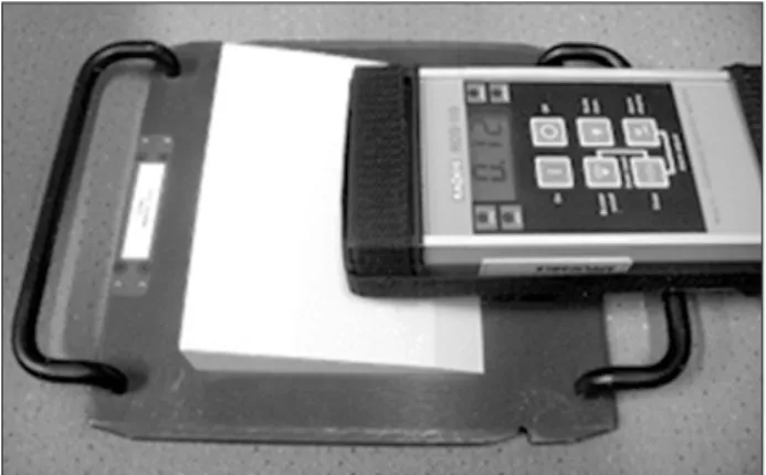
Fig. 1. Measurement of dose rate using GM detector and activated wedge filter.
Fig. 2. Measurement of energy spectrum using NaI scintillator detector.
Progress in Medical Physics Vol. 28, No. 2, June 2017 47
www.ksmp.or.kr
h after 6 min. The background radioactivity at the time of the experiment was 0.12 uSv/hr, and it was found that the radiation level was reduced to that of the background radiation after about 6 min. As shown in Fig. 3, the effective half-life was measured to be approximately 3.5 min.
Considering that the wedge is composed of an aluminum tray and steel body, and that the respective half-lives of Al-28 and Fe-53 are 2.2 min and 8.5 min, the effect of Al- 28 was found to be about 53% by solving the following effective half-life equation:
If 250 working days a year and 1 min per replacement work are assumed in the evaluation of the radiation dose for a radiation worker, a dose of 1 uSv/h for a maximum of 1 min is established. Assuming 20~50 patients per day and a maximum of 1 or 2 wedge replacements per single- patient treatment, the annual dose for the worker is 83~417 uSv, as shown in Table 1. However, this dose is received by the hand that is used in the replacement work, and the whole body dose can be considered smaller.
The energy spectrum using NaI is presented in Fig. 4.
The peak of Al-28 at 1,779 keV was observed from Fig. 4.
The measurement of Al-28 was almost identical when measured while mounted on the linear accelerator, measured near the linear accelerator, or measured outside the linear accelerator. The peak of Co-58 at 810.7 keV was measured near the linear accelerator, and was larger when
measured while mounted on the linear accelerator. The Co-58 peak was measured in the waveguide radioactivity measurement from Rosi’s study, and can be considered a result of radioactivity inside the linear accelerator.5) The peaks of Au-196 at 426.1 keV and Cd-105 at 648 keV were observed only when measured while mounted on the linear accelerator. The Au-196 peak was measured in the target and waveguide radioactivity measurement, and can be concluded to be a result of radioactivity inside the linear accelerator.5) The Cd-105 peak was measured in the cerroband radioactivity measurement in Borchard’s study, and is considered to be an effect of residual cerroband inside the linear accelerator.6)
Since the half-lives of Co-58 at 70.86 days, Au-190 at 6.2 days, and Cd-105 at 55.5 min are much longer than the half-life of Al-28 at 2.2 min and the effective half-life of 3.5 Table 1. Exposure dose to workers owing to removal of activated wedge after X-ray treatment.
Number of patients persons/day
Number of wedge remove times/patient
Exposure, uSv
20 1 83
2 167
30 1 125
2 250
40 1 167
2 333
50 1 208
2 417
Measureddoserate(uSv/hr)
0 1.5 1.0
0.5
Time (min) 0
0x05x5 25x25
6 5 4 3 2 1
Fig. 3. Measured dose rate of activated wedge after 5-Gy irradia- tion at various field sizes.
Normalizedcounts
0 101
Energy (keV)
Header Linac room Control room
2,000 102
103 104
105
1,800 1,600 1,400 1,200 1,000 800 600 400 200
196 105 58 54 28
Au: 426.1 keV Cd: 607,648 Co: 810.7 Mn: 834.8 AI: 1,779
keV keV keV keV
28AI
Fig. 4. Measured energy spectrum from activated wedge after 5-Gy irradiation.
Geum-mun Back, et al:Radiation Dose from Radioactivated Wedge
48
www.ksmp.or.kr
min measured by the GM detector, they were not reflected in the evaluation of the effective half-life.
Conclusion
The radioactivity from photo neutrons generated from the nuclear reaction between a 10-MV proton beam and a wedge was initially measured at around 1 uSv/hr, and the effective half-life was about 3.5 min. The annual doses additionally received by the radiation worker during wedge replacement were estimated to be 0.08−xs0.4 mSv.
Acknowledgements
This work was supported by the Nuclear Safety Research Program through the Korea Foundation of Nuclear Safety (KOFONA) and the Nuclear Safety and Security Commission (NSSC), Republic of Korea (Grant No. 1305033).
Conflicts of Interest
The authors have nothing to disclose.
Availability of Data and Materials
All relevant data are within the paper and its Supporting Information files.
References
1. NCRP Report 79. Neutron contamination from medical
electron accelerator, 1984.
2. AAPM TG Report 27. Neutron measurements around high energy X-ray radiotherapy machines. 1986.
3. Chilbani O, Ma C. Photoneutron dose calculations for high energy photon beams from Siemens and Varian LINACS.
Med Phys 1990–2000, 2003;30.
4. Gudowska I, Brahme A, Andreo P, et al. Calcluation of absorbed dose and biological effectiveness from photoneutron reactions in bremsstrahlungbeam of end point 50 MeV. Phys in Med Biol. 1999;44:2099.
5. Roig M, Panettieri V, Ginjaume M, et al. Photonuclear isotope characterization of a Siemens KDS 18 MeV LINAC head. Phys in Med Biol. 2004;49: N242-6.
6. Borchardt IM, Patterson JR, Beddoe AH. An investigation of photonuclear reactions in Cerrobend eutectic material with an 18 MV LINAC. Phys in Med Biol. 1991;36:649-53.
7. Pet rov ic N, K rest ic-Vesov ic J, Stojanov ic D, et a l.
Contribution of activation products to occupational exposure following treatment using high energy photons in radiotherapy. Rad Prot Dos. 2011;143:109-12.
8. Fischer HW, Tabot BE, Poppe B. Activation processes in a medical linear accelerator and spatial distribution of activation products. Phys Med Biol. 2006;51:N461.
9. Fischer HW, Tabot B, Poppe B. Comparison of activation products and induced dose rates in diferent high-energy medical linear accelerators. Health Physics. 2008;94:272-8.
10. Israngkul-Na-Ayuthaya I, Suriyapee S, Pengvanich P.
Evaluation of equivalent dose from neutrons and activation products from a 15 MV x-ray LINAC. J of Rad Research.
2015;56:919-26.