• 검색 결과가 없습니다.

Expression of Lily mottle virus Coat Protein and Preparation of IgY Antibody against the Recombinant Coat Protein

N/A
N/A
Protected

Academic year: 2021

Share "Expression of Lily mottle virus Coat Protein and Preparation of IgY Antibody against the Recombinant Coat Protein"

Copied!
6
0
0

로드 중.... (전체 텍스트 보기)

전체 글

(1)

Research Report

Expression of Lily mottle virus Coat Protein and Preparation of IgY Antibody against the Recombinant Coat Protein

Ha Na Yoo and Yong-Tae Jung

*

Department of Microbiology, Dankook University, Cheonan 330-714, Korea

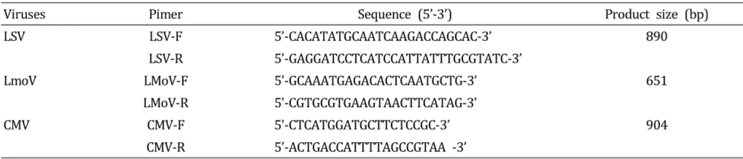
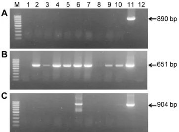
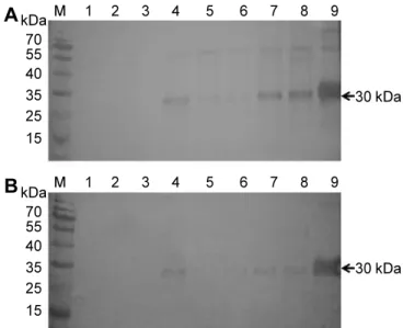
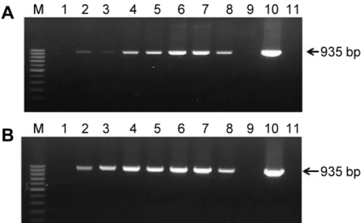
Abstract: Lily symptomless virus (LSV), Lily mottle virus (LMoV), and Cucumber mosaic virus (CMV) are the most prevalent viruses infecting lilies in Korea. Leaf and bulb samples showing characteristic symptoms of virus infection were collected in 2012, and 80 field samples were analyzed by reverse transcription polymerase chain reaction (RT-PCR). The infection frequencies were 79% for LMoV, 5% for LSV, and 3% for CMV. The LMoV coat protein gene was amplified and cloned into the pET21d(+) expression vector to develop serological diagnostic tools to detect LMoV. The resulting carboxy-terminal His-tagged coat proteins were expressed in Escherichia coli strain BL21 (DE3) by induction with IPTG. The recombinant proteins were purified using Ni-NTA agarose beads and used as an antigen to produce polyclonal antibodies in laying hens. The resulting egg yolk immunoglobulin (IgY) specifically recognized LMoV from infected plant tissues in immunoblotting assays and had comparable sensitivity to that of a mammalian antibody. In addition, method of immunocapture RT-PCR using this IgY was developed for sensitive, efficient, and rapid detection of LMoV. Based on these results, large-scale bulb tests and detection of LMoV in epidemiological studies can be performed routinely using this IgY. This is the first report of production of a polyclonal IgY against a plant virus and its use for diagnosis.

Additional key words:

egg yolk immunoglobulin, IC-RT-PCR

*Corresponding author: yjung@dankook.ac.kr

※ Received 22 October 2013; Revised 20 March 2014; Accepted 2 April 2014. This study was supported by the Center for Research and Development of Lilium, Ministry for Food, Agriculture, Forestry, and Fisheries, Republic of Korea.

Ⓒ 2014 Korean Society for Horticultural Science

Introduction

Lily symptomless virus (LSV), Lily mottle virus (LMoV), and Cucumber mosaic virus (CMV) are found in Korea.

LMoV is widespread, whereas little spread of CMV has been reported (Lim et al., 2009). These viruses occur in the field as single or mixed infections within lily plants.

The symptoms of lily viruses vary from vein clearing, leaf mottling, leaf mosaicism, chlorosis and yellow streaking, leaf curling, and brownish-necrotic spots to milder leaf symptoms. The development of mild symptoms in many cultivars prevents efficient removal of diseased plants, which may lead to an unacceptably high incidence of viruses in bulbs (Asjes, 2000). LMoV is a member of the Potyvirus genus within the Potyviridae family that has a linear single strand-positive sense-strand RNA genome of 9.6 kb containing a single large predicted open reading frame (Zheng et al.,

2003). Symptoms of LMoV range from fairly mild to severe compared to those of LSV, which range from symptomless to mild. If LMoV-infected plants are also infected with LSV, symptoms can be more severe.

Conventional reverse transcription polymerase chain

reaction (RT-PCR), real-time RT-PCR and enzyme-linked

immunosorbent assays (ELISA) have been developed to

detect lily viruses in infected leaves and bulbs. In earlier

studies, RT-PCR was used to identify and detect lily viruses

(Ha et al., 2008; Lim et al., 2009). Real-time RT-PCR has

been used to detect and quantify plant viruses (Beuve et

al., 2007; Boonham et al., 2009; Lim et al., 2010; Mortimer-

Jones et al., 2009). Among these detection methods, ELISA

is cost effective and suitable for rapid detection of a large

number of samples. Various ELISA alternatives based on

polyclonal or monoclonal antibodies have been used widely

to improve detection limits and broaden strain specificity

(2)

Table 1. PCR primers for the detection of Lily viruses from infected leaves.

Viruses Pimer Sequence (5’-3’) Product size (bp)

LSV LSV-F 5’-CACATATGCAATCAAGACCAGCAC-3’ 890

LSV-R 5’-GAGGATCCTCATCCATTATTTGCGTATC-3’

LmoV LMoV-F 5’-GCAAATGAGACACTCAATGCTG-3’ 651

LMoV-R 5’-CGTGCGTGAAGTAACTTCATAG-3’

CMV CMV-F 5’-CTCATGGATGCTTCTCCGC-3’ 904

CMV-R 5’-ACTGACCATTTTAGCCGTAA -3’

(Kong et al., 2009; Wang et al., 2010; Wu et al., 2011;

Zein et al., 2007). High viral concentrations can be detected by ELISA, but it is not as useful for detecting low viral concentrations (Kim et al., 19 9 5, 2012; Niimi et al., 19 9 9 ).

Preparation of a good quality antibody is important to achieve a highly sensitive ELISA, but large-scale production of mammalian antibodies is expensive and difficult.

Chicken egg yolk immunoglobulin (IgY) production has several advantages over conventional mammalian antibody production; chickens produce larger amounts of antibodies, IgY isolation is fast and simple, and egg collection is non- invasive (Dias et al., 2010; Schade et al., 2005; Spillner et al., 2012). IgY is the major antibody produced by laying hens. After immunization, IgY is secreted into the blood and transported to the egg yolk. Three immunoglobulins (IgM, IgA, and IgY) have been identified in chickens. IgM and IgA are undetectable in egg yolk, but IgY concentrations are 50-100 mg/egg yolk. One hen can produce 20 eggs per month and it is possible to obtain > 20 g IgY/year, as hens continually produce eggs for at least 10 months.

The use of IgY may help lower costs of immunological testing. IgY has been used in several studies for diagnostics and therapy (Dai et al., 2013; Wen et al., 2012).

In this study, we determined whether IgY could recognize the coat protein (CP) present in leaves. LMoV CP was expressed in Escherichia coli using the pET-21d(+) expression system and then used as an immunogen for producing chicken egg yolk IgY. IgY functioned well in Western blot analyses and immunocapture RT-PCR (IC-RT-PCR).

Materials and Methods Collection of Field Samples

Leaf and bulb samples with characteristic symptoms of lily virus infection were collected in different regions of Korea in 2012. Samples known to be infected with LMoV were obtained from the National Institute of Horticultural

& Herbal Science (Suwon, Korea) and stored at -80°C for use as positive controls.

Conventional RT-PCR

Infected leaves and bulbs were ground to a fine powder in liquid nitrogen using a mortar and pestle. Total RNA was extracted using Trizol reagent (Invitrogen, Carlsbad, CA, USA). Primers for specific detection of LSV, LMoV, and CMV were designed based on the alignment of the CP sequence of each pathogen available in the GenBank database (Table 1). RT and subsequent PCR reactions were conducted using a MJ Research PTC-150 Thermal Cycler (PE Applied Biosystems, Foster City, CA, USA) with the Maxime RT-PCR PreMix kit (Intron Biotechnology, Daejeon, Korea). The thermal cycling scheme was 42°C for 45 min, followed by 30 cycles of 95°C for 30 sec, 50°C for 1 min, and 72°C for 30 sec, with a final extension at 72°C for 7 min. The PCR products were examined by electrophoresis on 1.5%

agarose gels. RT-PCR products were cloned into the pGEM-T Easy plasmid vector (Promega, Madison, WI, USA) to construct the recombinant plasmids.

Cloning and Prokaryotic Expression of the CP Gene

The LMoV CP1 forward primer (5′-CCATGGCAAATGAGA

CACTTAAC-3′, NcoI restriction site is underlined), and the

LMoV CP2 reverse primer (5′-CTCGAGCATAGAAATTCCAA

GTAA-3′, XhoI restriction site is underlined) were used

to amplify the LMoV CP gene. The RT-PCR products were

cloned into a pGEM-T Easy plasmid vector (Promega). The

target fragment was released from the cloned pGEM-T Easy

plasmid using NcoI/XhoI and inserted into the corresponding

sites of the pET21d(+) expression vector (Novagen, Gibbstown,

NJ, USA). The recombinant plasmid was transformed in

E. coli strain BL21 (DE3). Protein expression was induced

at an absorbance of 0.3 at 600 nm using isopropyl-1-thio-β

-D-galactoside (IPTG) in a final concentration of 1 mM, and

the culture was incubated for 5 h with shaking at 37°C.

(3)

A

B

C

M 1 2 3 4 5 6 7 8 9 10 11 12

890 bp

651 bp

904 bp

Fig. 1. Reverse transcription polymerase chain reaction (RT- PCR)-based detection of Lily symptomless virus (LSV: A), Lily mottle virus (LMoV: B), and Cucumber mosaic virus (CMV:

C) in lily plants. Leaf samples with characteristic symptoms of lily virus infection were collected in different regions of Korea in 2012. The lengths of the PCR products were 890 bp (A), 651 bp (B), and 904 bp (C), respectively. Lane M, molecular size markers (100 bp ladder, Fermentas, Pittsburgh, PA, USA); lane 1, extract from healthy lily plant (‘Sorbonne’);

lanes 2-10, extracts from a diseased lily plant (lanes 2, 3, and 7 ‘Sorbonne’; lanes 4-6 ‘Ivory’; lane 8 ‘Yelloween’; lanes 9 and 10 ‘Siberia’); lane 11, cloned plasmid as a positive control; lane 12, water as a negative control.

The supernatant containing 6X His-fused CP was purified using Ni-NTA agarose beads (Qiagen, Hilden, Germany). The purified protein solution was quantified by standardization with bovine serum albumin and estimated by its molar extinction coefficient at 595 nm.

Immunization of Chickens

The purified recombinant CP was used as an immunogen and injected into 28-week-old ISA-brown chickens. Chickens were immunized on weeks 0, 2, and 4. The antigen was emulsified with an equal volume of Freund’s complete adjuvant (Sigma-Aldrich, St. Louis, MO, USA) for the first immunization. Freund’s incomplete adjuvant was similarly used for subsequent boosting immunizations. Eggs were collected 28 days from the initial immunization

Western Blot Analysis

Leaf samples from healthy or virus-infected plants were homogenized in sodium dodecyl sulfate (SDS) extraction buffer. The samples were denatured by boiling for 3 min and then separated on 12% SDS-polyacrylamide gel electro- phoresis (SDS-PAGE) gels. The proteins were then transferred to nitrocellulose membranes. The membranes were blocked in Tris-buffered saline Tween-20 (TBS-T) buffer containing 5% skim milk powder at 4°C overnight and incubated with primary polyclonal antibodies (IgG and IgY) specific to LMoV at room temperature for 2 h. After three washes in TBS-T, the membranes were incubated with horseradish peroxidase-conjugated goat anti-rabbit IgG (KOMAbiotech, Seoul, Korea) or mammalian antibody and AP-conjugated rabbit anti-chicken IgY (1:10,000) (Bethyl Laboratories, Montgomery, TX, USA) at room temperature for 1 h.

IC-RT-PCR

Leaf samples from lily plants showing disease symptoms were collected, and sap was extracted from the leaf samples using extraction buffer. The sap was centrifuged at 13,000 rpm for 1 min and the supernatants were kept on ice. A 200 µL aliquot of each diluted primary polyclonal antibody (IgG and IgY) was added to PCR tubes and incubated at 37°C for 4 h. After three washes in TBS-T, 200 μL of leaf sap was dispensed into the PCR tubes and incubated overnight at 4°C. After three washes in TBS-T, the virions were disrupted at 80°C for 10 min and used for RT-PCR.

The LMoV forward primer (5′-GCGTTCCAGGCAAATGAG-3′), and the LMoV reverse primer (5′-GTAAACCACACTATAACA CAAAGAG-3′) were used to amplify the LMoV gene.

Results

Conventional RT-PCR Assay

Leaf samples and bulbs showing characteristic symptoms of virus infection were collected from Gang-won and Chung- nam province, Korea in 2012. LSV, LMoV, and CMV were detected by RT-PCR (Fig. 1). Eighty field samples showing characteristic symptoms of virus infection were identified;

four plants with LSV, 63 plants with LMoV, and two plants with CMV. LMoV predominated in our tests, whereas little spread of LSV and CMV was found. The nucleotide sequences of eight isolates were identified (six LMoV; GenBank accession numbers KC430329 to KCKC430332, KC243417, JX908883:

one LSV; JX962776: one CMV, JX913865). The CP regions were determined and compared with the corresponding regions of LMoV (AJ564636), LSV (AJ516059), and CMV (AJ296154). The nucleic acid identities between reference viruses and eight isolates were 95-99%.

Cloning and Expression of LMoV CP

The entire LMoV cp gene was cloned from genomic RNA

(4)

M 1 2 3 4 5 6 7

30 kDa 70

55 40 35 25 15 kDa

Fig. 2. Purification of recombinant His-coated Lily mottle virus (LMoV) coat protein (CP) after Ni-NTA affinity chromatography.

The LMoV CP gene was cloned into pET21d(+) expression vector and the recombinant plasmid was transformed in E. coli strain BL21(DE3). Protein expression was induced at an absorbance of 0.3 at 600 nm using IPTG in a final concentration of 1 mM, and the culture was incubated for 5 h with shaking at 37°C. The supernatant containing 6X His-fused CP was purified using Ni-NTA agarose beads. Lane M, protein ladder; lane 1, flow- through; lanes 2- 3, column washing; lanes 4-6, elution of recombinant CP from the column.

A

B

kDa 70 55 40 35 25 15

kDa 70 55 40 35 25 15

30 kDa

30 kDa M 1 2 3 4 5 6 7 8 9

M 1 2 3 4 5 6 7 8 9

Fig. 3. Western blot detection of the Lily mottle virus (LMoV) coat protein in healthy and infected lily samples using IgY (A) and IgG (B) antibodies. The proteins were transferred to nitrocellulose membranes and the membranes were incubated with primary polyclonal antibodies (IgY and IgG) specific to LMoV. After washes, the membranes were incubated with AP-conjugated rabbit anti-chicken IgY(A) and horseradish peroxidase-conjugated goat anti-rabbit IgG(B). Lane M, protein marker; lane 1, extract from healthy lily plant as negative control; lanes 2-8 (lane 2, ‘Le Reve’; lane 3, ‘Siberia’; lanes 4- 7, ‘Medusa’; lane 8, unknown) extracts from a diseased lily plant; lane 9, purified His- tag coat protein of LMoV as positive control.

extracted from virus-infected lily by RT-PCR, and the sequence was confirmed using an automated DNA sequencing system.

The amplified fragments were inserted between the NcoI and XhoI sites in pET-21d(+) to produce a C-terminal His-tagged CP fusion protein. The resulting plasmid was designated pET-21d(+)-LMoV-CP. SDS-PAGE revealed that the recombinant LMoV CP band with a molecular mass of 30 kDa was expressed successfully in IPTG-induced pET-21d(+)-LMoV-CP(–)-transformed bacteria but not in pET-21d(+)-transformed control bacteria. Recombinant His-tagged LMoV-CP was purified from the soluble bacteria fractions through an Ni-NTA agarose column by elution with 300 mM imidazole buffer. The eluted fraction contained a major band of the 30 kDa recombinant CP and was used to induce production of polyclonal antiserum in rabbit and chicken (Fig. 2).

Detection of LMoV by Western Blot Using PAbs

To evaluate the suitability of PAbs to LMoV, antibodies recovered from rabbit and chicken were diluted to 1:10,000 for Western blotting. Figure 3 shows that both the IgY and IgG antibody reacted with LMoV-infected plant tissue extracts and the recombinant His-LMoV CP but not with healthy samples. IgY produced slightly stronger signals than those of IgG. Although some samples (Fig. 3, lanes 2 and 3) were RT-PCR positive, the CP band was not visible

in the Western blot.

IC-RT-PCR for Detecting LMoV

The IC-RT-PCR method was used to test for the presence of LMoV in field samples. PAb (100 μL) diluted at 1:100 in coating buffer was used to coat the PCR tubes, and 100 μL of leaf sap was used for detection. LMoV primers produced a single 935 bp band from LMoV-infected lily plants, but no band from healthy lily samples was amplified by IC-RT-PCR. In our Western blotting experiments, some samples were not detected with either the IgY or IgG antibody, but the same samples were amplified by IC-RT-PCR.

Discussion

LMoV is widespread in Korea and causes serious yield

losses in lily plants. Therefore a simple, reliable, and sensitive

technique to control the viral disease is required. Real-time

RT-PCR has been used to rapidly detect LMoV, but this

method is not suitable for large numbers of samples (Lim

(5)

A

B

M 1 2 3 4 5 6 7 8 9 10 11

M 1 2 3 4 5 6 7 8 9 10 11

935 bp

935 bp

Fig. 4. Immunocapture-reverse transcription polymerase chain reaction (IC-RT-PCR) detection of the Lily mottle virus (LMoV) coat protein in healthy and infected lily samples using IgY (A) and IgG (B) antibodies. A aliquot of each diluted primary polyclonal antibody (IgG and IgY) was added to PCR tubes.

After washes, leaf sap was dispensed into the PCR tubes.

The virions were disrupted and used for RT-PCR. Lane M, Molecular size markers (100 bp ladder, Fermentas, Pittsburgh, PA, USA); lanes 1 and 9, extract from a healthy lily plant;

lanes 2-8 (lane 2, ‘Le Reve’; lane 3, ‘Siberia’; lanes 4-7,

‘Medusa’; lane 8, unknown), extracts from a diseased lily plant; lane 10, PCR positive control; lane 11, PCR negative control.

et al., 2010). ELISA with PAbs or monoclonal antibodies is more convenient and practical than real-time RT-PCR (Edwards and Cooper, 1985; Liebenberg et al., 2009;

Tatineni et al., 2013). However, it is not easy to diagnose mixed infections with ELISA and it is not as useful for detecting low viral concentrations. In addition, preparation of a good qualitative and highly specific antibody is critical for obtaining reliable results.

In this study, the nucleic acid homologies between the different LMoV isolates were 95-99% and such high sequence conservation allows LMoV CP to be a suitable immunogen to produce IgY for detecting different LMoV isolates. The conventional method of antigen preparation is virion puri- fication following virus propagation but LMoV is difficult to purify due to its fragility and uneven distribution. In addition, lilies are often infected simultaneously by two or three different viruses. Using recombinant CP expression in a prokaryotic system does not cause these problems because it is simple, fast, and economical. Specific antiserum prepared using recombinant CP and its application to serological diagnostic tests has been successfully demonstrated for a number of plant viruses (Gulati-Sakhuja et al., 2009;

Kim et al., 2012; Wu et al., 2013).

IgY, the predominant serum immunoglobulin in birds, reptiles, and amphibians, is transferred from serum to egg yolk to confer passive immunity to embryos and baby chicks.

In recent years, specific IgY has received considerable attention in passive immunotherapy for infectious disease due to a number of advantages (Dai et al., 2013; Wen et al., 2012). IgY can be extracted in high yield and recovered from immunized hens over prolonged periods. The phylogenic distance of birds from mammals makes production of antibodies against antigens that may be non-immunogenic in mammals more successful in chickens. In addition, IgY binds neither the mammalian Fc receptor nor the rheumatoid factor. These differences have allowed for increasing interest in a wide variety of applications such as xenotransplantation, immunodiagnostics, and immunotherapy. Compared to antibody production in rabbits, IgY technology is economic, and very low quantities of antigen are required to obtain long-lasting and high IgY titers (Dias et al., 2010; Schade et al., 2005; Spillner et al., 2012).

In this study, the LMoV CP was expressed in E. coli, and two PAbs were produced in rabbits and chickens. A single protein band with a molecular mass close to LMoV was specifically detected by Western blotting in virus-infected leaf samples, although the CP bands were not detected in some leaf samples (Fig. 3). These findings indicate that the PAbs prepared in this study were highly specific for

LMoV and IgY and that they have comparable sensitivity to a mammalian antibody. We also developed an IC-RT-PCR procedure that offers sensitive, specific, and rapid detection of LMoV in field samples. This method avoided RNA extraction and was carried out easily in a single tube. IC-RT-PCR methods have been developed for detecting many other plant viruses (Ganesh Selvaraj et al., 2009; Kim et al., 2012;

Wu et al., 2011). We found that a single 9 35-bp band could be amplified from LMoV-infected leaf sap using the IC-RT-PCR assay, whereas no DNA band was amplified from healthy leaf sap (Fig. 4). IgY against CP has been used successfully to detect LMoV infection by IC-RT-PCR. In addition, IC-RT-PCR detected viruses in lily samples that were negative by Western blot. We speculate that a low virus concentration in some samples may have caused detection problems.

In conclusion, we successfully produced a large amount

of IgY against LMoV by immunizing laying hens with

recombinant LMoV CP. Using this PAb, IC-RT-PCR was

established for detecting lily viruses in field samples. This

method can be used for virus surveillance to help reduce

economic losses in the lily industry. Our results suggest

that IgY may offer a new large-scale source of low-cost

antibodies.

(6)

Literature Cited

Asjes, C.J. 2000. Control of aphid-borne Lily symptomless virus and Lily mottle virus in Lilium in the Netherlands. Virus Res.

71:23-32.

Berniak, H., T. Malinowski, and M. Kaminska. 2010. Characterization of polyclonal antibodies raised against two isolates of Cucumber mosaic virus. J. Plant Pathol. 92:231-234.

Beuve, M., L. Sempé, and O. Lemaire. 2007. A sensitive one-step real-time RT-PCR method for detecting Grapevine leafroll- associated virus 2 variants in grapevine. J. Virol. Methods 141:117-124.

Boonham, N., L. Laurenson, R. Weekes, and R. Mumford. 2009.

Direct detection of plant viruses in potato tubers using real-time PCR. Methods Mol. Biol. 508:249-258.

Dai, Y.C., X.F. Zhang, M. Tan, P. Huang, W. Lei, H. Fang, W.

Zhong, and X. Jiang. 2013. A dual chicken IgY against rotavirus and norovirus. Antiviral Res. 97:293-300.

Dias da Silva, W. and D.V. Tambourgi. 2010. IgY: A promising antibody for use in immunodiagnostic and in immunotherapy.

Vet. Immunol. Immunopathol. 135:173-180.

Edwards, M.L. and J.I. Cooper. 1985. Plant virus detection using a new form of indirect ELISA. J. Virol. Methods 11:309-319.

Ganesh Selvaraj, D., R. Pokorný, and L. Holková. 2009. Comparative analysis of ELISA, one step RT-PCR and IC-RT-PCR for the detection of bean yellow Mosaic virus in gladiolus. Commun.

Agric. Appl. Biol. Sci. 74:853-859.

Gulati-Sakhuja, A., J.L. Sears, A. Nuñez, and H.Y. Liu. 2009.

Production of polyclonal antibodies against Pelargonium zonate spot virus coat protein expressed in Escherichia coli and application for immunodiagnosis. J. Virol. Methods 160:29-37.

Ha, C., S. Coombs, P.A. Revill, R.M. Harding, M. Vu, and J.L.

Dale. 2008. Design and application of two novel degenerate primer pairs for the detection and complete genomic characterization of potyviruses. Arch. Virol. 153:25-36.

Kim, J.H., H.N. Yoo, E.H. Bae, and Y.T. Jung. 2012. Development of an indirect ELISA and immunocapture RT-PCR for Lily virus detection. J. Microbiol. Biotechnol. 22:1776-1781.

Kim, J.Y., S.T. Choi, and H.T. Hsu. 1995. Comparision of detection efficiency of lily symptomless virus in lily bulbs by ELISA, DBIA and tissue blotting method. J. Kor. Soc. Hort. Sci.

36:560-566.

Kong, B., J. Yu, H. Chen, D. Li, C. Han, and J. Wang. 2009.

Production of antiserum to recombinant coat protein for detecting Lily mottle virus in Yunnan, China. J. Phytopathol.

157:362-369.

Liebenberg, A., M.J. Freeborough, C.J. Visser, D.U. Bellstedt, and J.T. Burger. 2009. Genetic variability within the coat protein gene of Grapevine fanleaf virus isolates from South Africa and the evaluation of RT-PCR, DAS-ELISA and ImmunoStrips as virus diagnostic assays. Virus Res. 142:28-35.

Lim, J.H., E.H. Bae, Y.J. Lee, S.H. Park, K.J. Lee, S.R.M. Kim, and Y.T. Jung. 2009. Detection of Lily symptomless virus, Lily

mottle virus and Cucumber mosaic virus from Lilium grown in Korea by RT-PCR. Kor. J. Microbiol. 45:251-256.

Lim, J.H., E.H. Bae, S.H. Park, K.J. Lee, S.R.M. Kim, E.Y. Kwon, Y.J. Lee, and Y.T. Jung. 2010. Development of a real time RT-PCR with SYBR Green dye for the detection of several viruses from the bulbs and leaves of lily. Acta Virol. 54:319-321.

Mortimer-Jones, S.M., M.G. Jones, R.A. Jones, G. Thomson, and G.I. Dwyer. 2009. A single tube, quantitative real-time RT-PCR assay that detects four potato viruses simultaneously. J. Virol.

Methods 161:289-296.

Niimi, Y., T. Gondaira, Y. Kutsuwada, and H. Tsuji. 1999. Detection by ELISA and DIBa tests of Lily symptomless virus (LSV), Tulip breaking virus-lily (TBV-L) and Cucumber mosaic virus (CMV) in Lilium spp. grown in the field. J. Japan. Soc. Hort.

Sci. 68:176-183.

Schade, R., E.G. Calzado, R. Sarmiento, P.A. Chacana, J. Porankiewicz- Asplund, and H.R. Terzolo. 2005. Chicken egg yolk antibodies (IgY-technology): A review of progress in production and use in research and human and veterinary medicine. Altern. Lab.

Anim. 33:129-154.

Spillner, E., I. Braren, K. Greunke, H. Seismann, S. Blank, and D. du Plessis. 2012. Avian IgY antibodies and their recombinant equivalents in research, diagnostics and therapy. Biologicals.

40:313-322.

Tatineni, S., G. Sarath, D. Seifers, and R. French. 2013. Immuno- detection of Triticum mosaic virus by DAS- and DAC-ELISA using antibodies produced against coat protein expressed in Escherichia coli: Potential for high-throughput diagnostic methods.

J. Virol. Methods. 189:196-203.

Wang, R., G. Wang, Q. Zhao, Y. Zhang, L. An, and Y. Wang.

2010. Expression, purification and characterization of the Lily symptomless virus coat protein from Lanzhou Isolate. Virol.

J. 34:1-7.

Wen, J., S. Zhao, D. He, Y. Yang, Y. Li, and S. Zhu. 2012. Preparation and characterization of egg yolk immunoglobulin Y specific to influenza B virus. Antiviral Res. 93:154-159.

Wu, J., C. Meng, H. Shang, S. Rong, C. Zhang, J. Hong, and X. Zhou. 2011. Monoclonal antibody-based triple antibody sandwich-enzyme-linked immunosorbent assay and immunocapture reverse transcription-polymerase chain reaction for Odontoglossum ringspot virus detection. J. Virol. Methods 171:40-45.

Wu, J., Y. Ni, H. Liu, L. Rao, Y. Zhou, and X. Zhou. 2013.

Development and use of three monoclonal antibodies for the detection of rice black-streaked dwarf virus in field plants and planthopper vectors. Virol. J. 10:114.

Zein, H.S., M. Nakazawa, M. Ueda, and K. Miyatake. 2007.

Development of serological procedures for rapid and reliable detection of Cucumber mosaic virus with dot-immunobinding assay. World J. Agric. Sci. 3:430-439.

Zheng, H.Y., J. Chen, M.F. Zhao, L. Lin, J.P. Chen, J.F. Antoniw,

and M.J. Adams. 2003. Occurrence and sequences of Lily mottle

virus and Lily symptomless virus in plants grown from imported

bulbs in Zhejiang province, China. Arch. Virol. 148:2419-2428.

수치

Table  1.  PCR  primers  for  the  detection  of  Lily  viruses  from  infected  leaves.
Fig.  1.  Reverse  transcription  polymerase  chain  reaction  (RT-  PCR)-based  detection  of  Lily  symptomless  virus  (LSV:  A),  Lily  mottle  virus  (LMoV:  B),  and  Cucumber  mosaic  virus  (CMV:
Fig.  2.  Purification  of  recombinant  His-coated  Lily  mottle  virus (LMoV) coat  protein  (CP)  after  Ni-NTA  affinity  chromatography
Fig.  4.  Immunocapture-reverse  transcription  polymerase  chain  reaction  (IC-RT-PCR)  detection  of  the  Lily  mottle  virus  (LMoV)  coat  protein  in  healthy  and  infected  lily  samples  using  IgY  (A)  and  IgG  (B)  antibodies

참조

관련 문서

The object of this study is to analyse the bacteria of periapical bascess and fascial space abscess using multiplex real-time polymerase chain reaction (MRT-PCR) before

치아발생과 같은 기관의 발생과 석회화(mi neral i zati on)와 관련이 있는 BMPR-IB와 EF-handcal ci um bi ndi ng protei n은 APi n의 과발현을 유도한 경우에 는

The expression of Dynamin II protein expressed strongly at advanced bell stage in cytoplasm of odontoblast. Dynamin II protein localized in adjacent the nuclear

Inhibitory effects of the concentration of methanol extracts from Plantago Asiatica against the COX-2 protein expression and iNOS expression of the MDA-MB-231 human

qRT-PCR for VSMC synthetic phenotype protein, cellular retinol binding protein 1(CRBP-1) and connexin 43, from thoratic aorta showed that CRBP-1 expression was increased by 70%

 Predictions of chromatographic behavior based on product properties.  Prediction

Distribution of Citrus Tristza Virus or Satsusma Dwarf Virus at various parts of virus infected Satsuma mandarin

Our previous study verified that treatment of Nicotiana benthamiana plants with dsRNAs targeting helper component‑proteinase (HC-Pro) and nuclear inclusion b (NIb) genes