305 The abducens nerve passes through the deepest part of the
cranial base. Duplication of the nerve has sometimes been discovered during surgery for petroclival tumors or at autopsy. During the embryological development of human embryos, the abducens nerve is in the form of two aberrant branches.1 The failure of these nerve branches to fuse could lead to the duplication of the abducens nerve.1 There has been no report of associated eye movement in a patient with duplicated abducens nerve. Here, we present magnetic resonance imaging (MRI) findings of duplicated abducens nerves and associated eye movement.
Case Report
A 24-year-old woman was referred to us for the evaluation of eye movement from the otolaryngology department where she had undergone brain MRI for the evaluation of tinnitus.
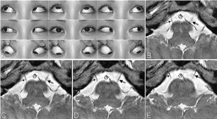
The 0.7-mm-thick MR images obtained with a T2-weighted 3D fast field echo technique in the axial plane at the level of the brainstem2 showed a duplicated left abducens nerve.
One right abducens nerve and two left abducens nerves were observed, otherwise there was no abnormal findings in the
brain (Fig. 1). She was born after an uneventful full-term pregnancy and delivery was normal. Data collection for this study conformed with Korean law.
On ophthalmologic evaluation, her best corrected visual acuities were 20/20 OU. She was orthotropic in 5 cardinal positions, and ductions and versions were full (Fig. 1). The anterior segment and fundi were normal.
Discussion
The abducens nerve can be reliably observed by MRI,2 thus MRI can be very helpful for accurate diagnosis as well as providing additional insight into the pathogenesis of various diseases related with congenitally anomalous abducens nerves, such as Duane's retraction syndrome,2-4 synergistic divergence,5 and congenital fibrosis syndrome.5
During the course of the abducens nerve between the brainstem and the lateral rectus muscle, the abducens nerve usually travels forward as a single trunk, but it is not uncommon for the nerve to split into two branches. Autopsy studies have described an incidence rate of the duplication of the abducens nerve from 5 to 28.6%,1,6 and MR images have also showed double rootlets of the abducens nerve in 25.2%
of the study's subjects.7 However, details of associated eye movement have not previously been reported in the literature.
In this case report, we present the findings of a duplicated abducens nerve with completely normal eye movement.
Normal Abduction in a Patient with Duplicated Abducens Nerve
Jae Hyoung Kim, MD1, Jeong-Min Hwang, MD2
Department of Radiology1, Department of Ophthalmology2, Seoul National University College of Medicine, Seoul National University Bundang Hospital, Seongnam, Korea
Purpose: To our knowledge, there has been no report of ophthalmologic findings related with a duplicated abducens nerve in the ophthalmic literature. This study reports such findings.
Methods: An ophthalmologic examination was performed in one patient with a duplicated abducens nerve, revealed with thin-sectioned magnetic resonance imaging (MRI) across the brainstem level.
Results: The MRI disclosed a duplicated left abducens nerve. The patient was orthotropic in five cardinal positions, and her ductions and versions were full.
Conclusions: One patient with a duplicated abducens nerve showed orthotropia and normal ocular movement. Korean Journal of Ophthalmology 19(4):305-306, 2005
Key Words: Duplicated abducens nerve, Magnetic resonance imaging, Ocular movement
Received: August 3, 2005 Accepted: September 15, 2005
Reprint requests to Jeong-Min Hwang, MD. Department of Ophthal- mology, Seoul National University College of Medicine, Seoul National University Bundang Hospital, #300 Gumi-dong, Bundang-gu, Seongnam-si, Gyeonggi-do 463-707, Korea. Tel: 82-31-787-7372, Fax:
82-31-787-4057, E-mail: [email protected]
*There is no financial conflict of interest regarding the subject matter in this manuscript.
Kor J Ophthalmol Vol.19, No.4, 2005
306
References
1. Iaconetta G, Tessitore E, Samii M. Duplicated abducent nerve and its course: microanatomical study and surgery- related considerations. J Neurosurg 2001;95:853-8.
2. Kim JH, Hwang JM. Presence of the abducens nerve according to the type of Duane's retraction syndrome.
Ophthalmology 2005;112:109-13.
3. Kim JH, Hwang JM. Magnetic resonance imaging in patients with abduction deficit found after head trauma. J Neurol 2005;252:224-6.
4. Kim JH, Hwang JM. Usefulness of MR imaging in children
without characteristic clinical findings of Duane's retraction syndrome. AJNR Am J Neuroradiol 2005;26:702-5.
5. Kim JH, Hwang JM. Hypoplastic oculomotor nerve and absent abducens nerve in congenital fibrosis syndrome and synergistic divergence with magnetic resonance imaging.
Ophthalmology 2005;112:728-32.
6. Ozveren MF, Sam B, Akdemir I, et al. Duplication of the abducens nerve at the petroclival region: an anatomic study.
Neurosurgery 2003;52:645-52.
7. Alkan A, Sigirci A, Ozveren MF, et al. The cisternal segment of the abducens nerve in man: three-dimensional MR imaging. Eur J Radiol 2004;51:218-22.
Fig. 1. (A) Ocular versions demonstrating normal eye movement in both eyes. (B-E) Four 0.7-mm-thick slice images obtained with a T2-weighted 3D fast field echo MRI sequence show a single right abducens nerve (arrows), and a duplicated left abducens nerve (double arrows) emerging from the pontomedullary junction and coursing in a superior oblique direction toward the clivus. Before entering the clivus, the left nerves appear to be fused.