pISSN 1976-1457 eISSN 2005-6168
Prophylactic effect of aqueous extract of Sesamum indicum seeds on ethanol-induced toxicity in male rats
Oyinloye, B.E.
1,2§, Nwozo, S.O.
2, Amah, G.H.
3, Awoyinka, A.O.
4, Ojo, O.A
1, Ajiboye, B.O.
1and Tijani, H.A.
2,51
Department of Biochemistry, College of Sciences, Afe Babalola University, Ado Ekiti, Nigeria
2
Nutritional and Industrial Research Laboratories, Department of Biochemistry, Faculty of Basic Medical Sciences, College of Medicine, University of Ibadan, Ibadan, Nigeria
3
Department of Biochemistry, School of Medicine, Babcock University, Ilishan-remo, Nigeria
4
Department of Medical Biochemistry, College of Medicine, Ekiti State University, Ado Ekiti, Nigeria
5
Department of Science Technology, Federal Polytechnic, Ado Ekiti, Nigeria
Abstract
The liver is vulnerable to alcohol-related injury because it is the primary site of alcohol metabolism. Additionally, a number of potentially dangerous by-products are generated as alcohol is broken down in the liver. However, dietary supplements may prevent or relieve some of alcohol’s deleterious effects. Therefore, this study was conducted to evaluate the prophylactic effect of aqueous extract of Sesamum indicum (SI) on ethanol induced toxicity in rats. Male Wistar albino rats were divided into control, ethanol, pre-treatment, simultaneous and post-treatment groups. In the prophylactic experiment, Sesamum indicum, (200 mg/kg body weight) was administered by oral gavage for 28 days; two hours before, simultaneously with or two hours after ethanol exposure. Toxicity was induced by administering 45% ethanol (4.8 g/kg bw) by oral gavage. Lipid peroxidation (TBARS) and reduced glutathione (GSH) levels and catalase (CAT), glutathione peroxidase (GPx), superoxide dismutase (SOD) and gluthathione-S-transferase (GST) activities were then determined in the liver, serum triglyceride (TG) levels, alanine aminotransferase (ALT) and aspartate aminotransferase (AST) activities were monitored and histological examination was carried out. The results revealed that ethanol administration led to significant elevation of TBARS level while depleting in the level of GSH as well as CAT, GPx, SOD and GST activities. Similarly, TG level and ALT and AST activities were elevated. The SI pre-treated group significantly inhibited TBARS, restored GSH level, enhanced CAT, GPx, SOD and GST activities and significantly decreased the elevated level of serum TG, ALT and AST activities. SI treatment (simultaneously with ethanol) exhibited similar effects to those of the SI pre-treated groups, while the SI post-treated group did not show the same protection as the Pre-treated group.
S. indicum possesses antioxidant and hepatoprotective properties, that eliminate the deleterious effects of toxic metabolites of ethanol.
Key Words: Antioxidant, ethanol, hepatoprotective, prophylactic, Sesamum indicum.
Introduction
8)Metabolism of drugs and xenobiotics primarily occurs in the liver; therefore the liver is prone to xenobiotics-induced injury as a result of its pivotal role in xenobiotics metabolism. Alcohol is metabolized in the liver, which results in generation of a number of potentially dangerous by-products such as acetal- dehyde and highly reactive free radicals that contribute to alcohol-induced liver damage [1,2]. Several studies have attempted to identify the molecular pathways, directly and indirectly affected by alcohol exposure in the liver. These pathways range from oxidative stress, metabolism-related effects, and inflam- mation to apoptosis. Induction of oxidative stress and activation of the inflammatory cascade have been identified as key elements in the pathophysiology of alcohol liver disease (ALD) [3-5].
The popularity of herbal remedies is increasing globally and
at least one quarter of patients with liver diseases use ethnobo- tanicals [6]. Several plants and natural compounds isolated from plants have been shown to have hepatoprotective activities [7]
and used to prevent the oxidative challenges against the liver during alcohol metabolism. It has been proposed that their efficacy is due to their free radical scavenging ability, which is believed to be associated with their bioactive and antioxidant constituents [8-10].
Sesamum indicum (SI), which is a plant with both medicinal and nutritive values, is popularly used as a herbal remedy against a wide range of ailments [11]. SI seed is not only a good source of edible nutrients, but is also widely considered to have medicinal value that includes antioxidant, anti-ageing, anti- mutagenic, antihypertensive, and anti-inflammatory activities, as well as the ability to inhibit cholesterol absorption from the intestine and synthesis in the liver. The seed is used as a duretic,
§Corresponding Author: Oyinloye, B.E., Tel. 234-803-5068-320, Fax. 234-2-810-3043, Email. [email protected]
Received:May 23, 2013, Revised: November 12, 2013, Accepted: November 12, 2013
ⓒ2014 The Korean Nutrition Society and the Korean Society of Community Nutrition
This is an Open Access article distributed under the terms of the Creative Commons Attribution Non-Commercial License (http://creativecommons.org/licenses/by-nc/3.0/)
which permits unrestricted non-commercial use, distribution, and reproduction in any medium, provided the original work is properly cited.
emollient, and a tonic for the liver and kidneys [12,13].
SI is an annual shrub with white bell-shaped flowers that is grown for the production of seeds, which are rich in oil content.
The plant is found in tropical, subtropical, and southern temperate areas of the world, particularly in India, China, South America and Africa [14]. This study was conducted to evaluate the prophylactic effects of aqueous extract of SI seeds on ethanol- induced toxicity in a rat model.
Materials and Methods
Animals
Thirty Male Wistar albino rats (weighing 180-200 g) were divided into control, ethanol, pre-treatment, simultaneous and post-treatment groups. The animals were allowed access to feed (obtained from Ladokun Feed Mill Nigeria Limited, Ibadan, Nigeria) and water ad libitum for fourteen days to allow their acclimatization prior to commencement of the experiment. A pilot study (unpublished data) showed that the aqueous extract was more potent than methanolic and hexane extracts at the tested concentrations (200 mg/kg bw). In the prophylactic experiment, SI (200 mg/kg bw) extract was administered by oral gavage for 28 days, two hours before, simultaneously with or two hours after ethanol exposure. Toxicity was induced by administering 45% ethanol (4.8 g/kg bw) by oral gavage. Animal experiments followed protocols established by the National Institute of Health (NIH) (NIH publication 85-23, 1985) for the Care and Use of Laboratory Animals and all procedures involving rats were conducted according to the ethical guidelines approved by the Animal Ethical Committee of Afe Babalola University (ABUAD- SCI03/13/09/002).
Preparation of the extract
SI seeds were purchased from Ojoo market in Ibadan, Nigeria and then identified and authenticated by the Botany Department, University of Ibadan. One kilogram of the air-dried seeds was subsequently pulverized into uniform powder using an electric blender (25-28℃). Pulverized seed (1 kg) was then defatted by mixing with n-hexane (2000 ml) using a magnetic stirrer at room temperature for 6 hours. The resulting slurry was filtered and the residue was air dried for 24 hour. Next the dried defatted residue (600 g) was extracted with 1500 ml of distilled water by maceration for 72 hours. The aqueous extract subsequently was filtered and the filtrate was concentrated using water bath (80℃) to yield a brown extract. This was carefully scraped into a clean sample bottle and stored in a refrigerator until further use. Prior to use, detailed phytochemical screening of SI was carried out according to the method described by Harborne [15].
Chemicals and biochemical assays
Randox alanine aminotransferase (ALT), aspartate aminotrans- ferase (AST), and triglyceride (TG) assay kits were purchased from ABJ Chemicals, Lagos (Nigeria). Adrenaline, thiobarbituric acid (TBA), Ellman’s reagent (DTNB), glutathione and bovine serum albumin (BSA) were purchased from Sigma Chemical (St.
Louis, MO, USA). All other chemicals were of the highest purity commercially available.
Lipid peroxidation (LPO) was assayed by measuring thiobar- bituric acid reactive substances (TBARS) as described by Varshney and Kale [16]. Catalase (CAT) activity was determined by measuring the rate of decomposition of hydrogen peroxide at 570 nm as described by Sinha [17]. SOD activity was determined as described by Misra and Fridovich [18].
Reduced glutathione (GSH) level was estimated using the method described by Beutler et al., [19] at 412 nm. GPx was determined by the method described by Hafeman et al. [20] based on the degradation of H
2O
2in the presence of GSH. Glutathione- S-transferase (GST) activity was determined according to Habig et al. [21]. Serum ALT and AST activites and TG levels were quantified spectrophotometrically using a Randox commercial assay kit.
Preparation of tissues for biochemical analyses and histological examination
Following daily exposure for 28 days, the animals were sacrificed 24 hours after the last dose by cervical dislocation.
Blood samples were then collected by retro-orbital puncture and allowed to coagulate at room temperature for half an hour, after which the serum was obtained by blood centrifugation at 3,000 rpm for 10 minutes and kept at 20℃ until analyses were done.
Liver samples were quickly excised and washed in ice-cold 1.15% KCl solution, dried using filter paper and weighed.
samples were then homogenized in 4 volumes of 56 mM Tris-HCl buffer (pH 7.4) containing 1.15% KCl, after which they were centrifuged at 10,000 g for 15 minutes.
The supernatant was collected and stored until needed for assays. Small pieces of liver sections were fixed in 10% formal saline, after which they were cut and stained with haematoxylin and eosin. The stained tissue sections were they observed under a light microscope (× 400 objective) for histological assessment.
Statistical analysis
All values were expressed as the mean ± S.D of six animals.
Data were analyzed by one-way analysis of variance (ANOVA)
followed by a post-hoc LSD test using the SPSS (10.0) statistical
software. A P < 0.05 was considered statistically significant.
Phytochemicals Flavonoids Saponins Tannins Phenols
Aqueous Extracts ++ +++ + +++
+++ = Abundance, ++ = Moderate, + = Trace
Table 1. Phytochemical screening of Sesamum indicum
Group TG (nmolL) ALT (UI/L) AST (UI/L)
Control 35.66 ± 6.82b 13.11 ± 0.38b 18.28 ± 0.33b Ethanol 54.52 ± 4.23a 19.05 ± 0.12a 31.21 ± 0.82a Pre-treatment 38.34 ± 6.62b 14.09 ± 0.27b 20.00 ± 0.36b Simultaneous 39.19 ± 5.61a,b 14.84 ± 0.36b 21.12 ± 0.27b Post-treatment 51.19 ± 7.20a 17.24 ± 0.23a 29.46 ± 0.30a Mean differences are significant (P< 0.05) when compared with: a control group, b ethanol group
Table 2. Effects of Sesamum indicum on serum TG, ALT and AST activities (mean ± S.D., n = 6)
Group GPx
(nmol/mgprotein/min) GSH
(nmol/mg protein) LPO (nmol/mg/protein) Control 48.65 ± 1.32b 36.80 ± 3.18b 38.46 ± 0.88b Ethanol 25.26 ± 1.10a 29.21 ± 1.41a 73.50 ± 1.77a Pre-treatment 38.24 ± 1.45 35.40 ± 2.14 47.22 ± 2.21 Simultaneous 36.17 ± 1.75 33.11 ± 1.16 49.14 ± 1.14a,b Post-treatment 28.65 ± 1.45 27.20 ± 2.02a 68.06 ± 1.74a Mean differences are significant (P< 0.05) when compared with: a control group, b ethanol group
Table 3. Effects of Sesamum indicum on hepatic GPx activity, GSH and LPO
levels (mean ± S.D., n = 6)
Group GST
(Units/mg protein) SOD
(Units/mg protein) CAT (Units/mg protein) Control 25.26 ± 2.40b 57.14 ± 0.73b 9.79 ± 0.52b Ethanol 21.00 ± 2.14a 43.60 ± 0.72a 5.57 ± 0.37a Pre-treatment 23.72 ± 2.02 56.16 ± 0.37b 8.63 ± 1.62 Simultaneous 23.21 ± 1.74 55.19 ± 0.21b 8.00 ± 0.74 Post-treatment 21.63 ± 2.94a 45.42 ± 0.06 6.53 ± 0.18 Mean differences are significant (P< 0.05) when compared with: a control group, b ethanol group
Table 4. Effects of Sesamum indicum on hepatic GST, SOD and CAT activities.
(mean ± S.D., n = 6)
(A) (B)
(C) (D)
(E)
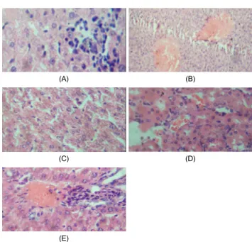
Fig. 1. Histological analysis of liver sections. Liver tissues were stained with H&E (×400). (A) Control: showing normal liver histology, no abnormalities was seen.
(B) Ethanol group: showing multiple foci of congestion, massive and severe sinusoid infiltration by inflammatory cells. (C) Pre-treatment group: showing very mild periportal cellular infiltration. (D) Simultaneous treatment group: showing very mild diffuse hydropic degeneration of hepatocytes. (E) Post-treatment group: showing mild accumulation of fats and infiltration of inflammatory cells.