Apoptosis of T Lymphocytes Isolated from Peripheral Blood of Patients with Acute Exacerbation
of Chronic Obstructive Pulmonary Disease
Sung Chul Lim, Jin Yung Ju, Su Young Chi, Hee Jung Ban, Yong Soo Kwon, In Jae Oh, Kyu Sik Kim, Yu Il Kim, and Young Chul Kim
Department of Internal Medicine, Chonnam National University Hospital, Gwangju, Korea.
Received: August 18, 2010 Revised: October 1, 2010 Accepted: October 14, 2010
Corresponding author: Dr. Sung Chul Lim, Department of Internal Medicine, Chonnam National University Hospital, 671 Jaebong-ro, Dong-gu, Gwangju 501-757, Korea.
Tel: 82-62-220-6570, Fax: 82-62-225-8578 E-mail: [email protected]
∙ The authors have no financial conflicts of interest.
© Copyright:
Yonsei University College of Medicine 2011 This is an Open Access article distributed under the terms of the Creative Commons Attribution Non- Commercial License (http://creativecommons.org/
licenses/by-nc/3.0) which permits unrestricted non- commercial use, distribution, and reproduction in any medium, provided the original work is properly cited.
Purpose: Chronic obstructive pulmonary disease (COPD) is characterized by chronic inflammation of the airways and progressive destruction of lung paren- chyma. Apoptosis is critical for the maintenance of normal tissue homeostasis and is in equilibrium with proliferation and differentiation. This study was under- taken to investigate relationship between apoptosis of peripheral blood lympho- cytes during exacerbation of COPD and inflammatory response that characterizes this condition. Materials and Methods: Seventeen patients with COPD exacer- bation, 21 stable COPD, and 12 control subjects were included. T lymphocytes were isolated from peripheral blood using MACS. Apoptosis of T lymphocytes was assessed with FACS using annexin V and 7-aminoactinomycin. Serum levels of interleukin (IL)-6, IL-8 and tumor necrosis factor (TNF)-α were determined by an immunoassay technique. Results: There was significantly increased percent- age of apoptotic lymphocytes, CD 4
+, and CD 8
+T cells in the peripheral blood of patients with exacerbation of COPD compared with stable COPD. Serum levels of IL-6, IL-8, and TNF-α were significantly increased in patients with exacerba- tion of COPD compared with stable COPD. Only TNF-α presented a positive correlation with apoptotic lymphocytes in patients with exacerbation of COPD.
Conclusion: Increased apoptotic lymphocytes may be associated with upregula- tion of TNF-α in the peripheral blood of patients with acute exacerbation of COPD.
Key Words: Chronic obstructive pulmonary disease, apoptosis, lymphocyte, tu- mor necrosis factor-α
INTRODUCTION
Chronic obstructive pulmonary disease (COPD) is characterized by an abnormal inflammatory response to respiratory pollutants, mostly from tobacco smoking.
This inflammation in the repair process can alter respiratory structure and func-
tion.
1,2In the control of repair process, apoptosis is an important mechanism that
MATERIALS AND METHODS
Study subjects
A total of 50 subjects were enrolled in the study from Janu- ary 2008 to March 2009. COPD was diagnosed on the basis of the Global Initiative for Chronic Obstructive Lung Dis- ease criteria.
17The inclusion criteria were a smoking history of at least 20 pack-years, a postbronchodilator forced expira- tory volume one-second (FEV
1)/forced vital capacity (FVC) ratio <70%, and an FEV
1<80% of the predicted values. Ex- clusion criteria were respiratory disorders other than COPD, pulmonary embolism, infectious diseases, malignancy, left ventricular dysfunction, recent surgery, and severe endocrine, hepatic, or renal diseases. The exacerbation of COPD was defined by any combination of the following major criteria:
aggravation of dyspnea, increase in sputum volume, and in- crease in sputum purulence; other minor criteria (upper respi- ratory tract infection in the last 5 days, fever, increased wheezing, increased cough) were also considered.
18All mea- surements were performed in patients with exacerbation of COPD during the first 24 hours of hospital admission. Stable COPD patients without a history of exacerbation for the pre- vious 2 months were recruited from our outpatient settings.
The control group consisted of current or former smokers with normal lung function. The inclusion criteria of control group were as follows: 1) older than 50 years of age in or- der to match the COPD group, 2) smoking history of at least 20 pack-years, 3) FEV
1/FVC ratio >70%, FEV
1>80%
of the predicted value, and 4) no other pulmonary disease by history and chest radiography.
This study protocol was approved by Chonnam National University Hospital Ethics Committee (CRI08058-1), and all subjects provided informed written consent before par- ticipation.
Isolation and culture of lymphocytes
Peripheral blood mononuclear cells (PBMC) were isolated by density gradient centrifugation on Lymphoprep (Axis- Shield; Oslo, Norway) from 50-60 mL of heparinized blood.
PBMCs were suspended in RPMl 1,640 culture medium with 10% fetal bovine serum supplemented with 0.2 L gluta- mine, 25 U/mL penicillin and 25 mg/mL streptomycin. T cells were, thereafter, separated in aliquots and cultured in sterile polypropylene tubes (Greiner; Frickenhausen, Germa- ny) at 37°C and 5% CO
2for 24 hours. The CD4
+and CD8
+T cells were enriched by magnetically activated cell sorting is related to the regulation of normal cell turnover in lungs.
Increasing evidence indicates that disturbance of the bal- ance between apoptosis and proliferation of lung tissue contributes to the pathogenesis of COPD.
3-7T lymphocytes play a central role in orchestrating cellular and humoral immune responses to agents such as bacteria, viruses and allergens. Activated T cells are increased in the airways in COPD and they could affect the development of the dis- ease.
3Apoptosis should remove these activated T cells at the end of an inflammatory response to maintain cellular homeostasis.
4,8Apoptosis can be initiated through two ma- jor pathways, one is a death receptor ligation and the other the release of cytochrome C from mitochondria.
8The death receptor pathway is triggered by the ligation of the tumor necrosis factor (TNF) family, such as TNF-α and Fas-ligand, to their respective ligands, TNF receptor 1, and Fas. These “death-inducing” receptors can be making an interaction with adaptor proteins such as Fas associated death domain protein (FADD). FADD affects caspase-8 activation which leads to either direct activation of cas- pase-3, or to cleavage of Bid, a member of the bcl-2 fami- ly, that targets mitochondria for cytochrome c release.
9,10Majo, et al.
5demonstrated increased apoptosis of lympho- cytes obtained from the airways by bronchoalveolar lavage of patients with COPD, and Segura-Valdez, et al.
6de- scribed an increase in endothelial cell apoptosis in lung tis- sue sections from COPD patients.
Acute exacerbations of COPD can contribute to continu- ous tissue damage and progressive bronchial obstruc- tion.
11,12Several cytokines such as interleukin (IL)-6, IL-8 and TNF-a which are involved in the local and systemic inflammation during exacerbation are also associated with the regulation of apoptosis.
13-15Schmidt-Ioanas, et al.
16re- ported a significant decrease of the apoptotic neutrophils during COPD exacerbation. However, little evidence ex- ists concerning the regulation of the circulating lympho- cytes apoptosis in patients with exacerbation of COPD.
Based on these findings, it is highly possible that the pat-
tern of apoptosis of the peripheral blood lymphocytes is re-
lated to the increased immune response in patients with ex-
acerbation of COPD. Cytokines involved in the local and
systemic inflammation during exacerbation are also associ-
ated with the regulation of apoptosis. The aim of the cur-
rent study was to investigate T lymphocytes apoptosis in
peripheral blood from patients with exacerbation of COPD
in order to investigate the relationship between lympho-
cytes apoptosis and cytokines.
Inc; Chicago, IL, USA) was used for all statistical analyses.
The data are presented as mean±SD for variables that were normally distributed, whereas they are expressed as the me- dian and interquartile range (IQR) for variables that were not normally distributed. Multiple comparisons were per- formed by one-way analysis of variance (ANOVA). When ANOVA revealed a significant difference, the Bonferroni correction was applied. The correlation between apoptotic lymphocytes and cytokine levels were evaluated by Spear- man’s coefficient. A p value of <0.05 was considered statis- tically significant.
RESULTS
Clinical characteristics
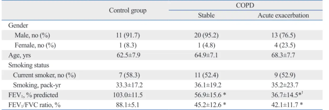
Table 1 presents the clinical and lung function variables of all participants. We included 17 patients with exacerbation of COPD, 21 patients with clinically stable COPD, and 12 con- trol groups in the study. Most subjects included in this study were male. The three groups of subjects were similar with re- gard to age, gender and smoking history. By design, the re- sults of pulmonary function test were normal in control groups. FEV
1was significantly decreased in patients with ex- acerbation of COPD compared to the stable COPD patients.
CD4
+and CD8
+T cells in peripheral blood
The percentage of CD8
+T cells was significantly increased in the peripheral blood of patients with exacerbation of COPD compared to stable COPD and control groups (exac- erbation COPD 30.8±11.7; stable COPD 21.1±9.5; control group 16.8±8.7, p=0.02), and there were significant differ- (MACS; Miltenyi Biotec, Bergisch Gladbach, Germany).
Usually, MACS did not alter structure, function, or activity status of labeled cells and did not influence flow cytometry.
Assessment of T lymphocytes apoptosis
To determine the apoptosis, we used flow cytometry (FACS Calibur; Becton Dickinson, UK) with double labeling with annexin V (Becton Dickinson; Plymouth, UK) and 7-amino- actinomycin (7 AAD) (Becton Dickinson; Plymouth, UK).
Briefly, the isolated lymphocytes were washed and centri- fuged in binding buffer [0.1 M HEPES/NaOH (pH 7.4), 1.4M NaCl, 25mM CaCl
2] and the pellet was resuspended in 5 mL of annexin V, 5 mL of 7AAD and 100 mL of binding buffer, followed by incubation for 15 minutes at room tem- perature in dark. Measurement was performed by flow cy- tometry within 15 minutes. We collected 20,000 cells in each sample using CellQuest software (Becton Dickinson; Moun- tain View, CA, USA), and results were expressed as a per- centage of cells exhibiting positive fluorescence.
Measurement of serum IL-6, IL-8 and TNF-α level For the measurement of IL-6, IL-8 and TNF-α, blood was collected in plastic tubes containing EDTA (Becton Dickin- son; Plymouth, UK) and was centrifuged within an hour (1,000×g for 10 min). The supernatants were stored at -70°C until analysis. Cytokines were measured by a commercially available sandwich enzyme-linked immunoassay (R&D systems; Minneapolis, MN, USA). The results were given as pg/mL.
Statistical analysis
A statistical software package (SPSS, version 17.0; SPSS
Table 1. Demographic Characteristics of the Subjects
Control group COPD
Stable Acute exacerbation
Gender
Male, no (%) 11 (91.7) 20 (95.2) 13 (76.5)
Female, no (%) 1 (8.3) 1 (4.8) 4 (23.5)
Age, yrs 62.5±7.9 64.9±7.1 68.3±7.7
Smoking status
Current smoker, no (%) 7 (58.3) 11 (52.4) 9 (52.9)
Smoking, pack-yr 33.3±17.2 36.1±19.2 35.2±23.7
FEV1, % predicted 103.0±11.5 56.9±15.6 * 36.7±14.5*†
FEV1/FVC ratio, % 88.1±5.1 45.2±12.6 * 42.1±11.7 *
Values are expressed as mean±SD.
FEV1, forced expiratory volume in one second; FVC, forced vital capacity.
*p<0.05 compared with control groups.
†p<0.05 compared with stable COPD.
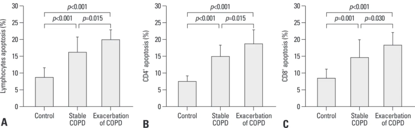
p=0.015). Apoptosis of CD4
+T cells was significantly in- creased in patients with exacerbation of COPD compared with stable COPD (18.6±4.4% vs. 14.9±4.5%; p=0.015).
Also, apoptosis of CD8
+T cells was significantly increased in patients with exacerbation of COPD compared with sta- ble COPD (18.3±3.7% vs. 14.5±5.4%; p=0.030).
Serum levels of cytokines
Serum levels of IL-6 were significantly increased in patients with exacerbation of COPD compared with the stable COPD and control groups [median, IQR, expressed in pg/
mL: exacerbation COPD 19.67 (13.55-23.25); stable COPD 6.58 (3.19-9.54); control group 2.86 (2.35-4.62), ANOVA p<0.001] (Fig. 2A). Serum IL-8 levels were significantly higher in patients with exacerbation of COPD than in the stable COPD and control groups [median, IQR, expressed in pg/ml: exacerbation COPD 87.87 (70.31-91.51); stable COPD 30.38 (27.46-39.83); control group 27.36 (26.08- 32.20), ANOVA p<0.001] (Fig. 2B). However, no differenc- es were observed in IL-6 and IL-8 levels between the stable COPD and control groups. Serum levels of TNF-α were sig- ences between the stable COPD and control groups. The per-
centage of blood CD4
+T cells decreased in patients with ex- acerbation of COPD compared to stable COPD and control groups (exacerbation COPD 69.1±14.5; stable COPD 78.8±10.2; control group 83.1±15.8, p=0.03), although there was no significant difference between the stable COPD and control groups. The ratio of CD4
+/CD8
+T cells was signifi- cantly decreased in patients with exacerbation of COPD compared to stable COPD and control groups (exacerbation COPD 2.0±1.6; stable COPD 3.4±1.7; control group 4.7±3.9, p=0.01), and there was significant difference between the sta- ble COPD and control groups.
Quantitation of apoptosis by flow cytometry
Fig. 1 shows the percentage of T lymphocytes, CD 4
+and CD 8
+T cells in the three groups studied. There was signifi- cantly increased apoptosis of T lymphocytes, CD4
+and CD8
+T cells in COPD patients (stable or exacerbated COPD) compared with control groups. Apoptosis of lym- phocytes was significantly higher in patients with exacerba- tion of COPD than stable COPD (19.8±3.1% vs. 16.1±4.7%;
Fig. 1. Mean apoptosis percentage of peripheral blood T lymphocytes (A), CD 4+ T cells (B), and CD 8+ T cells (C) in control groups, stable COPD, and exacer- bation of COPD measured by flow cytometry. COPD, chronic obstructive pulmonary disease.
Fig. 2. The serum levels of IL-6 (A), IL-8 (B), and TNF-α (C) in control groups, stable COPD, and exacerbation of COPD. Serum cytokine levels are expressed as the median value and interquartile range. •, outlier extending >1.5 box-lengths from the edge of the box. IL-6, interleukin-6; IL-8, interleukin-8; TNF-α, tumor necrosis factor-alpha. COPD, chronic obstructive pulmonary disease.
0 0 0
5 5 5
10 10 10
15 15 15
20 20 20
25 25 25
30 30 30
Lymphocytes apoptosis (%) CD4+ apoptosis (%) CD8+ apoptosis (%)
Control Stable Control Control
COPD Stable
COPD Stable
Exacerbation COPD
of COPD Exacerbation
of COPD Exacerbation
of COPD
p<0.001 p<0.001 p<0.001
p<0.001 p=0.015 p<0.001 p=0.015 p=0.001 p=0.030
0
20 0
50 100 150 200 250 300 350
40 60 80 100 120
0 10 20 30 40 50 60 70
IL-6 (pg/mL) IL-8 (pg/mL) TNF-α (pg/mL)
Control Stable Control Control
COPD Stable
COPD Stable
Exacerbation COPD
of COPD Exacerbation
of COPD Exacerbation
of COPD p<0.001
p<0.001 p<0.001
p=0.788
p=0.981 p=0.021
p<0.001
p<0.001 p<0.001
A
A
B
B
C
C
apoptotic stimuli in the airways or from apoptotic stimuli in the peripheral blood. Secondary necrosis may result in- creased apoptotic T-cells in the lung that has a pro-inflam- matory effect. Furthermore, defective immune response to infective organisms can be caused by increased T-cell apop- tosis, which contributes to the high frequency of infections seen in COPD.
20However, increased apoptosis of peripher- al blood T lymphocytes does not explain clearly its causal relationship with the pathogenesis of COPD. Hodge, et al.
20demonstrated increased propensity of peripheral blood T cells in COPD to undergo apoptosis. Recent study shows 87% increase in the apoptosis of airway epithelial cells and 103% increase of T lymphocytes apoptosis in bronchial brushings and bronchoalveolar lavage in COPD patients compared to non-smoking controls.
21On the contrary, how- ever, others did not find a significant difference in apoptotic alveolar wall cells in the lungs from smokers without em- physema compared to smokers with emphysema.
5In the present study, we found increased apoptosis of T lympho- cytes from peripheral blood in COPD patients compared to control groups.
The frequent occurrence of exacerbations is an important feature of COPD. There is enhancement of both local air- way and systemic inflammation during exacerbations. Spu- tum, serum, urine and exhaled air samples have been ana- lyzed during acute exacerbations.
22-27In our study, serum levels of IL-6, IL-8 and TNF-α were significantly higher in patients with exacerbation of COPD compared to stable COPD patients. Apoptosis can be regulated by several cy- tokines which are involved in the local and systemic in- flammation during exacerbation.
11-15The report from Duni- can, et al.
14suggested that TNF-α stimulates neutrophils apoptosis, while prolonged exposure has an inhibitory ef- nificantly increased in patients with exacerbation of COPD
compared with stable COPD and control groups. Also in- creased levels of TNF-α were observed in stable COPD pa- tients compared with control groups [median, IQR, ex- pressed in pg/mL: exacerbation COPD 139.33 (105.98- 175.17); stable COPD 57.31 (29.38-109.03); control group 26.05 (21.67-29.07), ANOVA p<0.001] (Fig. 2C).
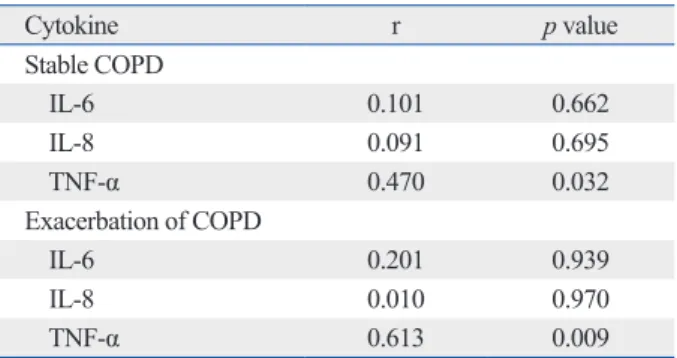
Correlation of apoptotic lymphocytes with serum cytokines
Only TNF-α presented a positive correlation with apoptotic lymphocytes in patients with stable COPD (r=0.470, p=
0.032) and exacerbation of COPD (r=0.613, p=0.009). Se- rum levels of IL-6 and IL-8 were not correlated with apop- totic lymphocytes in patients with stable and exacerbation of COPD (Table 2).
DISCUSSION
The pathophysiology of COPD includes inflammation, im- balance of protease and anti-protease, oxidative stress, and apoptosis. Abnormal apoptotic events have been demon- strated in epithelial and endothelial cells as well as in in- flammatory cells including neutrophils and lymphocytes in lungs with COPD patients. The main finding of our present study was that, compared to stable COPD, circulating apop- totic lymphocytes, CD 4
+and CD 8
+T cells were significant- ly increased in patients with exacerbation of COPD. TNF-α presented a positive correlation with apoptotic lymphocytes in patients with exacerbation of COPD.
COPD is currently regarded as a multi-component dis- ease with systemic manifestations in addition to local pul- monary inflammation. This inflammation leads to recurring cycles of injury and repair, and disorders in the repair pro- cess can lead to tissue remodeling with altered structure and function.
1,2Apoptotic cells are rarely seen in the normal hu- man lung,
19suggesting either low rates of cell death or a rapid removal process. This are increasing evidences to in- dicate that disturbance of the balance between apoptosis and proliferation in lung tissue contributes to the pathogen- esis of COPD.
3-7The increased numbers of T-cells may de- rived from local proliferation of T-cells in the lung of COPD or enhanced trafficking from the bloodstream. This process develops also from the airways to the bloodstream. There- fore, it is possible that increased apoptosis of T-cells in the peripheral blood in COPD may result either from local
Table 2. Correlation between the Percentage of Apoptotic Lymphocytes and Serum Levels of IL-6, IL-8, and TNF- α in Patients with Stable and Exacerbation of COPD
Cytokine r p value
Stable COPD
IL-6 0.101 0.662
IL-8 0.091 0.695
TNF-α 0.470 0.032
Exacerbation of COPD
IL-6 0.201 0.939
IL-8 0.010 0.970
TNF-α 0.613 0.009
IL-6, interleukin-6; IL-8, interleukin-8; TNF-α, tumor necrosis factor-alpha;
r, Spearman’s correlation coefficient; COPD, chronic obstructive pulmonary disease.
na A, et al. Activated T-lymphocytes and macrophages in bronchi- al mucosa of subjects with chronic bronchitis. Am Rev Respir Dis 1993;147:301-6.
4. Pabst R, Binns RM. Lymphocytes migrate from the bronchoalve- olar space to regional bronchial lymph nodes. Am J Respir Crit Care Med 1995;151:495-9.
5. Majo J, Ghezzo H, Cosio MG. Lymphocyte population and apop- tosis in the lungs of smokers and their relation to emphysema. Eur Respir J 2001;17:946-53.
6. Segura-Valdez L, Pardo A, Gaxiola M, Uhal BD, Becerril C, Sel- man M. Upregulation of gelatinases A and B, collagenases 1 and 2, and increased parenchymal cell death in COPD. Chest 2000;117:
684-94.
7. Saetta M, Di Stefano A, Turato G, Facchini FM, Corbino L, Mapp CE, et al. CD8+ T-lymphocytes in peripheral airways of smokers with chronic obstructive pulmonary disease. Am J Respir Crit Care Med 1998;157:822-6.
8. Degterev A, Boyce M, Yuan J. A decade of caspases. Oncogene 2003;22:8543-67.
9. Muzio M, Chinnaiyan AM, Kischkel FC, OʼRourke K, Shevchen- ko A, Ni J, et al. FLICE, a novel FADD-homologous ICE/CED-3- like protease, is recruited to the CD95 (Fas/APO-1) death--induc- ing signaling complex. Cell 1996;85:817-27.
10. Hirata H, Takahashi A, Kobayashi S, Yonehara S, Sawai H, Oka- zaki T, et al. Caspases are activated in a branched protease cascade and control distinct downstream processes in Fas-induced apopto- sis. J Exp Med 1998;187:587-600.
11. Gompertz S, O’Brien C, Bayley DL, Hill SL, Stockley RA.
Changes in bronchial inflammation during acute exacerbations of chronic bronchitis. Eur Respir J 2001;17:1112-9.
12. Song SH, Kim CH, Kwon SS, Kim YK, Kim KH, Moon HS, et al. Nuclear Factor-κB(NF-κB) activity and levels of IL-6, IL-8 and TNF-α in induced sputum in the exacerbation and recovery of COPD patients. Tuberc Respir Dis 2005;58:152-9.
13. Grutkoski PS, Graeber CT, Ayala A, Simms HH. Paracrine sup- pression of apoptosis by cytokine-stimulated neutrophils involves divergent regulation of NF-kappaB, Bcl-X(L), and Bak. Shock 2002;17:47-54.
14. Dunican AL, Leuenroth SJ, Grutkoski P, Ayala A, Simms HH.
TNFalpha-induced suppression of PMN apoptosis is mediated through interleukin-8 production. Shock 2000;14:284-8.
15. Biffl WL, Moore EE, Moore FA, Barnett CC Jr. Interleukin-6 sup- pression of neutrophil apoptosis is neutrophil concentration de- pendent. J Leukoc Biol 1995;58:582-4.
16. Schmidt-Ioanas M, Pletz MW, de Roux A, Lode H. Apoptosis of peripheral blood neutrophils in COPD exacerbation does not cor- relate with serum cytokines. Respir Med 2006;100:639-47 17. Rabe KF, Hurd S, Anzueto A, Barnes PJ, Buist SA, Calverley P, et
al. Global strategy for the diagnosis, management, and prevention of chronic obstructive pulmonary disease: GOLD executive sum- mary. Am J Respir Crit Care Med 2007;176:532-55.
18. Anthonisen NR, Manfreda J, Warren CP, Hershfield ES, Harding GK, Nelson NA. Antibiotic therapy in exacerbations of chronic obstructive pulmonary disease. Ann Intern Med 1987;106:196- 19. Kasahara Y, Tuder RM, Cool CD, Lynch DA, Flores SC, Voelkel 204.
NF. Endothelial cell death and decreased expression of vascular endothelial growth factor and vascular endothelial growth factor receptor 2 in emphysema. Am J Respir Crit Care Med 2001;163:
737-44.
fect mediated by the anti-apoptotic chemokine IL-8. A study of Biffl, et al.
15showed that IL-6 can also delay neu- trophil apoptosis. The uptake of apoptotic neutrophils may not only suppress the release of proinflammatory agents, such as IL-1β, IL-8, and TNF, but also increase macro- phage release of agents, such as transforming growth factor β, and prostaglandin E2, that have suppressive influences on the inflammatory response.
28,29A recent study revealed a significant reduction of percentage of apoptotic neutrophils at the development of COPD exacerbation. Apoptotic neu- trophils are not related to serum and sputum levels of IL-6, IL-8 and TNF-α.
16However, to our best knowledge, little ev- idence exists concerning the regulation of the circulating lymphocytes apoptosis in patients with exacerbation of COPD. Our present study revealed a significantly increased percentage of apoptotic circulating lymphocytes in patients with exacerbation of COPD compared to stable COPD pa- tients. Only TNF-α presented a positive correlation with apop- totic lymphocytes in patients with exacerbation of COPD.
In summary, this study demonstrated an increased per- centage of apoptotic lymphocytes, CD 4
+, and CD 8
+T cells from the peripheral blood in patients with exacerbation of COPD compared with stable COPD. Serum levels of IL-6, IL-8, and TNF-α were significantly increased in patients with exacerbation of COPD compared with stable COPD.
Only TNF-α showed a positive correlation with apoptotic lymphocytes in patients with exacerbation of COPD. Based on these findings, we postulate that increased apoptotic lym- phocytes may be associated with upregulation of TNF-α in the peripheral blood of patients with acute exacerbation of COPD.
ACKNOWLEDGEMENTS
This work was supported by the Chonnam National Univer- sity Hospital Research Institute of Clinical Medicine Grant CRI08058-1.
REFERENCES
1. Pauwels RA, Rabe KF. Burden and clinical features of chronic obstructive pulmonary disease (COPD). Lancet 2004;364:613-20.
2. Barnes PJ, Shapiro SD, Pauwels RA. Chronic obstructive pulmo- nary disease: molecular and cellular mechanisms. Eur Respir J 2003;22:672-88.
3. Saetta M, Di Stefano A, Maestrelli P, Ferraresso A, Drigo R, Pote-
COPD and death from pulmonary infection. Eur Respir J 1995;8:
1333-8.
26. Agustí AG, Villaverde JM, Togores B, Bosch M. Serial measure- ments of exhaled nitric oxide during exacerbations of chronic ob- structive pulmonary disease. Eur Respir J 1999;14:523-8.
27. Dekhuijzen PN, Aben KK, Dekker I, Aarts LP, Wielders PL, van Herwaarden CL, et al. Increased exhalation of hydrogen peroxide in patients with stable and unstable chronic obstructive pulmonary disease. Am J Respir Crit Care Med 1996;154:813-6.
28. Haslett C. Granulocyte apoptosis and its role in the resolution and control of lung inflammation. Am J Respir Crit Care Med 1999;
160:S5-11.
29. Fadok VA, Bratton DL, Konowal A, Freed PW, Westcott JY, Hen- son PM. Macrophages that have ingested apoptotic cells in vitro inhibit proinflammatory cytokine production through autocrine/
paracrine mechanisms involving TGF-beta, PGE2, and PAF. J Clin Invest 1998;101:890-8.
20. Hodge SJ, Hodge GL, Reynolds PN, Scicchitano R, Holmes M.
Increased production of TGF-beta and apoptosis of T lymphocytes isolated from peripheral blood in COPD. Am J Physiol Lung Cell Mol Physiol 2003;285:L492-9.
21. Hodge S, Hodge G, Holmes M, Reynolds PN. Increased airway epithelial and T-cell apoptosis in COPD remains despite smoking cessation. Eur Respir J 2005;25:447-54.
22. Bhowmik A, Seemungal TA, Sapsford RJ, Wedzicha JA. Relation of sputum inflammatory markers to symptoms and lung function changes in COPD exacerbations. Thorax 2000;55:114-20.
23. Crooks SW, Bayley DL, Hill SL, Stockley RA. Bronchial inflam- mation in acute bacterial exacerbations of chronic bronchitis: the role of leukotriene B4. Eur Respir J 2000;15:274-80.
24. Hurst JR, Donaldson GC, Perera WR, Wilkinson TM, Bilello JA, Hagan GW, et al. Use of plasma biomarkers at exacerbation of chronic obstructive pulmonary disease. Am J Respir Crit Care Med 2006;174:867-74.
25. Prescott E, Lange P, Vestbo J. Chronic mucus hypersecretion in