INTRODUCTION
Acute leukemia (AL), one of the most common malignant tu- mors in children, is classified as acute lymphoblastic leukemia (ALL) and acute myeloid leukemia (AML), among which ALL
accounts for 60–70% and AML for 30–40% of all AL cases.
1,2Due to aggravated environmental and air pollution, the incidence of childhood AL has increased in recent years.
3AL poses a seri- ous threat to the lives of patients, and its treatment remains an urgent problem in clinic practice.
microRNAs (miRNAs) are a class of small endogenous non- coding RNAs with 19–25 nucleotides in length that modulate gene expression at the post-transcriptional level via base pair- ing with 3'-untranslated regions (3'-UTRs) of their target genes.
4,5miRNAs play an extremely important role in biological pro- cesses of multiple tumors, such as cell proliferation, apopto- sis, migration, cycle and carcinogenesis.
6,7To date, numerous studies have reported that abnormal expressions of miRNAs are involved in the pathogenesis and development of AL. For example, Huang, et al.
8have reported that miR-519 promotes
microRNA-146a Promotes Growth of Acute Leukemia Cells by Downregulating Ciliary Neurotrophic Factor Receptor and Activating JAK2/STAT3 Signaling
Lei Wang
1, Hongyan Zhang
2, and Donghong Lei
31
Department of Pediatrics, Jinan Second Maternal and Child Health Hospital, Jinan, Shandong;
2
Department of Pediatrics, Jinan First People's Hospital, Jinan, Shandong;
3
Department of Pediatrics II, Yulin First Hospital, Suide, Shaanxi, China.
Purpose: Acute leukemia (AL) is classified as acute lymphoblastic leukemia (ALL) and acute myeloid leukemia (AML). This study aimed to investigate the effect of miR-146a on childhood AL and its underlying molecular mechanisms.
Materials and Methods: Bone marrow samples were obtained from 39 AL children and 10 non-cancer controls. The expressions of miR-146a and ciliary neurotrophic factor receptor (CNTFR) were detected by quantitative real-time polymerase chain reaction (qRT-PCR) in ALL and AML pediatric patients, as well as ALL (Jurkat) and AML (HL-60) cells. Correlations between miR-146a and clinical indicators were explored. A targeting relationship between miR-146a and CNTFR was detected by dual luciferase re- porter gene assay. Cell proliferation, apoptosis, migration, and invasion of Jurkat and HL-60 cells were measured by MTT assay, flow cytometry, and transwell assay, respectively. LIF expression was detected by qRT-PCR in Jurkat and HL-60 cells. The expression of p-JAK2, JAK2, p-STAT3, and STAT3 in HL-60 cells was measured by Western blot.
Results: miR-146a was increased in ALL and AML pediatric patients, while CNTFR was decreased. miR-146a expression was asso- ciated with immunophenotype, karyotype, fusion gene, and SIL-TAL1. CNTFR was a target gene of miR-146a. miR-146a could pro- mote cell proliferation, migration, and invasion, as well as inhibit cell apoptosis in Jurkat and HL-60 cells by downregulating CNT- FR. Meanwhile, miR-146a inhibited the expression of LIF and activated JAK2/STAT3 pathway by downregulating CNTFR.
Conclusion: miR-146a could promote the proliferation, migration, and invasion and inhibit the apoptosis of AL Jurkat and HL-60 cells by downregulating CNTFR and activating the JAK2/STAT3 pathway.
Key Words: miR-146a, acute leukemia, CNTFR, JAK/STAT pathway
pISSN: 0513-5796 · eISSN: 1976-2437
Received: May 7, 2019 Revised: July 17, 2019 Accepted: July 30, 2019
Corresponding author: Donghong Lei, BA, Department of Pediatrics II, Yulin First Hospital, No. 59, Wenhua Road, Suide, Shaanxi 718000, China.
Tel: 86-0912-5641024, Fax: 86-0912-5641024, E-mail: [email protected]
•The authors have no potential conflicts of interest to disclose.
© Copyright: Yonsei University College of Medicine 2019
This is an Open Access article distributed under the terms of the Creative Com- mons Attribution Non-Commercial License (https://creativecommons.org/licenses/
by-nc/4.0) which permits unrestricted non-commercial use, distribution, and repro- duction in any medium, provided the original work is properly cited.
Yonsei Med J 2019 Oct;60(10):924-934
https://doi.org/10.3349/ymj.2019.60.10.924
AML HL-60 cell proliferation via downregulating the expres- sion of RNA-binding protein human antigen R. miR-34a could enhance cell apoptosis and suppress autophagy by targeting HMGB1 in AML cells.
7miR-146a has been identified to play a dual oncogenic and anticancer role in leukemia,
9because it is overexpressed in childhood ALL and AML,
10but downregu- lated in adult AML,
11However, the precise roles of miR-146a in AL and its potential molecular mechanism remain to be fur- ther investigated.
Leukemic inhibitory factor (LIF), a member of the interleu- kin (IL)-6 family, is involved in the developmental processes of leukemia,
12Ciliary neurotrophic factor receptor (CNTFR) plays an important role in the pathogenesis of blood diseases by binding LIF.
12The downregulation of CNTFR could facili- tate Janus-activated kinases/signal transducer and activator of transcription (JAK/STAT) signaling.
13Ip, et al.
12indicated that miR-708 could exert oncogenic effects in leukemia by reg- ulating CNTFR and activating JAK/STAT pathway. However, whether miR-146a affects childhood AL through CNTFR-me- diated JAK/STAT pathway is unknown.
In this research, we investigated the effects of miR-146a on childhood AL and its related molecular mechanisms. Our re- sults suggested that miR-146a could promote the prolifera- tion, migration, and invasion of Jurkat and HL-60 cells, as well as inhibit their apoptosis, by downregulating CNTFR and acti- vating the JAK2/STAT3 pathway. The findings of our study may provide a new theoretical foundation for deeply exploring the treatment of childhood AL.
MATERIALS AND METHODS
Clinical samples
From January 2015 to December 2018, there were 39 newly di- agnosed AL pediatric patients (28 ALL and 11 AML) recruited for this study, and the inclusion criteria followed the AL di- agnosis criteria in the 2016 edition of the World Health Orga- nization.
14Bone marrow samples were obtained from 39 AL children and 10 non-cancer control children treated in the hematology department of Yulin First Hospital. The non-can- cer controls were children with no evidence of malignant he- matologic diseases. Monocytes were isolated from 5 mL of heparinized bone marrow by density gradient centrifugation at 500× g for 30 min using Ficoll Paque media (Robbins Scientif- ic, Sunnyvale, CA, USA). Subsequently, the monocytes were washed three times using Hank’s balanced salt solution (Ther- mo, Waltham, MA, USA). The project was approved by the Eth- ics Committee of Yulin First Hospital (2019-3), and it was per- formed in accordance with the Declaration of Helsinki. All recruited patients and participants provided signed informed consent.
Cell cultures
The human ALL cell line Jurkat, human AML cell line HL-60, and human embryonic kidney cell line HEK-293T were ob- tained from the American Type Culture Collection (ATCC, Manassas, VA, USA) and cultured in 5% CO
2at 37°C. Jurkat and HL-60 cells were cultured in RPMI-1640 (Gibco, Carlsbad, CA, USA) complemented with 10% fetal bovine serum (FBS, Gibco, Carlsbad, CA, USA) and 1% streptomycin/penicillin (Gibco). HEK-293T cells were grown in Dulbecco Modified Ea- gle Medium (DMEM, Gibco, Carlsbad, CA, USA) supplement- ed with 10% FBS and 1% streptomycin/penicillin.
Dual luciferase reporter gene assay
Using the TargetScan website, we found that CNTFR was a tar- get gene of miR-146a. We amplified the 3'-UTR of CNTFR con- taining the miR-146a binding site and then cloned the 3'-UTR fragment into Psi-CHECK2 reporter vector (Promega, Madison, WI, USA) to construct wild Psi-CHECK2-WT-CNTFR-3'-UTR (CNTFR-wt) and mutant Psi-CHECK2-MUT-CNTFR-3'-UTR (CNTFR-mut). For luciferase assay, miR-146a mimics or miR- 146a negative control mimics was co-transfected with report- ers plasmids into HEK-293T cells by using Lipofectamine 2000 (Invitrogen, Carlsbad, CA, USA). Based on the differences in transfection sequences, cells were grouped as follows: mutant- type (MT)+mimics group (transfected with mutant-type se- quences and miR-146a mimics), MT+negative control (NC) group (transfected with mutant-type sequences and miR-146a negative control mimics), wild-type (WT)+mimics group (trans- fected with wild-type sequences and miR-146a mimics), and WT+NC group (transfected with wild-type sequences and miR- 146a negative control mimics). The luciferase activity was mea- sured using dual luciferase kits (Promega) after 48 h transfec- tion.
Cell transfection assay
Jurkat and HL-60 cells were added to 6-well plates and incu-
bated at 37°C for 24 h. When cells reached 80% confluence in
the plate well, anti-miR-146a (antisense miR-146a oligonucle-
otide, Thermo), anti-miR-146a negative control (NC, Thermo),
CNTFR-siRNA (QIAGEN, Duesseldorf, Germany), CNTFR-siR-
NA negative control (QIAGEN), miR-146a mimics (GenePhar-
ma, Shanghai, China), miR-146a mimics negative control (Ge-
nePharma), and miR-146a inhibitor (GenePharma) were
cotransfected into Jurkat and HL-60 cells using Lipofectamine
®2000 Reagent (Invitrogen). The transfected Jurkat and HL-60
cells were randomly assigned to eight groups: Mock group (no
treatment), mimics-NC group (transfected with miR-146a mim-
ics negative control), miR-146a mimics group (transfected
with miR-146a mimics), miR-146a inhibitor group (transfect-
ed with miR-146a inhibitor), anti-miR-NC+siNC group (trans-
fected with anti-miR-146a NC and CNTFR-siRNA NC), anti-
miR-146a+siNC group (transfected with anti-miR-146a and
CNTFR-siRNA NC), anti-miR-NC+siCNTFR group (transfect-
ed with anti-miR-146a NC and CNTFR-siRNA), and anti-miR- 146a+siCNTFR group (transfected with anti-miR-146a and CNTFR-siRNA). Finally, all cells were cultured in 37°C incuba- tor for 48 h.
Cell proliferation assay
Cell proliferation of Jurkat and HL-60 cells was measured by 3-(4, 5-dimethylthiazol-2-yl)-2, 5-diphenyltetrazolium bro- mide (MTT) colorimetric assay (Sigma, St. Louis, MO, USA).
In brief, transfected Jurkat and HL-60 cells were seeded into 96-well plates at a density of 5×10
3cells/well. At different time points (0, 24, 48, and 72 h), the culture medium was removed, and 20 μL of MTT (5 mg/mL) was added into each well. After incubation at 37°C for 4 h, the MTT was removed, and absor- bance at 495 nm was measured on a microplate reader (Bio- Rad, Hercules, CA, USA).
Cell apoptosis assay
The apoptosis of Jurkat and HL-60 cells was detected by An- nexin V-FITC and propidium iodide apoptosis detection kits (Invitrogen). Briefly, at 48 h after transfection, the Jurkat and HL-60 cells were collected, washed three times with phosphate buffer saline, and re-suspended in 1×binding buffer. Then, Annexin V-FITC and propidium iodide were utilized to stain Jurkat and HL-60 cells for 15 minutes at room temperature. Fi- nally, apoptotic cells were analyzed using a flow cytometer (BD Biosciences, San Jose, CA, USA).
Transwell assay
Transwell assay was conducted using transwell chambers (Corning, New York, NY, USA) pre-coated with Matrigel (BD Biosciences). The transfected Jurkat and HL-60 cells (1×10
5cells/well) were collected and inoculated to the upper cham- ber. Then, 500 μL of RPMI-1640 containing 20% FBS was add- ed into the lower chamber. After incubation for 24 h at 37°C, the non-migratory cells were carefully removed. Then, the mi- grated cells were fixed with 4% paraformaldehyde for 20 min and stained with 0.5% crystal violet dye (Sigma) for 30 min.
The number of migrating cells was counted under an optical microscope at 200× magnification.
Real-time fluorogenic PCR assay
As recommendation of the supplier, total RNA of bone mar- row tissue from children with ALL and AML was extracted by using TRIzol (Invitrogen). In addition, total RNA of Jurkat and HL-60 cells was extracted. Then, 500 ng of RNA was reverse- transcribed into cDNA by Revert Aid First Strand cDNA Syn- thesis Kits (Thermo) and measured using quantitative real- time polymerase chain reaction (qRT-PCR) (Bio-Rad) with SYBR green qPCR Master Mix (Thermo). β-actin was em- ployed as the internal control in the quantitative analysis of CFTFR and LIF expressions and U6 as the internal control in the quantitative analysis of miR-146a expression. Primers
used for qRT-PCR analysis were as follows: miR-146a (for- ward) : 5'-CCGCCGTGAGAACTGAATTCCA-3', (reverse):
5'-GTGCAGGGTCCGAGGT-3'; U6 (forward): 5'-CTCGCTTC- GGCAGCACA-3', (reverse): 5'-AACGCTTCACGAATTTGC- GT-3'; CNTFR (forward): 5'-AAGGACCCAGCCCTCAAGAA-3', (reverse): 5'-TGCTCTAGAGCAATACAGCAAGTTACC-3'; LIF (forward): 5'-CAGCACCACTGAATCACAGA-3', (reverse):
5'-AGACACGTAAGGAAAACGCATTA-3'; β-actin (forward):
5'-ACACCTTCTACAATGAGCTG-3', (reverse): 5'-CTGCTT- GCTGATCCACATCT-3'.
Western blot analysis
Total proteins were extracted by lysis buffer. Protein samples (50 μg) were subjected to 10% sodium dodecyl sulfate-poly- acrylamide gel electrophoresis and then transferred to polyvi- nylidene difluoride membrane. The membranes were blocked with 5% skimmed milk, followed by overnight incubation at 4°C with primary antibody (1:500; JAK2, 1:1000; p-JAK2, 1:500;
STAT3, 1:1000; p-STAT3, 1:500; GAPDH, 1:3000, Abcam, Cam- bridge, MA, USA). Afterwards, peroxidase-labeled secondary antibody (anti-rabbit IgG, 1:5000, Cell Signal, Danvers, MA, USA) was used for incubation for 1 h at room temperature. The protein blots were visualized with an enhanced chemilumi- nescence kit. Finally, the densities of Western blot bands were analyzed using Quantity One 1-D Analysis Software (Bio-Rad).
Statistical analysis
All statistical analyses were performed using SPSS 22.0 soft- ware (IBM Corp., Armonk, NY, USA). The results are present- ed as means±standard deviations. Differences among multiple groups were analyzed by one-way ANOVA followed by Tukey’s post hoc test, and data for two groups were compared using Student’s t test. All experiments were repeated three times. p<
0.05 was considered to be statistically significant.
RESULTS
Expression of miR-146a upregulated and correlated with poor outcomes in children with AL
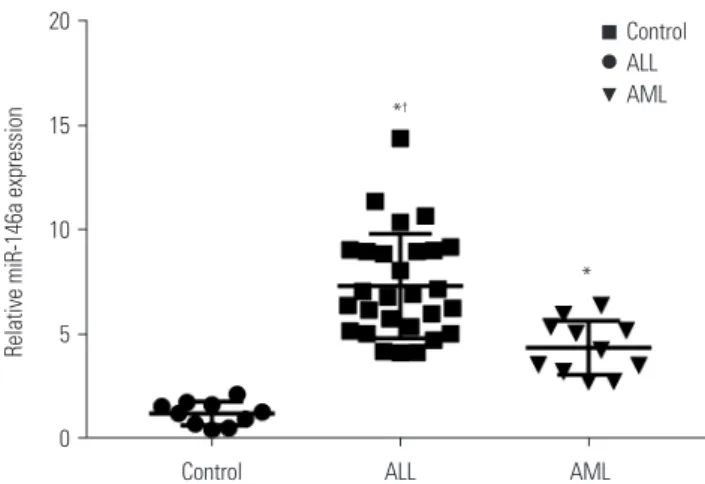
The results of qRT-PCR showed that the mRNA expression of miR-146a was significantly increased in ALL and AML chil- dren, compared with controls (p<0.05). In addition, miR-146a mRNA expression in the ALL group was higher than that in the AML group (p<0.05) (Fig. 1). These results suggested that miR- 146a is highly expressed in children with AL.
Correlations between the expression of miR-146a and clini-
cal indicators in children with AL are shown in Table 1. The
results revealed that the expression of miR-146a is associated
with clinical outcomes, such as immunophenotype, karyotype,
fusion gene, and SIL-TAL1 gene expression (p<0.05). Other
clinic factors, such as age, sex, initial white blood cell counts,
MLL gene rearrangement, TEL-AML1, and initial lactate de-
hydrogenase levels had no significant associations with miR- 146a expression (p>0.05).
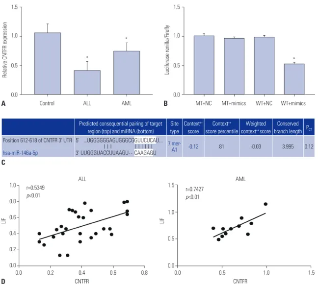
CNTFR a target gene of miR-146a
As shown in Fig. 2A, the mRNA expression levels of CNTFR were remarkedly lower in both ALL and AML groups than in the Control group (p<0.05). In addition, TargetScan predicted that the binding site of miR-146a to CNTFR was the 3'-UTR re- gion (Fig. 2B). According to luciferase reporter assay, we found that luciferase activity in the WT+mimics group was signifi- cantly lower than that in the WT+NC group, while the differ- ence in luciferase activity between the MT+NC and MT+mimics groups was not statistically significant (p<0.05) (Fig. 2C). In addition, we also found the expression of LIF was positively correlated with CNTFR expression in ALL and AML children (p<0.01) (Fig. 2D). Thus, we could confirm that miR-146a di- rectly regulates CNTFR expression.
miR-146a promotes cell proliferation and suppresses cell apoptosis of Jurkat and HL-60 cells via
downregulating CNTFR
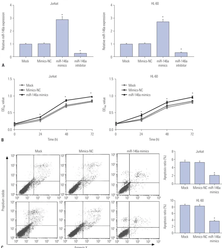
As shown in Fig. 3A, when compared with Mock and the mimics-NC group, the expression of miR-146a in both Jurkat and HL-60 cells was significantly increased in the miR-146a mimics group (p<0.05) and decreased in the miR-146a inhibi- tor group (p<0.05), suggesting that the transfection was suc- cessful. MTT assay showed that the proliferation ability of Jurkat and HL-60 cells in the miR-146a mimics group was markedly higher than that in the Mock and mimics-NC groups (p<0.05) (Fig. 3B). We also found that the apoptosis ability of Jurkat and HL-60 cells in miR-146a mimics group was lower than that in Mock and the mimics-NC group (p<0.05) (Fig. 3C).
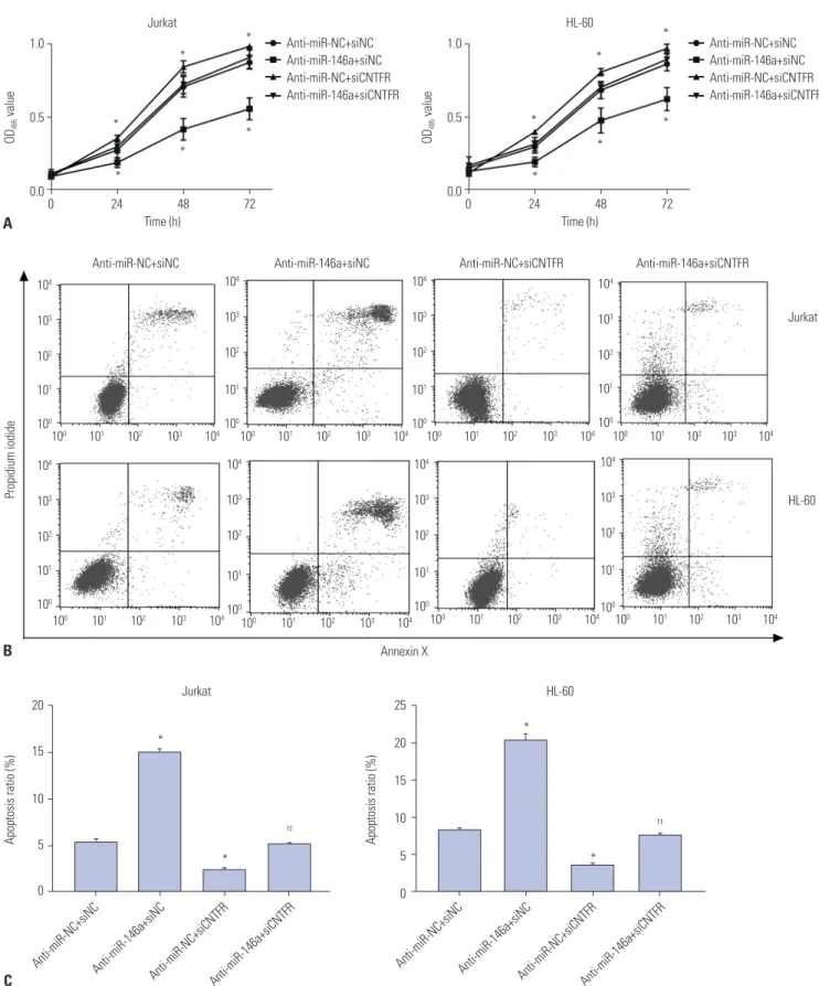
Moreover, Fig. 4A results showed that the cell proliferation ability of Jurkat and HL-60 was significantly decreased in the anti-miR-146a+siNC group, compared with the anti-miR-
NC+siNC group (p<0.05). When compared with the anti-miR- NC+siNC group, the cell proliferation abilities of Jurkat and HL- 60 cells were markedly increased in the anti-miR-NC+siCNTFR group (p<0.05). However, no significant difference was found between the anti-miR-146a+siCNTFR and anti-miR-NC+siNC groups (p>0.05). All results above indicated that miR-146a could promote cell proliferation of Jurkat and HL-60 cells via downregulating CNTFR.
The results of cell apoptosis assay showed that the cell apop- tosis of Jurkat and HL-60 was significantly increased in the an- ti-miR-146a+siNC group, compared with the anti-miR-NC+siNC group (p<0.05). However, the cell apoptosis of Jurkat and HL-60 was dramatically lower in the anti-miR-NC+siCNTFR group Fig. 1. The expression of miR-146a in acute lymphoblastic leukemia
(ALL) and acute myeloid leukemia (AML) pediatric patients. Data are presented as a mean±standard deviation. Experiments were performed three times. *p<0.05, vs. Control group;
†p<0.05, vs. AML group.
Table 1. Correlation between the Expression of miR-146a and Clinical Indicators in Children with AL
Clinical indicators Case
number miR-146a
expression p value
Sex 0.122
Male 20 5.80±2.29
Female 19 7.08±2.77
Age (yr) 0.220
<1 2 7.06±2.92
1–10 33 6.64±2.58
>10 4 4.30±1.98
Initial WBC (×10
9/L) 0.293
<50 19 5.93±1.89
50–100 7 7.55±3.69
>100 13 6.60±2.86
Immunophenotype 0.001
ALL 28 7.10±2.59
AML 11 4.46±1.23
Karyotype <0.001
Normal karyotype 24 7.65±2.50
Abnormal karyotype 15 4.46±1.08
Fusion gene 0.001
Non-detected 14 4.69±1.45
Abnormally detected 25 7.40±2.59
MLL gene rearrangement 0.420
Negative 27 6.20±2.75
Positive 12 6.94±2.18
SIL-TAL1 gene <0.001
Negative 31 5.55±1.88
Positive 8 9.83±2.39
TEL-AML1 gene 0.078
Negative 22 5.78±2.00
Positive 17 7.12±3.05
Initial LDH level 0.380
<500 U/L 23 6.12±2.58
≥500 U/L 16 6.87±2.61
AL, acute leukemia; WBC, white blood cell; ALL, acute lymphoblastic leuke- mia; AML, acute myeloid leukemia; LDH, lactate dehydrogenase.
Values are presented as mean±standard deviation unless otherwise indicated.
20
15
10
5
0
Relative miR-146a expression
Control ALL AML
*
†*
Control
ALL
AML
than in the anti-miR-NC+siNC group (p<0.05). When compared with anti-miR-146a+siNC, the cell apoptosis of Jurkat and HL- 60 cells was significantly suppressed in the anti-miR-146a+
siCNTFR group (p<0.05). In addition, the cell apoptosis of Jur- kat and HL-60 cells was markedly higher in the anti-miR-NC+
siCNTFR group than in the anti-miR-NC+siCNTFR group (p<
0.05) (Fig. 4B and C). All the results suggested that miR-146a could promote cell proliferation and suppress cell apoptosis of Jurkat and HL-60 cells via downregulating CNTFR.
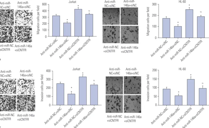
miR-146a increases cell migration and invasion of Jurkat and HL-60 cells by downregulating CNTFR The impact of miR-146a on cell migration and invasion of Jur- kat and HL-60 cells was assessed by transwell assay (Fig. 5).
From the results, we observed that counts of migratory and in- vasive Jurkat and HL-60 cells in the anti-miR-146a+siNC group were both significantly lower than those in the anti-miR-NC+
siNC group (p<0.05). In addition, dramatically higher counts of migratory and invasive Jurkat and HL-60 cells in the anti-miR- NC+siCNTFR group were found, compared with those in the anti-miR-NC+siNC group (p<0.05). Compared with anti-miR- 146a+siNC, the counts of migratory and invasive Jurkat and HL-60 cells were significantly increased in the anti-miR- 146a+siCNTFR group (p<0.05). Moreover, the counts of mi- gratory and invasive Jurkat and HL-60 cells was markedly low- er in the anti-miR-NC+siCNTFR group than in the anti-miR- NC+siCNTFR group (p<0.05). All of these suggested miR-146a could stimulate the migration and invasion of Jurkat and HL- 60 cells via downregulating CNTFR.
miR-146a inhibits the expression of LIF in Jurkat and HL-60 cells by downregulating CNTFR
The results of qRT-PCR showed that the mRNA expression of LIF in Jurkat and HL-60 cells was significantly greater in anti- 1.5
1.0
0.5
0.0
1.5
1.0
0.5
0.0
Control ALL AML MT+NC MT+mimics WT+NC WT+mimics
Relative CNTFR expression Luciferase renilla/Firefly
A B
1.0 0.8 0.6 0.4 0.2 0.0
1.5
1.0
0.5
0.0
0.0 0.2 0.4 0.6 0.8 0.0 0.5 1.0 1.5 r=0.5349
p<0.01 r=0.7427
p<0.01
CNTFR CNTFR
ALL AML
LIF LIF
D C
*
*
*
l Predicted consequential pairing of target
region (top) and miRNA (bottom) Site type
Context
++score
Context
++score percentile
Weighted context
++score
Conserved branch length P
CTPosition 612-618 of CNTFR 3' UTR
hsa-miR-146a-5p
5' ...UGGGGGGAGUGGGCGGUUCUCAU...
I I I I I I I I I 3' UUGGGUACCUUAAGU -- CAAGAGU
7 mer-
A1 -0.12 81 -0.03 3.995 0.12
Fig. 2. Ciliary neurotrophic factor receptor (CNTFR) is a target of miR-146a. (A) The expression of CNTFR was detected by quantitative real-time poly-
merase chain reaction (qRT-PCR) in acute lymphoblastic leukemia (ALL) and acute myeloid leukemia (AML) pediatric patients. (B) The binding target of
CNTFR and miR-146a was predicted by TargetScan. (C) Luciferase activity was measured by dual luciferase reporter gene assay. (D) The correlation of
leukemic inhibitory factor (LIF) and CNTFR in ALL and AML pediatric patients. Data are presented as a mean±standard deviation. Experiments were
performed three times. *p<0.05, vs. Control group and wild-type (WT)+negative control (NC) group. MT, mutant-type.
miR-146a+siNC group than in the anti-miR-NC+siNC group (p<0.05). On the contrary, LIF mRNA expression in Jurkat and HL-60 cells in the anti-miR-NC+siCNTFR group was markedly lower than that in the anti-miR-NC+siNC group (p<0.05). When
compared with anti-miR-146a+siNC, LIF mRNA expression in Jurkat and HL-60 cells was significantly lower than that in the anti-miR-146a+siCNTFR group (p<0.05). Furthermore, mRNA expression of LIF in Jurkat and HL-60 cells was markedly high-
Fig. 3. miR-146a promotes the proliferation and suppresses the apoptosis of Jurkat and HL-60 cells. (A) miR-146a expression was detected by quantita- tive real-time polymerase chain reaction (qRT-PCR) in Jurkat and HL-60 cells. (B) Proliferation of Jurkat and HL-60 cells measured by MTT assay. (C) Apoptosis of Jurkat and HL-60 cells detected by flow cytometry. Data are presented as a mean±standard deviation. Experiments were performed three times. *p<0.05, vs. Mock and mimics-negative control (NC) group. OD, optical density.
4
3
2
1
0
4
3
2
1
Mock 0 Mock
Jurkat HL-60
Mimics-NC miR-146a Mimics-NC
mimics miR-146a
mimics miR-146a
inhibitor miR-146a
inhibitor
Relative miR-146a expression Relative miR-146a expression
A
* *
* *
1.5
1.0
0.5
0.0
1.5
1.0
0.5
0.0
0 24 48 72 0 24 48 72
Time (h) Time (h)
Jurkat HL-60
OD
495value OD
495value
B
Mock Mimics-NC miR-146a mimics Mock
Mimics-NC miR-146a mimics
*
*
*
* *
*
8 6 4 2 0
10 8 6 4 2 0
Mock Mimics-NC
Mock Mimics-NC miR-146a
mimics
miR-146a mimics
Apoptosis ratio (%) Apoptosis ratio (%)
Mock Mimics-NC miR-146a mimics
Annexin X
Propidium iodide
10
010
110
210
310
410
010
110
210
310
410
010
110
210
310
410
010
110
210
310
410
010
110
210
310
410
010
110
210
310
4Jurkat
HL-60
*
*
C 10
410
310
210
110
010
410
310
210
110
010
410
310
210
110
010
410
310
210
110
010
410
310
210
110
010
410
310
210
110
01.0
0.5
0.0
1.0
0.5
0 24 48 72 0.0 0 24 48 72
Time (h) Time (h)
Jurkat HL-60
OD
495value OD
495value
A
Anti-miR-NC+siNC Anti-miR-146a+siNC Anti-miR-NC+siCNTFR Anti-miR-146a+siCNTFR
Anti-miR-NC+siNC Anti-miR-146a+siNC Anti-miR-NC+siCNTFR Anti-miR-146a+siCNTFR
* *
*
*
*
*
* *
*
*
*
*
Annexin X
Propidium iodide
10
010
110
210
310
410
010
110
210
310
410
010
110
210
310
410
010
110
210
310
410
010
110
210
310
410
010
110
210
310
410
010
110
210
310
410
010
110
210
310
4Jurkat
HL-60 Anti-miR-NC+siNC Anti-miR-146a+siNC Anti-miR-NC+siCNTFR Anti-miR-146a+siCNTFR
B
20
15
10
5
0
25 20 15
10 5 0
Anti-miR-NC+siNC Anti-miR-146a+siNC Anti-miR-NC+siCNTFR Anti-miR-146a+siCNTFR Anti-miR-NC+siNC Anti-miR-146a+siNC Anti-miR-NC+siCNTFR Anti-miR-146a+siCNTFR
Apoptosis ratio (%) Apoptosis ratio (%)
C
* *
* *
†‡ †‡
Jurkat HL-60
10
410
310
210
110
010
410
310
210
110
010
410
310
210
110
010
410
310
210
110
010
410
310
210
110
010
410
310
210
110
010
410
310
210
110
010
410
310
210
110
0Fig. 4. miR-146a promotes the proliferation and suppresses the apoptosis of Jurkat and HL-60 cells by downregulating ciliary neurotrophic factor re-
ceptor (CNTFR). (A) Proliferation of Jurkat and HL-60 cells measured by MTT assay. (B) Apoptosis of Jurkat and HL-60 cells detected by flow cytom-
etry. (C) Quantitative data of cell apoptosis ratio in Jurkat and HL-60 cells. Data are presented as a mean±standard deviation. Experiments were per-
formed three times. *p<0.05, vs. anti-miR-NC+siNC;
†p<0.05, vs. anti-miR-146a+siNC;
‡p<0.05, vs. anti-miR-NC+siCNTFR. NC, negative control; OD, optical
density.
er in the anti-miR-NC+siCNTFR group than in the anti-miR- NC+siCNTFR group (p<0.05) (Fig. 6), suggesting that miR- 146a could inhibit the expression of LIF in Jurkat and HL-60 cells by downregulating CNTFR.
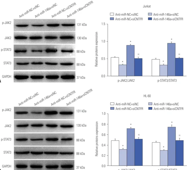
miR-146a activates JAK2/STAT3 signaling by downregulating CNTFR in Jurkat and HL-60 cells The results of Western blot assay (Fig. 7) showed that the ex- pressions of p-JAK2/JAK2 and p-STAT3/STAT3 in Jurkat and
600 400 300 200 100 0
400 300 200 100 0
300
200
100
0
200 150 100 50 0
Migration cells per field Invasion cells per field Migration cells per field Invasion cells per field
A
B Anti-miR- NC+siNC
Anti-miR- NC+siNC
Anti-miR- NC+siNC
Anti-miR- NC+siNC Anti-miR-
146a+siNC
Anti-miR- 146a+siNC
Anti-miR- 146a+siNC
Anti-miR- 146a+siNC Anti-miR-NC
+siCNTFR
Anti-miR-NC +siCNTFR
Anti-miR-NC +siCNTFR
Anti-miR-NC +siCNTFR Anti-miR-146a
+siCNTFR
Anti-miR-146a +siCNTFR
Anti-miR-146a +siCNTFR
Anti-miR-146a +siCNTFR Anti-miR-NC+siNC
Anti-miR-NC+siNC
Anti-miR-NC+siNC
Anti-miR-NC+siNC Anti-miR-146a+siNC
Anti-miR-146a+siNC
Anti-miR-146a+siNC
Anti-miR-146a+siNC Anti-miR-NC+siCNTFR
Anti-miR-NC+siCNTFR
Anti-miR-NC+siCNTFR
Anti-miR-NC+siCNTFR Anti-miR-146a+siCNTFR
Anti-miR-146a+siCNTFR
Anti-miR-146a+siCNTFR
Anti-miR-146a+siCNTFR
* *
*
* *
*
†‡
*
*
†‡
†‡
†‡
Jurkat
Jurkat
HL-60
HL-60
Fig. 5. miR-146a increases the migration and invasion of Jurkat and HL-60 cells by downregulating ciliary neurotrophic factor receptor (CNTFR). Cell mi- gration (A) and invasion (B) of Jurkat and HL-60 cells detected by transwell assay after crystal violet staining. Data are presented as a mean±standard deviation. Experiments were performed three times. Original magnification: ×100. *p<0.05, vs. anti-miR-NC+siNC;
†p<0.05, vs. anti-miR-146a+siNC;
‡p<0.05, vs. anti-miR-NC+siCNTFR. NC, negative control.
Fig. 6. miR-146a inhibits the expression of leukemic inhibitory factor (LIF) in Jurkat and HL-60 cells by downregulating ciliary neurotrophic factor re- ceptor (CNTFR). (A) The expression of LIF in Jurkat cells detected by quantitative real-time polymerase chain reaction (qRT-PCR). (B) The expression of LIF in HL-60 cells detected by qRT-PCR. Data are presented as a mean±standard deviation. Experiments were performed three times. *p<0.05, vs.
anti-miR-NC+siNC;
†p<0.05, vs. anti-miR-146a+siNC;
‡p<0.05, vs. anti-miR-NC+siCNTFR.
Relative LIF expression Relative LIF expression
2.0
1.5
1.0
0.5
0.0
2.0
1.5
1.0
0.5
0.0
Anti-miR-NC+siNC Anti-miR-146a+siNC Anti-miR-NC+siCNTFR Anti-miR-146a+siCNTFR Anti-miR-NC+siNC Anti-miR-146a+siNC Anti-miR-NC+siCNTFR Anti-miR-146a+siCNTFR
* *
* *
†‡ †‡
Jurkat HL-60
A B
DISCUSSION
AL, a hematopoietic stem cell malignant clonal disease, often occurs in children under 15 years of age.
15-17Children with re- fractory AL have a low survival rate.
18To date, chemotherapy remains the primary treatment method for leukemia; however, a large number of patients experience poor responses to che- motherapy.
19,20Thus, it is urgent to explore and discover new molecular approaches in order to better understand this disease and to identify new therapeutic targets. In this study, we dem- onstrated that miR-146a could promote the proliferation, migra- tion, and invasion and inhibit the apoptosis of Jurkat and HL- 60 cells by downregulating CNTFR and activating JAK2/STAT3 signaling.
Accumulating evidence indicates that a variety of miRNAs could play oncogenic or anticancer roles in AL.
21Wang, et al.
22reported that overexpression of miR-125b suppresses AML HL-60 cells were significantly decreased in the anti-miR-146a+
siNC grouped, compared with the anti-miR-NC+siNC group (p<0.05). However, p-JAK2/JAK2 and p-STAT3/STAT3 expres- sion in Jurkat and HL-60 cells in the anti-miR-NC+siCNTFR group was markedly increased, compared with the anti-miR- NC+siNC group (p<0.05). When compared with anti-miR- 146a+siNC, p-JAK2/JAK2 and p-STAT3/STAT3 expression in Jurkat and HL-60 cells was significantly higher in the anti-miR- 146a+siCNTFR group (p<0.05). Furthermore, the expressions of p-JAK2/JAK2 and p-STAT3/STAT3 in Jurkat and HL-60 cells were markedly lower in the anti-miR-NC+siCNTFR group than in the anti-miR-NC+siCNTFR group (p<0.05). Altogether, our results revealed that miR-146a may activate the JAK2/STAT3 pathway by downregulating CNTFR in Jurkat and HL-60 cells.
Fig. 7. miR-146a activates the JAK2/STAT3 pathway by downregulating ciliary neurotrophic factor receptor (CNTFR) in Jurkat and HL-60 cells. (A) The expression of p-JAK2, JAK2, p-STAT3 and STAT3 in Jurkat cells detected by western blot. (B) The expression of p-JAK2, JAK2, p-STAT3 and STAT3 in HL-60 cells detected by western blot. Data are presented as a mean±standard deviation. Experiments were performed three times. *p<0.05, vs. anti- miR-NC+siNC;
†p<0.05, vs. anti-miR-146a+siNC;
‡p<0.05, vs. anti-miR-NC+siCNTFR. GAPDH, glyceraldehyde-3-phosphate dehydrogenase.
p-JAK2
JAK2
p-STAT3
STAT3
GAPDH p-JAK2
JAK2
p-STAT3
STAT3
GAPDH
131 kDa
130 kDa
88 kDa
88 kDa
37 kDa 131 kDa
130 kDa
88 kDa
88 kDa
37 kDa
1.5
1.0
0.5
0.0
1.0
0.8
0.6
0.4
0.2
0.0
p-JAK2/JAK2
p-JAK2/JAK2
p-STAT3/STAT3
p-STAT3/STAT3 Relative proteins expression Relative proteins expression
Anti-miR-NC+siNC Anti-miR-NC+siNC
Anti-miR-146a+siNC Anti-miR-146a+siNC
Anti-miR-NC+siCNTFR Anti-miR-NC+siCNTFR
Anti-miR-146a+siCNTFR Anti-miR-146a+siCNTFR
*
*
*
*
*
*
*
*
†‡
†‡
†‡
†‡