583
DOI 10.4070 / kcj.2008.38.11.583 Copyright ⓒ 2008 The Korean Society of Cardiology
An Overview of Myocardial Bridging With a Focus on Multidetector CT Coronary Angiographic Findings
Sung-Min Ko, MD
Department of Radiology, Konkuk University Hospital, Konkuk University School of Medicine, Seoul, Korea ABSTRACT
Myocardial bridging (MB) is a common anatomical variant rather than a congenital anomaly, and it is usually considered benign. It is generally confined to the mid-portion of the left anterior descending coronary artery.
Atherosclerotic plaques are often located in the segment proximal to the bridged segment, although the tunneled segment is typically spared. Conventional coronary angiography is the gold standard for detection, but it is inva- sive and may not be sensitive enough to detect a thin bridge. The prevalence of MB reported in multidetector CT (MDCT) coronary angiographic series has ranged from 3.5% to 30.5% in patients with chest pain or with suspected or known coronary artery disease. Today, MDCT coronary angiography is an alternative noninvasive imaging tool that allows for easy and accurate evaluation of MB. (Korean Circ J 2008;38:583-589)
KEY WORDS: Coronary arteries; Myocardial bridging; Computerized tomography.
Introduction
Myocardial bridging (MB) is defined as a segment of a major epicardial coronary artery that proceeds intra- murally through the myocardium beneath the muscle bridge (Fig. 1A and B). It is generally confined to the midportion of the left anterior descending coronary artery (LAD). The incidence of MB varies substantially between angiographic series (0.5-4.5%) in the general population1-4) and autopsy specimens (15-85%).1)2)5) Al- though MB is clinically silent in most cases, it has been associated with myocardial ischemia, myocardial infarc- tion, arrhythmia, and sudden death.1)2)6-9) A diagnosis of MB is made possible via the tunneled segment and its systolic compression as seen on conventional coro- nary angiography (CCA)(Fig. 1C and D) and as detected by intravascular ultrasound (IVUS) and intracoronary Doppler ultrasound (ICD). CCA is the gold standard for detection, but it is invasive and may not be sensi- tive enough to detect a thin bridge.1)2) Recently, rapid technical developments in multidetector CT (MDCT) have made possible the clear detection of the entire runn- ing courses of coronary arteries and the MB itself. MD- CT is accepted as a reliable and sensitive tool for the diagnosis of MB and the evaluation of associated intra-
coronary hemodynamics.10-21) The purpose of this ar- ticle is to briefly review the literature and to analyze in- formation concerning the prevalence, anatomic features, pathophysiologic mechanisms, clinical manifestations, and therapeutic options associated with MB. We also review the classical appearance on various imaging mo- dalities, with a particular emphasis on MDCT coronary angiography using 64-slice MDCT and dualsource CT.
Prevalence and Morphology
The real prevalence of MB is still unknown, ranging from 0.5% to 4.9% in angiographic series and from 15% to 85% in autopsies. This is not just because most patients with MB have unrelated overt symptoms and are rarely referred for CCA, but also because thin bridges cause little compression on CCA. CCA with provoca- tion testing enhances systolic myocardial compression, and hence the rate of MB rises to 40%. Furthermore, atherosclerotic plaques located in the segment proxi- mal to the tunneled segment cause underdiagnosis on CCA.1)2) A high prevalence of MB has been reported in patients with hypertrophic cardiomyopathy2)22) and in heart transplant recipients.1)23)
Since MDCT has been widely used for the noninva- sive evaluation of coronary artery disease (CAD),24)25) radiologists have encountered MB more frequently than previously reported.
The incidence of MB reported in MDCT coronary angiographic series has ranged from 3.5% to 30.5% in
Correspondence: Sung-Min Ko, MD,Department of Radiology, Konkuk University Hospital, Konkuk University School of Medicine, 4-12 Hwayang- dong, Gwangjin-gu, Seoul 143-729, Korea
Tel: 82-2-2030-5578, Fax: 82-2-447-8726 E-mail: [email protected]
patients with chest pain or with suspected or known CAD.15)17)19) MB is generally confined to the mid-por- tion of the LAD, even though it may be located in any coronary artery. Ferreira et al.5) defined two types of bridging: 1) superficial bridges (75% of cases) crossing the LAD transversely towards the apex of the heart at an acute angle or perpendicularly, and 2) deep bridges (25%
of cases) arising from the right ventricular apical tra- beculae that cross the LAD transversely, obliquely, or he- lically before terminating in the interventricular septum.
The “Protective” Role of Myocardial Bridging
Atherosclerotic plaques are often located in the seg- ment proximal to the bridged segment, although the tunneled segment is typically spared (Fig. 2). This “pro-
tective” role of MB has been further confirmed by recent studies using MDCT. This phenomenon can be explained by pathologic studies, which found that the intima beneath the tunneled segment always consisted solely of contractile-type smooth muscle cells. The in- tima lacked synthetic-type smooth muscle cells, which produce collagen fibrils and elastic fibers in the intima as atherosclerosis progresses. In addition, low wall shear stress proximal to the tunneled segment promotes ath- erosclerotic plaque formation. However, the high wall shear stress seen in the tunneled segment is thought to exert a protective effect against atherosclerosis.1)2)
Mechanism of Ischemia and Clinical Significance
Since 85% of coronary blood flow normally occurs
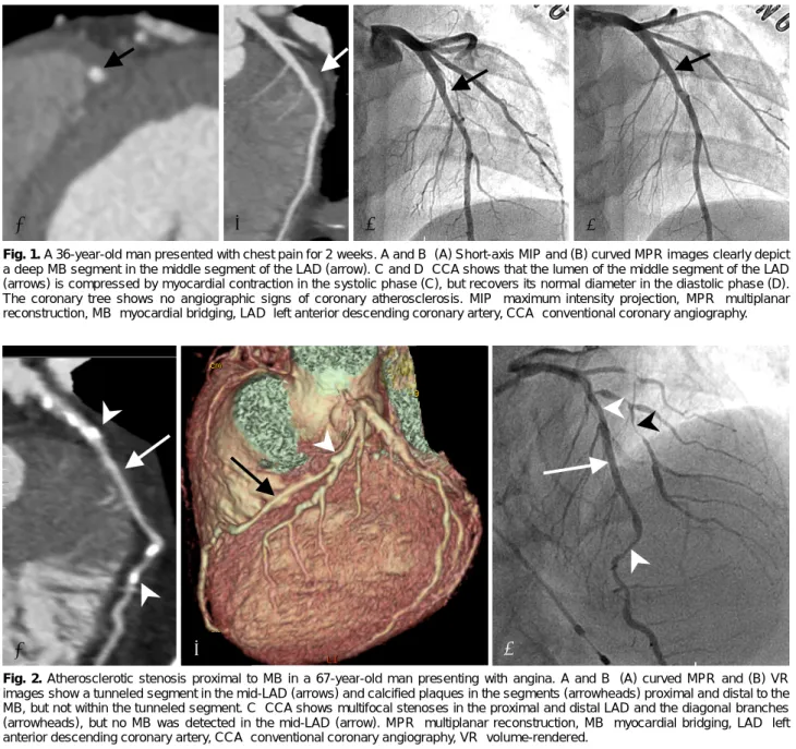
Fig. 1. A 36-year-old man presented with chest pain for 2 weeks. A and B: (A) Short-axis MIP and (B) curved MPR images clearly depict a deep MB segment in the middle segment of the LAD (arrow). C and D: CCA shows that the lumen of the middle segment of the LAD (arrows) is compressed by myocardial contraction in the systolic phase (C), but recovers its normal diameter in the diastolic phase (D).
The coronary tree shows no angiographic signs of coronary atherosclerosis. MIP: maximum intensity projection, MPR: multiplanar reconstruction, MB: myocardial bridging, LAD: left anterior descending coronary artery, CCA: conventional coronary angiography.
A B C D
A B C
Fig. 2. Atherosclerotic stenosis proximal to MB in a 67-year-old man presenting with angina. A and B: (A) curved MPR and (B) VR images show a tunneled segment in the mid-LAD (arrows) and calcified plaques in the segments (arrowheads) proximal and distal to the MB, but not within the tunneled segment. C: CCA shows multifocal stenoses in the proximal and distal LAD and the diagonal branches (arrowheads), but no MB was detected in the mid-LAD (arrow). MPR: multiplanar reconstruction, MB: myocardial bridging, LAD: left anterior descending coronary artery, CCA: conventional coronary angiography, VR: volume-rendered.
in diastole, systolic compression of the tunneled segment alone cannot sufficiently explain ischemia and associated symptoms. Myocardial ischemia and its associated symp- toms are related to the length and depth of the tun- neled segment, the degree of systolic dysfunction, stress- and exercise-induced sympathetic drive, increased local shear force, and local coronary endothelial dysfunction.1)2) Although MB is clinically silent in most cases, it has been associated with angina, myocardial ischemia, myo- cardial infarction, left ventricular dysfunction, myocar- dial stunning, paroxysmal atrioventricular (AV) blockade, exercise-induced ventricular tachycardia, and sudden
cardiac death.1)2)6-9) MB is frequent in patients with hy- pertrophic cardiomyopathy, representing a potential in- creased risk of sudden death in young patients (Fig. 3).
MB must be considered in symptomatic young patients at low risk for coronary atherosclerosis.1) Occasionally, MB has caused technical problems during coronary by- pass surgery (Fig. 4).26)
Imaging Findings on Conventional Coronary Angiography, Intracoronary
Ultrasound, and Intracoronary Doppler Ultrasound
The typical angiographic findings of the “milking effect” and the “step down-step up” phenomena are in- duced by systolic compression of the tunneled segment (Fig. 1C and D). CCA is likely to underestimate the prevalence of MB by delivering a visualization that is limited to the vessel lumen, thus necessitating that in- vestigators rely on indirect signs. Even though demon- stration of systolic compression and the milking effect are considered diagnostic, these signs are rather insen- sitive in the superficial type of MB that demonstrates minimal systolic compression or none at all. Similarly, the step down-step up phenomenon may be absent in superficial MB. CCA has gradually relinquished its position as the gold standard for diagnosing MB be- cause deeply located tunneled segments usually appear to be sufficiently compressed during systolic phase, and atherosclerotic stenosis proximal to the MB may hamper identification of the tunneled segment.1)2) IVUS de-
A B
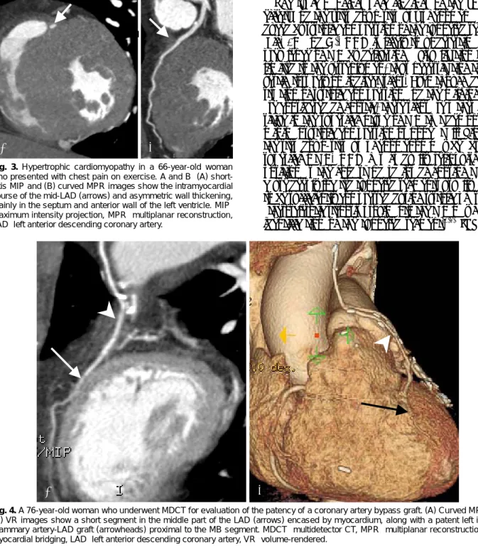
Fig. 4. A 76-year-old woman who underwent MDCT for evaluation of the patency of a coronary artery bypass graft. (A) Curved MPR and (B) VR images show a short segment in the middle part of the LAD (arrows) encased by myocardium, along with a patent left internal mammary artery-LAD graft (arrowheads) proximal to the MB segment. MDCT: multidetector CT, MPR: multiplanar reconstruction, MB:
myocardial bridging, LAD: left anterior descending coronary artery, VR: volume-rendered.
Fig. 3. Hypertrophic cardiomyopathy in a 66-year-old woman who presented with chest pain on exercise. A and B: (A) short- axis MIP and (B) curved MPR images show the intramyocardial course of the mid-LAD (arrows) and asymmetric wall thickening, mainly in the septum and anterior wall of the left ventricle. MIP:
maximum intensity projection, MPR: multiplanar reconstruction, LAD: left anterior descending coronary artery.
A B
monstrates the “half-moon phenomenon” in the tun- neled segment. ICD with pullback of the Doppler wire reveals a characteristic flow pattern: the “fingertip ph- enomenon” or the “spike-and-dome pattern”. IVUS is a sensitive method for assessing wall thickness and ves- sel size prior to stenting the MB to avoid coronary per- foration. Even though CCA, IVUS, and ICD can reveal the morphological and functional features of MB, these procedures are invasive.1)2)27)
Imaging Findings on Multidetector CT Coronary Angiography
The recent advent of MDCT scanners using 16, 40, and 64 slices has enabled clear and reliable visualiza- tion of the coronary arteries.10-21) The latest 64-slice MDCT scanners render 64 slices per rotation and of- fer a maximum temporal resolution of 165 ms and an unprecedented spatial resolution of isotropic 0.4 mm3. Multiplanar reformatted images assessed by MDCT provide information about the lumen and walls of the coronary arteries and the myocardium in any plane.
Therefore, MB can be demonstrated on MDCT coro- nary angiography, regardless of the thicknesses and directions of muscle bundles in the MB. Furthermore, MDCT coronary angiography is effective in the evalua- tion of concomitant atherosclerotic coronary lesions.
According to recent studies, MDCT coronary angio- graphy is a reliable noninvasive imaging modality for the diagnosis of MB that allows for evaluation of the real incidence and anatomical characteristics of MB in vivo. The differences in the prevalence (3.5-30.5%) of MB using MDCT can be explained by the better tem- poral and spatial resolution obtained using the latest 64-slice MDCT scanners, by differences in postproces- sing techniques used to delineate MB, and by inclu-
sion or exclusion of superficial MB.20) The location of MB varies in MDCT coronary angiographic series even though MBs are principally confined to the mid-segment of the LAD (Fig. 5).13)15)17) Konen et al.15) defined three useful anatomical patterns of MB in the LAD accord- ing to the depth and course of the tunneled segment on CT: the “superficial” type (Fig. 6), seen in 29% of all intramuscular LAD segments, in which the tunneled segment had a superficial course along the interventri- cular septum and was covered by a thin layer of tissue (<1 mm thick); the “deep” type (Fig. 7), seen in 41% of all tunneled LAD segments, in which the tunneled seg- ment penetrated the interventricular septum at a depth between 1 and 6.2 mm; and the “right ventricular” type (Fig. 8), seen in 29% of all tunneled LAD segments, in which the tunneled segment crossed through the right ventricular anterior wall adjacent to the interventri-
A B
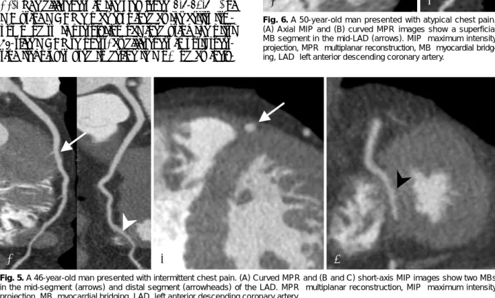
Fig. 5. A 46-year-old man presented with intermittent chest pain. (A) Curved MPR and (B and C) short-axis MIP images show two MBs in the mid-segment (arrows) and distal segment (arrowheads) of the LAD. MPR: multiplanar reconstruction, MIP: maximum intensity projection, MB: myocardial bridging, LAD: left anterior descending coronary artery.
A B
Fig. 6. A 50-year-old man presented with atypical chest pain.
(A) Axial MIP and (B) curved MPR images show a superficial MB segment in the mid-LAD (arrows). MIP: maximum intensity projection, MPR: multiplanar reconstruction, MB: myocardial bridg- ing, LAD: left anterior descending coronary artery.
C
cular septum.
In general, MB may be a coincidental finding, but MB has been implicated as a causative factor in symp- tomatic patients. The deep tunneled segment could com- promise coronary diastolic flow and result in ischemia, compared with the superficial tunneled segment.1)2)5)27) It is known that the degree of tunneled artery obstruc- tion is dependent on such factors as location, thickness, length of the muscle bridges, and degree of cardiac con- tractility. Leschka et al.18) demonstrated that the degree of systolic MB compression on CCA was significantly correlated with tunneled segment depth, but not length.
However, they did not investigate the relationship be- tween MB and clinical symptoms.
Measurement of the systolic compression of the tun- neled segment with 64-slice MDCT may be inaccurate because the limited temporal resolution of CT scanners
does not allow for reliable coronary anatomical deline- ation during the end-systolic phase. Dual-source CT, with 2 arrays consisting of an X-ray tube and detectors arranged at 90 degrees to each other, and with a gantry rotation time of 330 ms, allows for a temporal resolution of 83 ms at the center of the rotation when half-scan im- age reconstruction algorithms are used. The high tem- poral resolution enables visualization of the vessel lumen during most of the cardiac cycle. Hence, the milking ef- fect can be observed in the 4D reconstruction (Fig. 9).21)
Treatment
Asymptomatic MB warrants no treatment. Sympto- matic MB may be treated medically or surgically. Med- ical treatment as a first-line therapy includes nitrates, beta-blockers, and calcium antagonists. In patients with severe angina and clinically relevant ischemia, surgical treatments such as myotomy and coronary artery bypass grafting are considered.1)2) Coronary stent implanta- tion may be the treatment of choice for patients whose conditions are complicated by infarction or recalci- trance.1)2)29)30)
Conclusion
MB is a common anatomical variant characterized by myocardial encasement of a coronary artery seg- ment. Even though this variant is considered a benign condition, early diagnosis and treatment are important due to the complications associated with MB. Diag- nosis of MB is made possible with the tunneled segment and the systolic compression seen on various imaging modalities. MDCT coronary angiography is accepted as a reliable non-invasive method for the diagnosis of
A B C
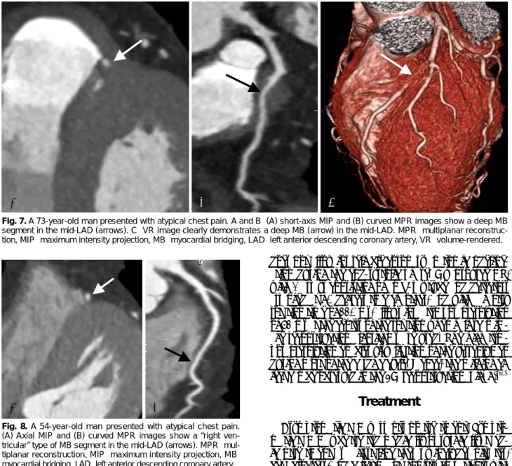
Fig. 7. A 73-year-old man presented with atypical chest pain. A and B: (A) short-axis MIP and (B) curved MPR images show a deep MB segment in the mid-LAD (arrows). C: VR image clearly demonstrates a deep MB (arrow) in the mid-LAD. MPR: multiplanar reconstruc- tion, MIP: maximum intensity projection, MB: myocardial bridging, LAD: left anterior descending coronary artery, VR: volume-rendered.
A B
Fig. 8. A 54-year-old man presented with atypical chest pain.
(A) Axial MIP and (B) curved MPR images show a “right ven- tricular” type of MB segment in the mid-LAD (arrows). MPR: mul- tiplanar reconstruction, MIP: maximum intensity projection, MB:
myocardial bridging, LAD: left anterior descending coronary artery.
MB because MDCT directly depicts the length and dep- th of the tunneled segment, along with its degree of compression during systole.
REFERENCES
1) Mohlenkamp S, Hort W, Ge J, Erbel R. Update on myocardial bridging. Circulation 2002;106:2616-22.
2) Alegria JR, Herrmann J, Holmes DR Jr, Lerman A, Rihal CS.
Myocardial bridging. Eur Heart J 2005;26:1159-68.
3) Noble J, Bourassa MG, Petitclerc R, Dyrda I. Myocardial bridg- ing and milking effect of the left anterior descending coronary artery: normal variant or obstruction? Am J Cardiol 1976;37:
993-9.
4) Rossi L, Dander B, Nidasio GP, et al. Myocardial bridges and ischemic heart disease. Eur Heart J 1980;1:239-45.
5) Ferreira AG Jr, Trotter SE, Konig B Jr, Decourt LV, Fox K, Olsen EG. Myocardial bridges morphological and functional aspects. Br Heart J 1991;66:364-7.
6) Kramer JR, Kitazume H, Proudfit WL, Sones FM Jr. Clinical significance of isolated coronary bridges: benign and frequent condition involving the left anterior descending artery. Am Heart J 1982;103:283-8.
7) Uhm JS, Park CS, Kim TS, et al. A case of acute myocardial infarction caused by coronary thrombus associated with a myo- cardial bridge and slow coronary flow. Korean Circ J 2005;
35:639-42.
8) Lee JH, Bae Y, Lee HS, et al. A case of myocardial infarction in a patient with myocardial bridge and atrial fibrillation. Korean Circ J 2004;34:319-22.
9) Kim IW, Jeung SM, Won TK, et al. Clinical observation of myocardial bridge. Korean Circ J 2001;31:637-44.
10) Amoroso G, Battolla L, Gemignani C, et al. Myocardial bridging on left anterior descending coronary artery evaluated by multi- detector computed tomography. Int J Cardiol 2004;95:335-7.
11) Goitein O, Lacomis JM. Myocardial bridging: noninvasive diag- nosis with multidetector CT. J Comput Assist Tomogr 2005;29:
238-40.
12) Ko SM, Kim KS. Multidetector-row CT coronary angiographic finding of myocardial bridging. Br J Radiol 2007;80:e196-200.
13) Kantarci M, Duran C, Durur I, et al. Detection of myocardial bridging with ECG-gated MDCT and multiplanar reconstruction.
AJR Am J Roentgenol 2006;186(6 suppl 2):S391-4.
14) Zeina AR, Odeh M, Blinder J, Rosenschein U, Barmeir E. Myo- cardial bridge: evaluation on MDCT. AJR Am J Roentgenol 2007;
188:1069-73.
15) Konen E, Goitein O, Sternik L, Eshet Y, Shemesh J, Di Segni E.
The prevalence and anatomical patterns of intramuscular co- ronary arteries. J Am Coll Cardiol 2007;49:587-93.
16) Kawawa Y, Ishikawa Y, Gomi T, et al. Detection of myocardial bridge and evaluation of its anatomical properties by coronary multislice spiral computed tomography. Eur J Radiol 2007;61:
130-8.
17) Ko SM, Choi JS, Nam CW, Hur SH. Incidence and clinical significance of myocardial bridging with ECG-gated 16-row MDCT coronary angiography. Int J Cardiovasc Imaging 2008;24:
445-52.
18) Leschka S, Koepfli P, Husmann L, et al. Myocardial bridging:
depiction rate and morphology at CT coronary angiography: com- parison with conventional coronary angiography. Radiology 2008;
246:754-62.
19) Hazirolan T, Canyigit M, Karcaaltincaba M, et al. Myocardial bridging on MDCT. AJR Am J Roentgenol 2007;188:1074-80.
20) Konen E, Goitein O, Di Segni E. Myocardial bridging, a com- mon anatomical variant rather than a congenital anomaly. Se- min Ultrasound CT MR 2008;29:195-203.
21) Lu GM, Zhang LJ, Guo H, Huang W, Merges RD. Comparison of myocardial bridging by dual-source CT with conventional coronary angiography. Circ J 2008;72:1079-85.
22) Sorajja P, Ommen SR, Nishimura RA, Gersh BJ, Tajik AJ, Hol- mes DR. Myocardial bridging in adult patients with hypertro- phic cardiomyopathy. J Am Coll Cardiol 2003;42:889-94.
23) Wymore P, Yedlicka JW, Garcia-Medina V, et al. The incidence of myocardial bridges in heart transplants. Cardiovasc Intervent Radiol 1989;12:202-6.
24) Kopp AF, Schroeder S, Kuetner A, et al. Non-invasive coronary
A B C
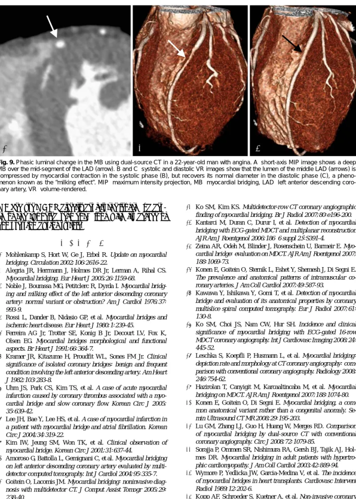
Fig. 9. Phasic luminal change in the MB using dual-source CT in a 22-year-old man with angina. A: short-axis MIP image shows a deep MB over the mid-segment of the LAD (arrow). B and C: systolic and diastolic VR images show that the lumen of the middle LAD (arrows) is compressed by myocardial contraction in the systolic phase (B), but recovers its normal diameter in the diastolic phase (C), a pheno- menon known as the “milking effect”. MIP: maximum intensity projection, MB: myocardial bridging, LAD: left anterior descending coro- nary artery, VR: volume-rendered.
angiography with high resolution multidetector-row computed to- mography: results in 102 patients. Eur Heart J 2002;23:1714-25.
25) Schoenhagen P, Halliburton SS, Stillman AE, et al. Noninvasive imaging of coronary arteries: current and future role of multi- detector row CT. Radiology 2004;232:7-17.
26) Ochsner JL, Mills NL. Surgical management of diseased intra- cavitary coronary arteries. Ann Thorac Surg 1984;38:356-62.
27) Ge J, Jeremias A, Rupp A, et al. New signs characteristic of myo- cardial bridging demonstrated by intracoronary ultrasound and Doppler. Eur Heart J 1999;20:1707-16.
28) Ge J, Erbel R, Gorge G, Haude M, Meyer J. High wall shear stress proximal to myocardial bridging and atherosclerosis: in- tracoronary ultrasound and pressure measurements. Br Heart J 1995;73:462-5.
29) Ng E, Jilaihawi H, Gershlick AH. Symptomatic myocardial bridg- ing: a niche indication for drug-eluting stents? Int J Cardiol 2005;
99:463-4.
30) Choi SH, Shim SJ, Byun KH, Choi DH, Shim WH. A case of coronary stenting in the management of myocardial ischemia caused by myocardial bridging. Korean Circ J 2001;31:940-4.