Korean Circulation Journal
Introduction
Acute coronary syndrome (ACS) as a presentation of coronary atherosclerosis is associated with inflammatory mechanisms
involved in the development of atherosclerotic plaque and subsequent rupture and thrombosis.
1)Inflammation-based markers have been used to detect high-risk patients and their prognosis.
2)Leukocytes have important roles in inflammatory processes.
3)Increased white blood cell (WBC) count has been shown to be a predictor of clinical outcomes of patients with ACS.
4)Besides leukocytes, platelets have been reported to have substantial effect on the development of cardiovascular events through inflammatory mechanisms.
5)Mean platelet volume (MPV) as a marker of platelet activation is another inflammatory marker that has been demonstrated to be a prognostic marker in ACS setting.
6)Metabolic syndrome (MetS) is a combined cardiovascular risk factors phenomenon. It includes visceral obesity, dysglycemia, hypertension, elevated triglycerides, and decreased high density lipoprotein.
7)MetS has become a serious public health problem due to increases in its prevalence and risk of developing type 2 diabetes mellitus and cardiovascular atherosclerotic diseases.
8)However,
Print ISSN 1738-5520 • On-line ISSN 1738-5555
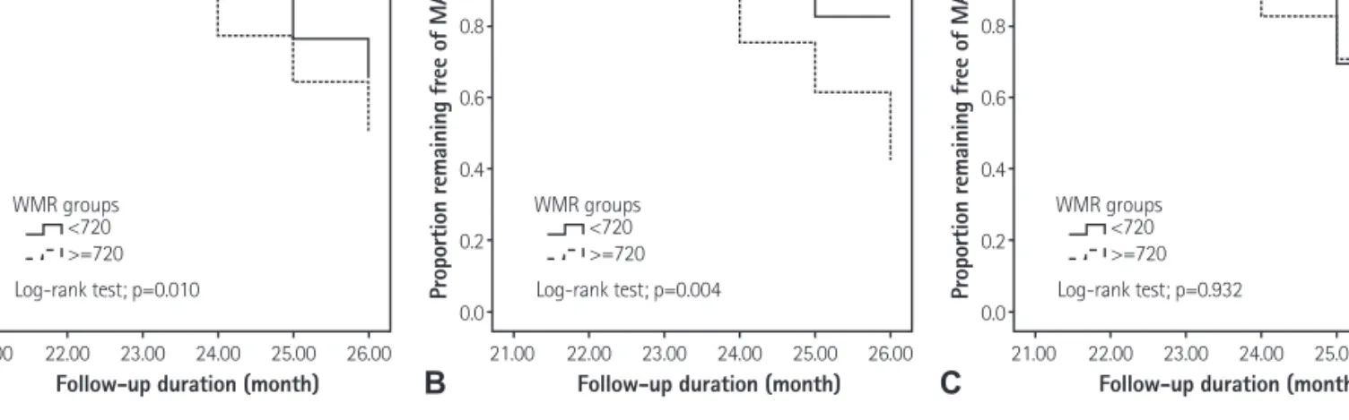
White Blood Cell Count to Mean Platelet Volume Ratio Is a Prognostic Factor in Patients with Non-ST Elevation Acute Coronary Syndrome with or without Metabolic Syndrome
Mohammad Reza Dehghani, MD 1 , Yousef Rezaei, MD 2 , Sanam Fakour, MD 3 , and Nasim Arjmand, MD 3
1