Veterinary Science
http://dx.doi.org/10.4142/jvs.2012.13.1.93
Received: 12 Jan. 2011, Revised: 19 Jan. 2012, Accepted: 20 Jan. 2012
Original Article
*Corresponding author: Tel: +39-051-2097531; Fax: +39-051-2097593; E-mail: [email protected]
ⓒ 2012 The Korean Society of Veterinary Science.
This is an Open Access article distributed under the terms of the Creative Commons Attribution Non-Commercial License (http://creativecommons.org/licenses/by-nc/3.0) which permits unrestricted non-commercial use, distribution, and reproduction in any medium, provided the original work is properly cited.
Biceps femoris muscle transposition for treatment of cranial cruciate ligament rupture in small breed dogs
Roberto Tamburro*, Stefania Pinna, Anna Maria Tribuiani, Alessandra Panacea, Fabio Carli, Antonio Venturini
Department of Veterinary Medical Sciences, University of Bologna, 40064 Ozzano dell´Emilia (Bologna), Italy
The purpose of this study was to evaluate a new extracapsular surgical technique for the treatment of cranial cruciate ligament rupture in small breed dogs. Nine small breed dogs (seven females and two males) weighing ≤ 15 kg were treated with biceps femoris muscle transposition (BFT).
The duration of the BFT procedure was 20 min. Each patient underwent a standard clinical protocol and a questionnaire for the owners. Follow-up (at 1, 3, and 12 months postoperative) confirmed significant improvement in all patients, especially at 1 month postoperatively (p < 0.01) and again after complete stifle joint assessment at 3 months postoperatively. After 12 months, only two patients showed a slight increase in osteoarthritis. According to our results, BFT is a simple extracapsular surgical technique that can be used for the treatment of cranial cruciate ligament rupture in small breed dogs.
Keywords: biceps femoris, cranial cruciate ligament, small breed dog, stifle, transposition
Introduction
Cranial cruciate ligament (CrCL) rupture is the most common cause of lameness in dogs [13,14,21]. In large breed dogs, many surgical treatments have been proposed;
however, few reports exist in the literature regarding CrCL rupture in small breed dogs in which, conservative management was the preferred method of treatment. Along this same line, Vasseur [31] showed that conservative management has beneficial effects in 85.7% of patients.
Due to the long recovery period of conservative management techniques, however, surgery is now preferred for the treatment of CrCL rupture in small breed dogs [17].
The goal of surgery is to stabilize the stifle joint, preserve
range of motion, and prevent osteoarthritis (OA) [22].
Intracapsular, extracapsular, and tibial osteotomy procedures have been described [2,5,6,8,12,16-18,26-28,33]. Indeed, many surgical methods have been proposed, but no specific procedure is considered optimal [1].
Gait evaluation of dogs is generally obtained using qualitative analysis methods through direct inspection examination and/or video recording [20]. Further, gait analysis can be carried out using a system featuring multimodal (kinetic, kinematic and electromyographic) evaluation and three-dimensional measurements [34].
Even though investigations obtained using force platforms are the most reliable, it is also possible to obtain important quantitative data supporting the clinical evaluation of patients by using specific questionnaires completed by the owners [11,30].
The aims of this paper were to describe a new surgical technique for the extracapsular stabilization of the CrCL rupture through transposition of a strip of biceps femoris and to objectively evaluate the results by using specific questionnaires focused on the stifle joint.
Materials and Methods
Clinical data were obtained from dogs that were presented in 2009. The inclusion criteria were based on the diagnosis of CrCL insufficiency in small breed dogs (weight ≤ 15 kg).
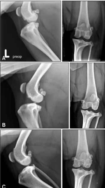
Each patient underwent the same protocol: orthopaedic evaluation, preoperative X-ray examination, surgery, and clinical follow-up carried out at 1, 3, and 12 months.
Postoperative radiographs were performed at 3 (nine patients) and 12 months (six patients). Medical records including all clinical data were compiled for each dog.
During the follow-up evaluation, a multiple-choice
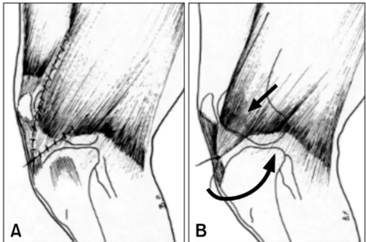
Fig. 1. (A) Preparation of a triangular flap taken from biceps femoris muscle. (B) Transposition in a distal and medial direction.
Fig. 2. (A) Flap is sutured on patellar ligament. (B) Transposed biceps muscle (small arrow) acts on the tibial tuberosity with a force directed caudally and externally (big arrow).
questionnaire was completed by the owners.
Surgical technique
All patients were anesthetized and placed in lateral recumbency with the affected side uppermost. A cranio- lateral approach to the stifle joint was carried out; the incision extended from the distal third of the femur to the proximal third of the tibia. The aponeurotic portion of the biceps femoris muscle was exposed, and the margin between the biceps femoris and fascia lata was recognized. This was followed by identification of the cranial insertion of the biceps femoris muscle, after which two incisions were made through the flap that would be used subsequently (Fig. 1A).
A triangular pedicle flap was prepared; one side was the cranial margin of the biceps muscle separated from the caudal margin of the vastus lateralis by a 3 cm incision, whereas the other side was formed by a 2 or 3 cm incision along the muscle fibers of the caudal portion of the distal biceps itself (Fig. 1B). The resulting flap was moved up, and by applying gradual traction in a cranial-medial-distal direction, it was transposed towards the tibial tuberosity and maintained extension of the limb. The flap was located next to the patellar ligament, where it was sutured with a 2-0 monofilament glycomer (Fig. 2A). In this manner, the biceps femoris muscle assumed a portion of its contractile fibers, and the slope was similar to that of the CrCL.
Intraoperatively, both tibial thrust and drawer tests were carried out to assess the degree of cranial shift of the tibia.
If the drawer sign was still evident, tension of the biceps femoris flap was increased so as to completely counter the cranial displacement of the tibia towards the femur. The flap was sutured to the lateral margin of the patellar ligament as close as possible to the tuberosity. Neither arthrotomy nor arthrocentesis were carried out, and routine suturing was performed. All patients underwent antibiotic
and analgesic therapy, and a soft bandage was placed over the wound. The owners were instructed to limit physical activity of the dogs to a minimum for approximately 15 days before allowing resumption of normal levels of activity.
Medical records and owner questionnaire
Medical records compiled by the clinician included various parameters: stifle pain, patellar-femoral crepitus, joint stability, range of motion, swelling, muscle mass, and lameness. These parameters were divided into three different subscales: visual examination, manual examination, and X-ray evaluation of OA.
Each owner completed a questionnaire [4] consisting of 24 questions divided into three subscales: pain, stiffness, and limb function. Five choices were given for each of the six subscales (three medical record and three questionnaire subscales) regarding the presence of clinical signs (4:
always, 3: obvious/often, 2: moderate/sometimes, 1:
mild/rarely, 0: absent/never).
Data processing
The data were normalized using a standardization test and were transformed into a score from 0 to 100.
The normalized score was calculated as shown below:
Normalized score = 100 − total score of each subscale × 100 / possible maximum score for the subscale
Our results were classified as: excellent (81∼100), good
(61∼80), poor (41∼60), or failed (0∼40). The score
obtained for each subscale was added to that of the same
subscale for each clinical case, and the mean values,
calculated at each postoperative examination, were plotted
on a graph to assess healing.
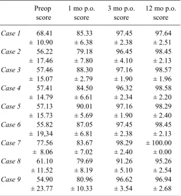
Table 3. Total patient score: for each patient, the mean value obtained from the six subscale scores
Preop score
1 mo p.o.
score
3 mo p.o.
score
12 mo p.o.
score
Case 1Case 2 Case 3 Case 4 Case 5 Case 6 Case 7 Case 8 Case 9
68.41
± 10.90 56.22
± 17.46 57.46
± 15.07 57.41
± 14.79 57.13
± 15.73 55.82
± 19,34 77.56 ± 8.06 61.10
± 11.52 54.90
± 23.77
85.33
± 6.38 79.18
± 7.80 88.30 ± 2.79 84.50 ± 6.61 90.01 ± 5.69 87.05 ± 6.81 83.67 ± 7.02 79.69 ± 8.19 80.96
± 10.33
97.45
± 2.38 96.45
± 4.10 97.16
± 1.90 96.32
± 2.34 97.16
± 1.90 97.45
± 2.38 98.29
± 2.40 91.26
± 5.10 96.62
± 3.54
97.64 ± 2.51 98.45 ± 2.13 98.57 ± 1.96 98.58 ± 2.20 98.29 ± 2.40 98.45 ± 2.13
± 100.00
± 0.00 95.26 ± 2.54 96.94 ± 2.68
Score evaluation: excellent (81∼100), good (61∼80), poor (41∼60), and failed (0∼4). All data are represented as the mean ± SD for score. Significant improvement was shown at 1 month (mo) p.o.; at 12 mo, follow-up showed excellent scores for all dogs.
Table 2. Preoperative (Preop) and postoperative (p.o.) normalized scores obtained from medical records and questionnaire
Preop score
1 mo p.o. score
3 mo p.o. score
12 mo p.o. score Medical records
Visual exam Manual exam X-ray evaluation
Questionnaire
Pain Stiffness Limb function48.15
± 10.02 66.60 ± 6.60
71.84
± 10.85 68.33 ± 12.50 48.41 ± 20.05
76.85 ± 5.55 88.22 ± 4.19 86.11
± 13.18 88.66
± 3.77 85.56
± 8.08 82.21
± 8.02
98.15
± 5.56 96.03
± 1.19
96.06
± 2.43 94.44
± 1.67 97.62
± 3.57
100.00
± 0.00 97.62 ± 1.84 83.33
± 20.41 97.92 ± 1.81 97.22
± 2.64 98.81
± 2.52
Score evaluation: excellent (81∼100), good (61∼80), poor (41∼60), and failed (0∼40). All data are represented as the mean ± SD.