© 2015 The Korean Ophthalmological Society
This is an Open Access article distributed under the terms of the Creative Commons Attribution Non-Commercial License (http://creativecommons.org/licenses /by-nc/3.0/) which permits unrestricted non-commercial use, distribution, and reproduction in any medium, provided the original work is properly cited.
Original Article
Hyperhomocysteinemia, a Biochemical Tool for Differentiating Ischemic and Nonischemic Central Retinal Vein Occlusion during
the Early Acute Phase
Kapil Deb Lahiri1, Somnath Mukherjee2, Sambuddha Ghosh3, Suman Mukherjee4, Jayanta Dutta4, Himadri Datta4, Harendra Nath Das5
1Department of Biochemistry, ESI-PGIMSR & ESIC Medical College Joka, Kolkata, India
2Department of Ophthalmology, N R S Medical College, Kolkata, India
3Department of Ophthalmology, North Bengal Medical College, Siliguri, India
4Department of Ophthalmology, Regional Institute of Ophthalmology, Kolkata, India
5Department of Biochemistry, RG Kar Medical College & Hospital, Kolkata, India
Purpose: The purpose of the study was to differentiate ischemic central retinal vein occlusion (CRVO) from non- ischemic CRVO during the early acute phase using plasma homocysteine as a biochemical marker.
Methods: Fasting plasma homocysteine, serum vitamin B12, and folate levels were measured in 108 consec- utive unilateral elderly adult (age >50 years) ischemic CRVO patients in the absence of local and systemic disease and compared with a total of 144 age and sex matched nonischemic CRVO patients and 120 age and sex matched healthy control subjects.
Results: Homocysteine level was significantly increased in the patients with ischemic CRVO in comparison with nonischemic CRVO patients (p = 0.009) and also in comparison with control subjects (p < 0.001). Analysis also showed that hyperhomocysteinemia was associated with increased incidence of ischemic CRVO (odds ratio, 18) than that for nonischemic CRVO (odds ratio, 4.5). Serum vitamin B12 and folate levels were significantly lower (p < 0.001) in CRVO patients compared to the control but were not significantly different between nonischemic and ischemic CRVO patients (p > 0.1).
Conclusions: Hyperhomocysteinemia can be regarded as useful in differentiating nonischemic and ischemic CRVO during the early acute phase in absence of local and systemic disease in the elderly adult (age >50 years) population.
Key Words: Central retinal vein occlusion, Homocysteine, Hyperhomocysteinemia, Ischemic central retinal vein occlusion, Nonischemic central retinal vein occlusion
Retinal vein occlusion is a common cause of vision loss in older individuals, and the second most common retinal
vascular disease after diabetic retinopathy [1]. There are two distinct types, which are classified according to the site of occlusion: in central retinal vein occlusion (CRVO), the occlusion is at, or proximal to, the lamina cribrosa of the optic nerve, where the central retinal vein exits the eye [2].
CRVO is further categorized as either perfused (nonisch- emic) or nonperfused (ischemic); each of which has differ-
Received: October 25, 2013 Accepted: July 8, 2014
Corresponding Author: Kapil Deb Lahiri, MD. 4/2 Shibchandra Sarb- abhowma lane, Baranagar, Kolkata, 700036 West Bengal, India. Tel: 91- 903-840-5409, E-mail: [email protected]
ent implications for prognosis and treatment [3]. The patho- genesis of CRVO is believed to follow the principles of Virchow’s triad for thrombogenesis, involving vessel dam- age, stasis and hypercoagulability [4]. The central retinal vein and artery share a common adventitial sheath at arte- riovenous crossings posterior to the lamina cribrosa so that atherosclerotic changes in the artery may compress the vein and precipitate CRVO [5]. Damage to the retinal vessel wall from atherosclerosis and compression alters rheologic properties in the adjacent central vein contributing to sta- sis, thrombosis, and thereafter, occlusion [6]. Nonischemic CRVO is more common, accounting for about 75% of all CRVO cases. Presentation of the disease is associated with sudden, unilateral blurred vision with visual acuity of 20 / 200 or better. Ischemic CRVO is characterized by rapid-on- set venous obstruction resulting in decreased retinal perfu- sion, capillary closure and retinal hypoxia with visual acui- ty less than 20 / 200.
Prior to this study, there were four functional tests (visual acuity, visual fields, relative afferent pupillary defect [RAPD], electroretinography) and two morphologic tests (ophthalmoscopy and fluorescein fundus angiography) available for the differentiation of ischemic CRVO from nonischemic CRVO during the early acute phase of the dis- ease. None of the six tests, however, had 100% sensitivity or specificity for differentiation during the early acute phase, and therefore, no singular test could be considered a
"gold standard" [7]. Moreover, there was no biochemical test available that could differentiate ischemic CRVO from nonischemic CRVO during the early acute phase.
Homocysteine (Hcys) is a sulfur-containing nonprotein amino acid, which is derived from dietary methionine and acts as a methyl group donor in the form of S-adenosyl me- thionine. When a methyl group is donated, S-adenosyl Hcys is formed, which is then converted to Hcys. Hcys is either metabolized to cystathionine through the transsulfu- ration pathway, requiring B6, or it is converted back to me- thionine by B12 and folate, requiring transmethylation [8].
Hyperhomocysteinemia (HHcys) can also occur; the cause of which is varied. Severe HHcys is due to rare genetic de- fects that result in deficiencies in the enzymes cystathi- onine β-synthase and methylenetetrahydrofolate reductase.
Mild HHcys can be caused by impaired function of en- zymes in a transmethylation pathway sometimes associated with deficiencies in nutrients such as B12 and folate [9].
It has been well established that HHcys is an independent
risk factor in CRVO [10,11]. In our previous study, we es- tablished the cut off value of HHcys in an Indian popula- tion [12]. It has also been suggested that the MTHFR C677T mutation is associated with HHcys in ischemic CRVO in the Chinese population [13]. However, no study has demonstrated the frequency of HHcys in either catego- ry of unilateral CRVO, in the absence of local and systemic disease, in the elderly (age >50 years) Indian population.
Therefore, a 2-year prospective study of elderly adults (age
>50 years) with unilateral CRVO, in the absence of local and systemic disease, was performed to evaluate whether HHcys could be used as a biochemical parameter in the differentiation of ischemic CRVO from nonischemic CRVO during the early acute phase of the disease.
Materials and Methods
A 2-year prospective case-control study of consecutive, unrelated, elderly adult (age >50 years) patients, with a di- agnosis of ischemic or nonischemic CRVO in the absence of any other local or systemic disease, was conducted at the Regional Institute of Ophthalmology, Kolkata and the De- partment of Biochemistry in the R G Kar Medical College
& Hospital, Kolkata, West Bengal, India. Patients experi- encing any confounding conditions, such as malignancy, sepsis, liver and renal failure, recent cardiovascular and cerebrovascular accidents (<6 months), previous thrombo- embolic events, inflammatory disorders, thyroid disorder, diabetes, hypertension, glaucoma, dyslipidemia, vitamin intake (B12 and folate), alcohol or drug use (methotrexate, fibrates), food faddists, age <50 years (autoimmune diseas- es causing CRVO at <50 years), elevated prothrombin time, elevated activated partial thromboplastin generation time and smoking, were excluded from the study population by detailed history, clinical examination and laboratory inves- tigation. The people who accompanied the ischemic and nonischemic CRVO patients were evaluated as controls, based on the same afore-mentioned inclusion and exclusion criteria. The institutional ethics committee of both hospi- tals approved the study and informed consent was obtained from all study participants, in accordance with the Decla- ration of Helsinki. Ophthalmic examinations, including vi- sual acuity, RAPD, electroretinography, fluorescent angi- ography and fundus examination of both eyes, were used for the clinical diagnosis of ischemic and nonischemic
CRVO. Statistical analysis was performed using the Stu- dent’s t-test and SPSS ver. 11.5 (SPSS Inc., Chicago, IL, USA).
Measurement of plasma homocysteine
Venous blood samples were drawn from fasting patients into vials containing ethylene diamine tetraacetic acid within 48 to 72 hours of disease onset. Immediately after collection, all samples were separated from blood cells by centrifugation at 1,000× g at 25°C for 3 minutes and stored tightly capped at 2°C to 8°C for up to 48 hours or frozen at -20°C if testing was delayed. Plasma Hcys was estimated by enzymatic method in a semiautoanalyzer (Accurex AT 112 Plus Biochemistry Analyzer) with a reagent kit, supplied by Lilac Clinical Chemistry Division(linearity extends to 50 μmol/L) [14]. In addition, serum vitamin B12 and folate levels were also measured (IMX Analyzer;
Abbott Laboratories Diagnostics Division, Abbott Park, IL, USA). Blood collected in plain vial (without anticoagulant) was kept in a tilted position for 30 to 45 minutes at room temperature and then centrifuged to separate serum. The IMX B12 assay was carried out, which is based on micro- particle enzyme immunoassay technology, whereas the IMX folate assay is an ion-capture assay technique. For these immunologic assays, inter and intra-assay coefficients of variation were <10%.
Results
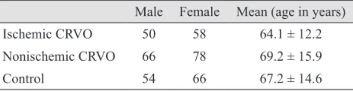
Based on the inclusion and exclusion criteria, 108 patients with unilateral ischemic CRVO (58 females and 50 males) were selected from a total of 248 ischemic CRVO patients who participated in the study. A total of 144 age- and sex- matched nonischemic CRVO patients (78 females and 66 males) from a total of 324 nonischemic CRVO patients, and a total of 120 age- and sex-matched healthy controls (66 females and 54 males) from a total of 202 people who had accompanied the ischemic and nonischemic CRVO patients to the hospital, were analyzed in this study based on a laboratory investigation separate from the initial questio- nnaire. The mean age of ischemic CRVO patients was 64.1
± 12.2 years and the mean ages of the nonischemic CRVO patients and healthy control participants were 69.2 ± 15.9 and 67.2 ± 14.6 years, respectively (Table 1).
In regards to clinical characteristics, peripheral areas of non-perfusion could not be evaluated accurately as many patients with either ischemic or nonischemic CRVO had experienced extensive hemorrhage in all four quadrants, which had gone undetected by both ophthalmoscopy and fluorescent angiography in the early stage of the disease.
Sixty-seven ischemic CRVO patients showed RAPD whereas only 17 nonischemic CRVO patients showed RAPD. Ninety-seven ischemic CRVO patients had visual acuity less than 20 / 200 where as only 47 nonischemic CRVO patients had visual acuity less than 20 / 200. Sixty- nine ischemic CRVO patients had diminished ‘b’ wave amplitude of less than 60% compared to a normal eye.
The plasma Hcys levels were significantly higher in patients with ischemic CRVO patients (mean Hcys, 20.43 ± 4.26 μmol/L) compared with that of the control subjects (mean Hcys, 12.53 ± 2.16 μmol/L; p < 0.001) and nonischemic CRVO patients (mean Hcys, 15.33 ± 5.38 μmol/L; p = 0.009) (Table 2). Serum vitamin B12 and folate levels were significantly lower (p < 0.001) in CRVO pa- tients compared to control subjects, but were not signifi- cantly different between the nonischemic and ischemic va- rieties of CRVO ( p > 0.1) (Table 2). Our analysis also showed that HHcys was more closely associated with isch- emic CRVO (odds ratio, 18) than it was to nonischemic CRVO (odds ratio, 4.5) (Table 3).
Discussion
The prognosis of ischemic CRVO is extremely poor due to macular ischemia. Rubeosis iridis develops in appro- ximately 50% of eyes, usually between 2 and 4 months of diagnosis (100-day glaucoma), and there is a high risk of neovascular glaucoma. Retinal neova scular ization occurs in about 5% of CRVO eyes. When possible, patients with ischemic CRVO should be seen monthly for 6 months to detect the onset of anterior segment neovas cularization.
Table 1. Baseline characteristics of study population (n = 372) Male Female Mean (age in years)
Ischemic CRVO 50 58 64.1 ± 12.2
Nonischemic CRVO 66 78 69.2 ± 15.9
Control 54 66 67.2 ± 14.6
CRVO = central retinal vein occlusion.
Subsequent reviews should continue for up to 2 years to detect significant ischemia and macular edema [3,5]. In nonischemic CRVO patients, an initial follow-up should take place after 3 months. Conversion of the disease to ischemic CRVO occurs in 15% of cases within 4 months and in 34% of cases within 3 years. In cases that do not subsequently become ischemic, the prognosis is reasonably good with return of vision to normal or near normal in about 50% of cases. Therefore, it is always essential to diagnose and differentiate the categories of perfused (nonischemic) and nonperfused (ischemic) CRVO during the early acute phase, because each diagnosis has different implications for prognosis and treatment [3].
In our previous study, we observed that Hcys signi- ficantly increased in retinal vein occlusion cases, in comparison with the control (p < 0.001), and the prevalence of plasma Hcys rose even more in cases of CRVO (odds ratio, 13) than in branch retinal vein occlusion (odds ratio, 5.03) in an Indian population [15]. Gao et al. [13] suggested that the MTHFR C677T mutation is associated with HHcys in ischemic CRVO within the Chinese population.
In this study, we observed a significant increase in Hcys
levels of patients with ischemic CRVO (mean Hcys, 20.43 ± 4.26 μmol/L) compared to patients with nonischemic CRVO (mean Hcys, 15.33 ± 5.38 μmol/L; p = 0.009) and control subjects (mean Hcys, 12.53 ± 2.16 μmol/L; p <
0.001) (Table 2). Hcys possibly exerts its toxic effect on the endothelium by decreasing the bioavailability of nitric oxide [16],altering the expression of various thrombotic factors, eliciting a mitogenic effect on arterial smooth muscle cells [17], and through the expression of acute stress-related genes [18].Moreover, the high pKa value of the sulfhydryl group (pKa, 10.0) of Hcys is responsible for the formation of stable disulfide bonds with protein cysteine residues, and in the process, it alters or impairs the function of many proteins. Albumin, fibronectin, transthyretin, annexin II, and factor V have now been identified as molecular targets for Hcys [19]. Metabolic conversion of Hcys to a chemically reactive metabolite, Hcys-thiolactone, is suggested to contribute to Hcys toxicity in humans (Hcys-thiolactone hypothesis) leading to endo- thelial dysfunction [20].
An odds ratio of 3.0 for fasting HHcys in patients with CRVO was reported by Lattanzio et al. [9]. Janssen et al.
[21] observed an overall OR of 8.9 for Hcys. This study showed an odds ratio of 18 for fasting HHcys in patients with ischemic CRVO in comparison to an odds ratio of 4.5 for nonischemic CRVO patients (Table 3) indicating ischemic CRVO was more often attributed to HHcys than nonischemic CRVO. The meta-analysis by Cahill et al. [22]
showed that raised plasma Hcys levels caused by low serum folate levels were associated with retinal vascular occlusion. In this study, serum vitamin B12and folate levels were significantly lower ( p < 0.001) in CRVO patients compared to the control, indicating a possible cause of HHcys in CRVO cases, however, there was no significant difference between the nonischemic and Table 3. OR of hyperhomocysteinemia in ischemic CRVO
and nonischemic CRVO patients in comparison with the con- trol (n = 372)
Parameter Ischemic
CRVO Nonischemic
CRVO Control
High Hcys 72 48 12
Normal Hcys 36 96 108
Total 108 144 120
Values are presented as number of participants; OR = 18 for isch- emic CRVO and OR = 4.5 for nonischemic CRVO.
OR = odds ratio; CRVO = central retinal vein occlusion; Hcys = homocysteine.
Table 2. Plasma homocysteine, serum folic acid and serum vitamin B12levels in CRVO, ischemic CRVO, nonischemic CRVO, and control subjects (n = 372)
Parameter CRVO (ischemic and
nonischemic) Ischemic CRVO Nonischemic CRVO Control
Homocysteine (μmol/L) 17.52 ± 5.5 20.43 ± 4.26*† 15.33 ± 5.38 12.53 ± 2.16
Folic acid (ng/mL) 7 ± 4 6.8 ± 3.8*‡ 7.1 ± 4.2 12.2 ± 3.6
Vitamin B12(pg/mL) 229 ± 75 224 ± 71*‡ 232 ± 74 401 ± 103
Values are presented as mean ± SD.
CRVO = central retinal vein occlusion.
*p < 0.001 in response to control; † p = 0.009 in response to nonischemic CRVO; ‡ p > 0.1 in response to nonischemic CRVO.
ischemic varieties (p > 0.1) (Table 2). A possible reason for the significant HHcys prevalence in ischemic CRVO over nonischemic CRVO could be explained by a role of Hcys as an acute phase reactant [23]. Due to this research, we conclude that HHcys may be regarded as a biochemical parameter to be used as a supplement to functional and morphological tests in the differentiation of nonischemic and ischemic CRVO during the early acute phase, in the absence of local and systemic disease, in the elderly adult (age >50 years) Indian population.
Conflict of Interest
No potential conflict of interest relevant to this article was reported.
Acknowledgements
This research was funded by the West Bengal University of Health Sciences. We would like to thank Prof. Ila Bhat- tacharje for her assistance and guidance in this research.
We would also like to thank Lilac Clinical Chemistry Divi- sion for providing reagents at a reduced cost.
References
1. Cugati S, Wang JJ, Knudtson MD, et al. Retinal vein occlu- sion and vascular mortality: pooled data analysis of 2 pop- ulation-based cohorts. Ophthalmology 2007;114:520-4.
2. Mitchell P, Smith W, Chang A. Prevalence and associations of retinal vein occlusion in Australia: the Blue Mountains Eye Study. Arch Ophthalmol 1996;114:1243-7.
3. Hayreh SS. Classification of central retinal vein occlusion.
Ophthalmology 1983;90:458-74.
4. Rogers S, McIntosh RL, Cheung N, et al. The prevalence of retinal vein occlusion: pooled data from population stud- ies from the United States, Europe, Asia, and Australia.
Ophthalmology 2010;117:313-9.e1.
5. Parodi MB, Bandello F. Branch retinal vein occlusion: clas- sification and treatment. Ophthalmologica 2009;223:298- 305.
6. Rehak J, Rehak M. Branch retinal vein occlusion: patho- genesis, visual prognosis, and treatment modalities. Curr
Eye Res 2008;33:111-31.
7. Hayreh SS, Klugman MR, Beri M, et al. Differentiation of ischemic from non-ischemic central retinal vein occlusion during the early acute phase. Graefes Arch Clin Exp Oph- thalmol 1990;228:201-17.
8. Finkelstein JD. Inborn errors of sulfur-containing amino acid metabolism. J Nutr 2006;136(6 Suppl):1750S-4S.
9. Lattanzio R, Sampietro F, Ramoni A, et al. Moderate hy- perhomocysteinemia and early-onset central retinal vein occlusion. Retina 2006;26:65-70.
10. Chua B, Kifley A, Wong TY, Mitchell P. Homocysteine and retinal emboli: the Blue Mountains Eye Study. Am J Ophthal- mol 2006;142:322-4.
11. Chua B, Kifley A, Wong TY, Mitchell P. Homocysteine and retinal vein occlusion: a population-based study. Am J Ophth- almol 2005;139:181-2.
12. Lahiri KD, Datta H, Das HN. Reference interval determi- nation of total plasma homocysteine in an Indian popula- tion. Indian J Clin Biochem 2014;29:74-8.
13. Gao W, Wang YS, Zhang P, Wang HY. MTHFR C677T mutation in central retinal vein occlusion: a case-control study in Chinese population. Thromb Res 2008;121:699-703.
14. Ueland PM, Refsum H, Stabler SP, et al. Total homocyste- ine in plasma or serum: methods and clinical applications.
Clin Chem 1993;39:1764-79.
15. Lahiri KD, Dutta J, Datta H, Das HN. Hyperhomocystein- emia, as an independent risk factor for retinal venous oc- clusion in an Indian population. Indian J Clin Biochem 2013;28:61-4.
16. Weiss N. Mechanisms of increased vascular oxidant stress in hyperhomocys-teinemia and its impact on endothelial function. Curr Drug Metab 2005;6:27-36.
17. Postea O, Krotz F, Henger A, et al. Stereospecific and re- dox-sensitive increase in monocyte adhesion to endothelial cells by homocysteine. Arterioscler Thromb Vasc Biol 2006;
26:508-13.
18. Jakubowski H. Pathophysiological consequences of homo- cysteine excess. J Nutr 2006;136(6 Suppl):1741S-9S.
19. Jacobsen DW, Catanescu O, Dibello PM, Barbato JC. Mo- lecular targeting by homocysteine: a mechanism for vascu- lar pathogenesis. Clin Chem Lab Med 2005;43:1076-83.
20. Selhub J. The many facets of hyperhomocysteinemia: stud- ies from the Framingham cohorts. J Nutr 2006;136(6 Sup- pl):1726S-30S.
21. Janssen MC, den Heijer M, Cruysberg JR, et al. Retinal vein occlusion: a form of venous thrombosis or a complica-
tion of atherosclerosis? A meta-analysis of thrombophilic factors. Thromb Haemost 2005;93:1021-6.
22. Cahill MT, Stinnett SS, Fekrat S. Meta-analysis of plasma homocysteine, serum folate, serum vitamin B(12), and thermolabile MTHFR genotype as risk factors for retinal
vascular occlusive disease. Am J Ophthalmol 2003;136:1136- 50.
23. Senaratne MP, Griffiths J, Nagendran J. Elevation of plas- ma homocysteine levels associated with acute myocardial infarction. Clin Invest Med 2000;23:220-6.