INTRODuCTION
Endoscopic sinus surgery (ESS) is well-established as the mainstay of surgical treatment for refractory chronic rhinosinusitis (CRS). Outcomes of ESS have improved due to advances in endoscopic systems, surgical techniques, and instruments.
1)However, ESS still carries a potential risk of injury to adjacent structures such as the orbital contents and skull base.
1)These complications may present as CSF leakage, orbital hematoma, optic nerve, or extraocular muscle injury
2)and are the leading cause of litigations in otorhinolaryngology.
3)However, oculomotor nerve palsy is extremely rare after ESS, and isolated oculomotor nerve palsies after ESS without orbital injury have never been reported. We report an unusual case of isolated complete oculomotor palsy after ESS along with a discussion of the potential mechanisms.
J Rhinol 21(2), 2014
- 134 -
www.ksrhino.or.kr
A Case of Isolated Complete Oculomotor Nerve Palsy Following Endoscopic Sinus Surgery
Nayeon Choi
1, Hyun-Jin Cho
1, Kyung-Ah Park
2and Sang Duk Hong
1Departments of 1Otorhinolaryngology-Head and Neck Surgery and 2Opthalmology, Samsung Medical Center, Sungkyunkwan University School of Medicine, Seoul, South Korea
ABSTRACT
Orbital complications after endoscopic sinus surgery (ESS), such as optic nerve or medial rectus injuries, are well known, but isolated complete oculomotor nerve palsy has never been reported. In this case, a 31-year-old male was transferred to our hospital after ESS. Physical examination showed complete left oculomotor nerve palsy, with a bony defect on the sellar floor, which had not fully recovered after more than 1 year. We hypoth- esized that blunt trauma could be the main cause of the oculomotor palsy. Surgeons performing ESS must keep in mind the possibility of oculomotor palsy due to blunt trauma, especially when operating around the sphenoid and posterior ethmoid sinus.
KEY WORDS : Endoscopic Sinus SurgeryㆍOrbital ComplicationㆍOculomotor Nerve Palsy.
Address correspondence and reprint requests to Sang Duk Hong, MD, Department of Otorhinolaryngology-Head and Neck Surgery, Samsung Medical Center, Sungkyunkwan University School of Medicine, 81 Ir- won-ro, Gangnam-gu, Seoul 135-710, Korea
Tel: +82-2-3410-3579 Fax: +82-2-3410-3879 E-mail: [email protected]
Received for publication on August 28, 2014 Accepted for publicatoin on October 22, 2014
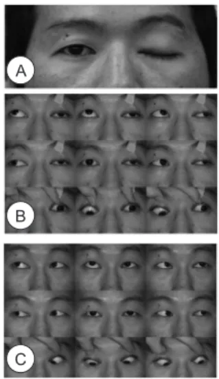
Fig. 1. (A) The patient suffered from left complete ptosis. (B) Composite photograph of eye movements 1 day post-oper- atively. We observed exotropia with ptosis in primary position (center middle panel) and significant limitation of movement of the left eye except the left lateral gaze. (C) Composite pho- tograph of eye movement 1 year after surgery. Ptosis was com- pletely resolved while some limitation in upward and downward gaze of left eye movement remained.
A
B
C
135 / J Rhinol 21(2), 2014
CASE REpORT
A 31-year-old male was referred to our hospital one day after bilateral ESS for CRS with nasal polyps from private clinic. The patient underwent ESS under local anesthesia and felt severe headache and complained of left orbital pain, left ptosis, and diplopia immediately after sphe- noidotomy (Fig. 1A).
The left pupil was dilated to 7 mm without direct or consensual response to light or accommodation with temporal deviation of the eye. Extraocular movements were impaired in all directions except for abduction, with complete ptosis of the eyelid (Fig. 1B). Visual acuity was 20/20 in both eyes. Anterior segment of eye ball and fun- dus examinations were normal.
In direct endoscopic visualization, there was bony defect at the sellar floor in the sphenoid sinus and optic canal in Onodi cells. However, there was no evidence of cerebrospinal fluid (CSF) leakage (Fig. 2). There was no evidence of direct injury to the orbit, any other site of the
skull base, or the brain stem on computed tomography (CT) and magnetic resonance imaging (MRI) (Fig. 3).
The patient received intravenous injections of dexam- ethasone (15 mg per day for 1 week) for treatment of his symptoms. Three months postoperatively, the left ptosis had completely recovered and pupillary reactivity had improved. Extraocular movements were recovered al- most completely, except for an upward gaze in the left eye. At 1 year post-operatively, however, the patient still complained of diplopia. On opthalmologic examination, he had nor mal balance in all directions of gaze except for the superior direction. He had left hypotropia of 8 prism diopters in upgaze and slight limitation of upgaze and downgaze, with abnormal adducting movement on at- tempted upgaze in the left eye (Fig. 1C). His left pupil was mildly dilated with slightly reduced reactivity.
DISCuSSION
ESS is currently the treatment of choice of for medi- cally intractable sinusitis. Its use has extended to various disease of the sinuses, orbit, and skull base.
4)The growing popularity and refinement of ESS techniques and advance- ments in instruments allow for constant and clear surgi- cal views, minimal intraoperative bleeding, and reduced surgical time and complications. However, complications associated with ESS are still a concern and demand at- tention from otorhinolaryngologists. Opthalmic injuries during ESS can involve the optic nerve, orbital vessels, extraoccular muscles, and the nasolacrimal system. How- ever, oculomotor nerve palsy after ESS is extremely rare.
There has only been one case report describing partial oc- ulomotor nerve palsy following penetrating injury to the lamina papyracea.
5)In that case, there was an offending bone fragment impinging on the medial rectus and op- tic nerve, and after removal of the bone chip, the patient gradually and fully recovered from oculomotor palsy.
However, the present case is the first report of complete oculomotor nerve palsy after ESS without direct orbital or nerve injury.
In this case, we performed both CT and MRI for evalua- tion of potential causes of nerve palsy and extent of injury, because CT may fail to detect midbrain damage and oculo- motor nerve root avulsion, MRI should be performed.
6)We found no evidence of neurovascular injury, bony disruption of the lamina papyracea, or brain stem abnormalities. We hypothesize that the mechanism of oculomotor nerve palsy in this case was focal stretching or contusion in the para- sellar segment of the oculomotor nerve by blunt trauma that occurred while performing sphenoidotomy by force, although this is extremely unlikely because the oculomo- tor nerve pathway is quite distant from the sellar floor.
Fig. 2. A nasal endoscopic view demonstrated a bony defect at the left sellar floor (black arrow) and the optic canal (white arrow) without evidence of cerebrospinal fluid leakage.
Fig. 3. CT and MRI were performed 1day after surgery. Axial (A) and coronal (B) CT scan showed hemorrhagic opacity, but there was no bony defect. T2 VISTA MRI (C) and T2 coronal MRI (D) demonstrated no abnormal finding at oculomotor nerve pathway, orbital contents and adjacent soft tissues.