INTRODUCTION
Gender differences in the development of cardiovascular diseases have been documented in both human and animal studies. The rate of incidence of cardiovascular disease is lower in premenopausal women than in men, but increases sharply in postmenopausal women (1). Among the clinical conse- quences of postmenopausal estrogen deficiency, the deaths associated with cardiovascular diseases represent the largest concern in public health. Many studies have demonstrated that estrogen replacement therapy reduces the risk of cardio- vascular disease in postmenopausal women (2). Animal stud- ies also demonstrate gender differences in the development of cardiovascular diseases (3-5). Thus, circulating endogenous estrogen is proposed to protect against cardiovascular disease.
However, mechanisms by which estrogen induces its protec- tive effects are not fully understood.
Various vasoactive substances and growth factors have been implicated in cardiac and vascular remodeling, as well as fur- ther degenerative transformation of these tissues (6). A criti- cal role of the cardiac renin-angiotensin system (RAS), among others, has been recognized (7, 8). Increased activity of the
cardiac RAS has been confirmed both in humans (9) and ani- mal models of cardiac failure (10, 11), whereas inhibition of the RAS decreased ventricular remodeling and improved car- diac function (12, 13).
Cardiac hypertrophy is known to be one of the most critical risk factors of heart diseases. It has been demonstrated that monocrotaline (MCT) treatment produces pulmonary hyper- tension and right ventricular (RV) hypertrophy in male rats but not in female rats (4, 14). We previously found enhanced gene expressions of the cardiac RAS in the hypertrophied RV of male rats (15). The present study evaluated 1) the impor- tance of the ovairan function and the RAS in the progression of the pulmonary hypertension, and 2) RV hypertrophy in MCT treated rats. We examined the cardiac expression of genes that contribute to the pathogenesis of cardiac hypertro- phy, such as the RAS components, TGF- 1, and endothelin- 1 as well as histological changes in the lung and heart. Al- though this MCT rat model has no human equivalent, as a study design, we used a system of MCT-induced cardiopul- monary dysfunction to investigate possible beneficial effects of estrogen and inhibition of angiotensin converting enzyme.
Byung Hoon Ahn*, Hwan Ki Park, Hyun Gug Cho�, Hae Ahm Lee, Young Man Lee�, Eun Kyoung Yang, Won Jung Lee
Department of Physiology, School of Medicine, Kyungpook National University, Daegu;
*Department of Otolaryngology, College of Medicine Keimyung University, Daegu;
�Department of Visual Optics, Kyungwoon University, Gumi; �Department of Physiology, School of Medicine, Catholic University of Daegu, Daegu, Korea
Address for correspondence Won-Jung Lee, M.D.
Department of Physiology, School of Medicine Kyungpook National University, 101 Dongin-dong, Jung-gu, Daegu 700-422, Korea
Tel : +82.53-420-6926, Fax : +82.53-424-3349 E-mail : [email protected]
*This work was supported by a research grant from the Korea Science and Engineering Foundation (R04-2001-00086).
641
Estrogen and Enalapril attenuate the Development of Right Ventricular Hypertrophy induced by Monocrotaline in Ovariectomized Rats
The present study evaluated the importance of ovarian functions and the renin- angiotensin system in the progression of the right ventricular (RV) hypertrophy. Female Sprague-Dawley rats were bilaterally ovariectomized (Ovx) and injected with monocro- taline (MCT, 60 mg/kg, sc). Four weeks after MCT-treatment, only the male and Ovx female rats showed marked RV hypertrophy. The hypertrophied RV of the male-MCT and Ovx-MCT rats exhibited remarkably elevated renin mRNA levels. Gene expres- sion levels of angiotensinogen, TGF- 1, and endothelin-1 in the hypertrophied RV also increased, but to the less degree than did the renin mRNA. To investigate ben- eficial effects of estrogen or enalapril on progression of the pulmonary hypertension and RV hypertrophy, histological changes of the lung and heart were examined. Sham- MCT female rats showed histological changes indicating pulmonary hypertension without RV hypertrophy. In contrast, Ovx-MCT rats showed marked RV hypertrophy with pathological changes, denoting severe pulmonary and myocardial injuries. Estro- gen- or enalapril-treated Ovx-MCT rats did not show RV hypertrophy, and showed remarkably ameliorated ultrastructural changes in the lung and RV. These results from this rat model suggest that both estrogen and inhibition of the renin-angiotensin system have protective functions against the development of the pulmonary hyper- tension and cardiac remodeling.
Key Words : Sex Difference; Cardiac Remodeling, Ventricular; Hypertension, Pulmonary; Renin-Angiotensin System
Received : 8 April 2003 Accepted : 9 June 2003
MATERIALS AND METHODS Ovariectomy and Development of RV Hypertrophy
Thirteen-week-old male and female Sprague-Dawley rats were used. Female rats were randomly divided, and a bilater- al ovariectomy or sham operation was performed using pen- tobarbital anesthesia (40 mg/kg, intraperitoneal, ip). After two weeks, the ovariectomized (Ovx) and the sham-operated female rats, as well as the intact male rats, were given a single subcutaneous injection of 60 mg MCT/kg (Sigma, St. Louis, MO, U.S.A.). MCT was dissolved in phosphate-buffered saline (PBS) and the pH adjusted to 7.4 with 0.5 N HCl. The con- trol rats were injected with saline.
Four weeks after the MCT treatment, all the rats were anes- thetized with ether, and the heart and lungs were removed from each rat. The heart was divided into the RV and left ventricle plus septum (LV+S), and each portion was separately weighed.
RV samples were rapidly frozen in liquid nitrogen and then stored at -80℃until total RNA isolation. The experiment was carried out following the guidelines for animal care pro- vided by Kyungpook National University in Korea.
RNA Extraction and RT-PCR
Total RNA was extracted according to the method of Chom- czynski and Sacchi (16) with slight modifications as described previously (17). Samples of RNA were stored at -80℃as a suspension in 70% ethanol. RNA was spectrophotometrical- ly quantified by measuring the optical density of samples at 260/280 nm.
The nucleotide sequence of the primers were used as pre- viously indicated (15). Total RNA (20 g) was primed with oligo (dT) primers, and the first strand cDNA was synthesized using Moloney murine leukemia virus reverse transcriptase (Promega, Madison, WI, U.S.A.) in a 50 L of reaction vol- ume for 90 min at 37℃. PCR cycles were performed in a DNA thermal cycler (PTC-100, M.J Research, Watertown, MA, U.S.A.) with the following profile: denaturation for 45 sec at 94℃, annealing for 45 sec at 56℃for GAPDH, renin, AT1A, AT1Band TGF- 1primers and 1 min extension step at 72
℃; denaturation for 45 sec at 94℃, annealing for 45 sec at 58℃for angiotensinogen primers and 1 min extension step at 72℃; denaturation for 45 sec at 94℃, annealing for 45 sec at 56℃for ET-1 primers and for 90 sec extension step at 72℃. After the end of the PCR, one-tenth reaction mixture was separated on a 1% agarose gel, containing 0.5 g/mL of ethidium bromide.
Densitometric Analysis
Polaroid film was scanned using an Epson (GT-9500) scan- ner with a resolution of 72 DPI. The resulting image was ana- lyzed using the NIH-Image analysis program (NIH, Bethe-
da, MD, U.S.A.). The scale of each band was expressed by multiplying the values of mean density and total area of the band. The resulting scale was used to quantify each band.
Histological Evaluation
Eighteen female rats (13 week old) were randomly divided into six groups, and were either sham-operated or ovariec- tomized. Two weeks after the operation, MCT (60 mg/kg, subcutaneous, s.c.) or vehicle was given to the Ovx and sham- operated rats, and 17 -estradiol (50 g s.c., twice/week) or enalapril (250 mg/L drinking water) was treated in Ovx-MCT rats for 4 weeks. Then the heart and lungs were removed and weighed, and small blocks of the lung and RV were fixed for morphological examination.
RV walls were fixed by immersion in cold 2.5% glutaralde- hyde solution (pH 7.4). After fixation, samples were rinsed and postfixed with 1% osmium tetroxide in 0.1 M PBS for 2 hr at room temprature, dehydrated through a graded series of ethanol to propylene oxide and embedded in epoxy resin.
Block sections, 60-70 nm thick, were cut, and then stained with uranyl acetate and lead citrate.
Lung tissues were cut into small sizes and fixed by 10% neu- tral buffered formalin. Then air was removed from the tissue by capping the sample vial and by applying a small vacuum with a 50 mL syringe. After fixation, samples were washed in running tap water, dehydrated through a graded series of ethanol to xylene, and embedded in paraffin. Paraffin block sections, 4 m thick, were cut and then stained with hema- toxylin and eosin.
Statistical Analysis
Data are expressed as mean±SE. The data were analyzed statistically using the SPSS for Windows statistical program.
Comparisons between the control and MCT-treated male rats were made with an unpaired Student’s t-test. One-way ANOVA with Tukey’s multiple test was used to compare the four female groups. Differences were considered statistically significant at a value of p<0.05.
RESULTS
Effect of Ovariectomy on the Development of RV Hypertrophy and Gene Expressions of the Cardiac RAS, TGF- 1and Endothelin-1
The results of the body, heart, and lung weights are shown in Table 1. The body weight (BW) of the Ovx rats was signif- icantly greater than that of the sham-operated rats through- out the experimental period. Treatment with MCT at a dose of 60 mg/kg caused significantly lower body weights in both the male and the Ovx female groups, compared with their
respective control groups. The lung and RV weights were simi- lar between the sham and the Ovx female groups. However, the MCT-treated males and the Ovx females showed signif- icantly higher weight ratios of lung/BW, RV/BW and RV/
(LV+S) than did their respective controls. The weight ratio of (LV+S)/BW in the MCT-treated males and the Ovx females was not different from their respective controls. Interesting- ly, the sham-MCT females showed a significantly increased lung weight without significant RV hypertrophy.
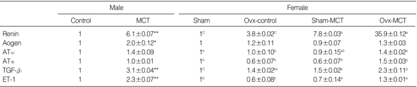
Changes in gene expressions of the cardiac RAS components, TGF- 1, and endothelin-1 are shown in Table 2. Renin mRNA level both in RV and LV was very low in a basal state (Fig. 1), but ovariectomy increased RV renin mRNA level 3.8-fold. The Ovx-MCT group showed 9.4-fold higher RV renin mRNA level than the Ovx-control group. The male- and sham-MCT groups showed approximately 6~8-fold higher RV renin mRNA levels than their respective control groups. LV renin mRNA expression in the MCT-treated male and Ovx rats also increased approximately 2-fold, but the increase was marked- ly lower than the RV level (data not shown). Neither the Ovx- control nor the sham-MCT group showed alteration in LV renin mRNA expression.
RV angiotensinogen mRNA expression increased two-fold in the male-MCT group, but was not significantly increased in MCT treated female groups. LV angiotensinogen mRNA expression was not significantly changed either in the MCT
treated male, sham or Ovx female groups (data not shown).
Gene expression of Ang II-AT1Aand AT1Breceptors in RV in- creased slightly but significantly only in the Ovx-MCT group.
Four-week treatment of MCT elevated RV TGF- 1mRNA expression 3-fold in male rats, but lesser degree in the sham and the Ovx groups. MCT-treatment slightly elevated TGF- 1 mRNA expression in LV to the lesser degree than that in the RV of the male and the Ovx rats (data not shown). The Ovx- control rats did not show significant increase in TGF- 1mRNA level either in RV or LV. Endothelin-1 mRNA levels increased significantly in the RV of male- and Ovx-MCT rats, but not in the Ovx or sham-MCT rats.
Male Female
Control MCT Sham Ovx-control Sham-MCT Ovx-MCT
Initial BW, g 298±7.5 295±7.6* 225±9.6 232±5.8 236±6.8 232±8.6
Final BW, g 410±14.6 354±8.7* 292±18bc 355±14.1a 280±4.5c 314±13.6b
RV/BW, ×10-4 6.8±0.2 13.5±1.6* 5.9±0.4bc 5.5±0.2c 7.0±0.7b 15.1±1.1a
(LV+S)/BW, ×10-4 20.5±1.1 20.6±0.3 22.0±1.2 20.0±0.7 22.1±0.5 22.5±0.9
RV/(LV+S) 0.33±0.01 0.65±0.07* 0.27±0.01b 0.28±0.01b 0.32±0.04b 0.67±0.04a Lung/BW, ×10-3 4.1±0.34 6.6±0.37* 4.6±0.24b 4.0±0.18b 6.9±0.60a 7.5±0.72a Values are mean±SE of six rats in each group. *p<0.01, vs. control in male rats by t-test. Different superscript letters in female rats show significant differences at p<0.05 by Tukey’s test. RV, right ventricle; LV, left ventricle; S, septum.
Table 1.Body weight (BW), the wet weights of hearts and lungs of the control and monocrotaline (MCT)-injected male rats, as well as the ovariectomized (Ovx) and the sham-operated female rats
Male Female
Control MCT Sham Ovx-control Sham-MCT Ovx-MCT
Renin 1 6.1±0.07** 1C 3.8±0.02C 7.8±0.03b 35.9±0.12a
Aogen 1 2.0±0.12* 1 1.2±0.11 0.9±0.07 1.3±0.03
AT1A 1 1.4±0.09 1b 1.0±0.10b 0.9±0.15ab 1.4±0.02a
AT1B 1 1.0±0.01 1b 0.6±0.07b 0.6±0.07b 1.5±0.03a
TGF- 1 1 3.1±0.04** 1C 1.4±0.02bc 1.5±0.02b 2.3±0.11a
ET-1 1 2.3±0.07** 1b 0.6±0.08b 0.7±0.14b 1.3±0.01a
Values are mean±SE of six rats in each group. *p<0.05, **p<0.01, vs. control in male rats and sham in female rats. Different superscript letters in female rats show significant differences at p<0.05 by Tukey’s test.
Table 2.Ratios of the densitometry readings of the RT-PCR amplification of renin, angiotensinogen (Aogen), angiotensin II receptor subtypes AT1A, AT1B, TGF- 1and endothelin-1 and GAPDH mRNAs in the right ventricle from the control and monocrotaline (MCT)- injected male rats, as well as the ovariectomized (Ovx) and the sham-operated female rats
Renin
MCT GAPDH
Con MCT Sham Ovx Sham Ovx
Fig. 1.Representative photographs of the RT-PCR products for renin mRNA in the right ventricle of the control and monocrotaline (MCT)-treated male rats, and the ovariectomized (Ovx) and the sham-operated female rats. GAPDH mRNA was amplified in par- allel for the internal control.
♂ ♀
Effect of Estrogen and Enalapril on the Progression of the Pulmonary Hypertension and RV Hypertrophy:
Histological Evaluation
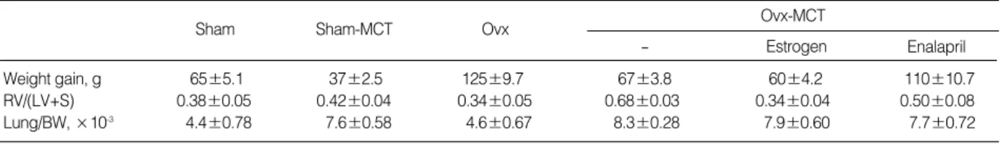
To substantiate the notion of beneficial effects of estrogen and inhibition of the renin-angiotensin system on the progres- sion of the RV hypertrophy, histological changes of the lung and RV were examined in female rats. RV hypertrophy was prominent in the Ovx-MCT rats, but not in the sham-MCT rats and estrogen or enalapril treated Ovx-MCT rats (Table
3). However, lung weight increased in all of the sham-MCT and the Ovx-MCT rats treated with or without estrogen and enalapril.
Histological changes of the lung and RV are shown in Fig.
2 and 3, respectively. Compared to the sham-operated female rats, the lungs of the Ovx rats showed increased alveolar size, partial atelectasis, thickened interalveolar septa, and slightly hypertrophied walls of the small arteries. Lungs of the sham- MCT rats showed prominent atelectasis and hypertrophied walls of the small arteries, denoting developed pulmonary hyper-
Fig. 2.Light microscopic findings of the lung tissues of the sham-operated (A) or the ovariectomized (Ovx) female rats (B) treated with or without monocrotaline (MCT). Hematoxylin and eosin stain, scale bar=50 m. The Ovx rats (B) show histological changes such as increased alveolar size, inflammatory cells (arrows), and mild bronchial obstruction. The sham-MCT rats (C) show hypertrophy of small arteries (white arrows) and atelectasis (arrow), denoting pulmonary hypertension. The Ovx-MCT (D) rats show severe pathological changes such as dif- fused atlectasis, fibrosis and hypertrophy of vascular smooth muscle. In contrast, estrogen (E) or enalapril (F) treatment to the Ovx-MCT rats markedly ameliorate the pathological findings, i.e., patent alveoli, and less prominent hypertrophy of vascular wall.
A
D
B
E
C
F Ovx-MCT
Sham Sham-MCT Ovx
- Estrogen Enalapril
Weight gain, g 65±5.1 37±2.5 125±9.7 67±3.8 60±4.2 110±10.7
RV/(LV+S) 0.38±0.05 0.42±0.04 0.34±0.05 0.68±0.03 0.34±0.04 0.50±0.08
Lung/BW, ×10-3 4.4±0.78 7.6±0.58 4.6±0.67 8.3±0.28 7.9±0.60 7.7±0.72
Values are mean±SE of 3 rats in each group.
Table 3.Body weight gain, and heart and lung weights of the sham-operated or the ovarictomized (Ovx) female rats treated with monocrotaline (MCT) with or without administration of estrogen or enalapril
tension. The Ovx-MCT rats, in comparison with the sham- MCT rats, showed very severe pathological changes such as obliteration of most alveoli, prominent fibrosis, infiltration of inflammatory cells, bronchial obstruction, and hypertrophied vessel walls. The histological findings were similar between the Ovx-MCT and the male-MCT rats (data not shown). How- ever, the treatment by using estrogen or enalapril to the Ovx- MCT rats markedly ameliorated the patholocial changes of the lungs; patent and almost intact alveoli, and less prominent hypertrophy of blood vessels.
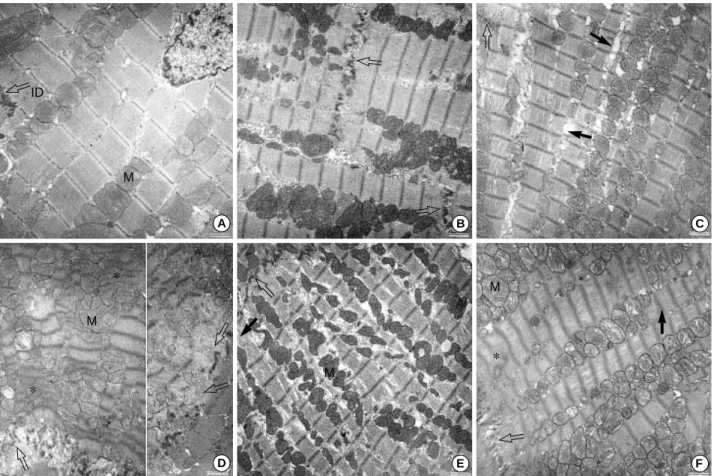
By the electron microscopy examination of the RV, the sham- MCT rats did not reveal abnormal findings of the cardiac myo- cytes, i.e., an intact intercalated disk and striated pattern, but slightly dialated intramembranous spaces of the diad system of the T-tuble. In contrast, the Ovx-MCT rats showed very severe injuries of the hypertrophied RV tissue, i.e., disruption of the intercalated disk, irregular pattern of cross-striation, formation of hypercontraction band, fraying of myofibrils, and aggregation of enlarged mitochondria. Treatment of estrogen
or enalapril to the Ovx-MCT rats remarkably ameliorated the ultrastructures of the RV myocytes toward the normal.
DISCUSSION
The present study shows that RV hypertrophy was not in- duced by MCT treatment in female rats, not like in male rats, as reported previously (4, 14), but was exacerbated after ovariec- tomy. Although the MCT-treated female rats did not show RV hypertrophy, they showed histological changes-denoting moderate pulmonary hypertension. The Ovx-MCT rats showed remarkable RV hypertrophy with very severe pathological changes in the lung and RV, and enhanced gene expressions of the RV RAS. Treatment by using estrogen or enalapril to the Ovx-MCT rats prevented the RV hypertrophy, and remark- ably ameliorated the ultrastructural changes in the lung and RV. These findings from this rat model suggest that estrogen and the inhibition of the renin-angiotensin system have pro-
Fig. 3.Electron microscopic findings of the right ventricular wall of the sham-operated (A) or the ovariectomized (Ovx) female rats (B) treat- ed with or without monocrotaline (MCT). Uranyl acetate and lead citrate stain, scale bar=2 m. The sham-MCT rats (C) show intact interca- lated disk (line arrow) and striated pattern, but dilated intramembranous spaces of the diad system of T-tubule and sarcoplasmic reticu- lum (arrows). The Ovx-MCT rats (D) show severe myocardial injuires such as disruption of the intercalated disk (line arrow), formation of a hypercontraction band (asterik), an irregular pattern of cross-striation, and an aggregation of enlarged mitochondria (M). The right side shows the disoriented myofilaments of the degenerated cardiac myocyte at the intercalated disk. Estrogen (E) or enalapril (F) treatment to the Ovx-MCT rats ameliorate the ultrastructure of the RV striated patterns, intercalated disc and mitochondria.
A
D
B
E
C
F M
M
M
*
*
ID
M
*
tective functions against the development of the pulmonary hypertension and cardiac remodeling.
The protective influences of estrogen on the development of RV hypertrophy in the present study could potentially result from both direct and indirect actions on the heart. Estrogen has been recognized to directly influence cardiac as well as vascular cells (18). Indeed, functional estrogen - and -recep- tors have been demonstrated in the cardiac myocytes and fi- broblasts as well as vascular endothelial and smooth muscle cells (19, 20). Several months after ovariectomy, abnormal cardiac mass, function, and biochemistry were produced in rats (21, 22). Estrogen treatment after ovariectomy prevented left ventricular hypertrophy in rats with spontaneously hyperten- sive heart failure (21), and improved cardiac performance fol- lowing global ischemia and reperfusion (23, 24). Estrogen also attenuated the cardiac hypertrophic response to pressure over- load by transverse aortic constriction in ovariectomized mice (25). The antihypertrophic effect was not mediated by the blood pressure-lowering effect of estrogen. Therefore, these results support the notion that estrogen has direct protecting effects on cardiac myocytes and heart.
Considering ultrastructural findings of the lung and RV in the present study, additional possibility is that the less degree RV hypertrophy in the sham-MCT female and the E2-treated Ovx-MCT rats is secondary to lower pulmonary hyperten- sion. Although pulmonary arterial pressure was not measured directly in the present study, more severe and prevalent hyper- trophy of the pulmonary arterial wall of the Ovx-MCT rats may suggest higher pulmonary hypertension. Considering the severe atelectasis and lung injury of the Ovx-MCT rats, it is possible that hypoxia also contributed to the observed arte- rial remodeling and pulmonary hypertension. Several studies suggested that ovariectomy augmented chronic hypoxia- induced pulmonary hypertension, RV hypertrophy and arte- rial remodeling (26), whereas estrogen attenuated the devel- opment of pulmonary hypertension induced either by chronic hypoxia (26) or MCT (27) in rats. In hypoxia-induced pulmonary hypertension, the protective effects of estrogen appear to be mediated in part by reduced pulmonary ET-1 expression (28) and associated arterial modeling (26), and decreased polycythemia (29). Direct beneficial effects of estrogen on the lung tissue have been reported that ovariec- tomy decreased gas-exchange surface area and increased alveolar size, whereas treatment of estrogen prevented the changes (30). Taken together, estrogen may attenuate the MCT-induced RV hypertrophy secondary to diminished pulmonary hypertension. To differentiate relative impor- tance of direct and indirect cardioprotective roles for estro- gen in this animal model, the development of RV hypertro- phy should be compared at similar levels of pulmonary hyper- tension.
Myocardial hypertrophy is the compensatory response to a chronic elevation in mechanical workload. It is a complex process that involves cardiac cell growth and accumulation
of extracellular matrix proteins (6). In cultured cardiac fibro- blast, estrogen inhibited cell growth, and collagen synthesis (31). In the pressure-overloaded heart, several vasoactive sub- stances and growth factors synthesized and released from the heart may be involved in the process of cardiac remodeling (6). A critical role of the cardiac RAS, among others, has been recognized (7). Actually, beneficial effects of an inhibition of RAS has been shown to decrease ventricular remodeling in- duced by chronic hypoxia (32, 33) and myocardial infarction (34, 35).
The present study showed enhanced gene expression of the renin-angiotensin system components in the hypertrophied RV of the male MCT rats, as we previously reported (15). Inter- estingly, although the sham-MCT female rats did not show RV hypertrophy, they showed a similar degree of increase in RV renin mRNA level as the male MCT rats. However, the Ovx-MCT rats showed remarkably greater increase in renin mRNA level in the hypertrophied RV. This result may suggest significant interactions between estrogen and cardiac RAS, and raise a question whether estrogen inhibit cardiac renin expression. Angiotensinogen mRNA level was also increased in the hypertrophied RV of the Ovx-MCT rats, but to a less degree. Although ACE mRNA was not measured in the present study, we previously reported increased ACE mRNA in the hypertrophied RV of MCT-male rats (15). Increased ACE activity and expression in the hypertrophied RV (36), and plasma levels of renin, ACE and angiotensin II (32) have also been demonstrated in rats with hypoxic pulmonary hyperten- sion. In contrast, AT1 receptors do not seem to be changed in the hypertrophied RV. Adamy et al. (37) also showed no change in AT1 and AT2 receptors in RV and LV of the MCT- treated male rats.
Elevated cardiac renin-angiotensin system suggests possi- ble contribution to the MCT-induced RV hypertrophy. This hypothesis was supported by the present result that treatment of enalapril, an ACE inhibitor, prevented RV hypertrophy and profoundly ameliorated ultrastructural changes of the lung and RV in the Ovx-MCT rats. Enalapril and estrogen produced very similar effects. Previous studies also demonstrated the benefit of ACE inhibitors and AT1 receptor antagonists against the development of MCT (38)- and hypoxia (32, 33)-induced pulmonary hypertnesion and RV hypertrophy in male rats.
Inhibitory effects of enalapril against the development of RV hypertrophy in the present study may also be consequences of direct cardiac action and secondary to diminished pulmonary hypertension. In addition, further studies are needed to eval- uate possible influence of enalapril-induced decrease in systemic blood pressure on the MCT-induced cardiopulmonary patho- genesis.
Increased gene expression of TGF- 1and endothelin-1 in the hypertrophied RV of the male-MCT and the Ovx-MCT rats in the present study confirms our previous findings (15).
Both TGF- 1and endothelin-1 are known to stimulate the production and accumulation of extracellular matrix proteins
in myocardial hypertrophy and fibrosis (39, 40). A blockade of endothelin-1 prevented MCT-induced pulmonary hyper- tension and RV hypertrophy (41, 42) suggesting that endoge- nous endothelin-1 contributes to the progression of cardiopul- monary alterations. On the other hand, TGF- 1mRNA expres- sion was slightly but significantly increased even in the unhy- pertrophied RV of the Ovx control and the sham-MCT female rats, but increased further in the hypertrophied RV of the Ovx- MCT rats. The result from this rat model may suggest a sup- pressive effect of estrogen on cardiac TGF- 1expression and cardiac remodeling.
In summary, our results demonstrate gender differences and beneficial effects of the ovarian function and the inhibition of the renin-angiotensin system on the development of MCT- induced pulmonary hypertension and RV hypertrophy. En- hanced gene expression of the cardiac renin-angiotensin sys- tem, TGF- 1and endothelin-1 in the hypertrophied RV of the Ovx-MCT rats may suggest the interaction of these cardiac vasoactive substances with estrogen. However, further inves- tigations are required to understand underlying mechanisms of the interactions in the pathogenesis of heart disease.
ACKNOWLEDGMENTS
This work was supported by a research grant from the Korea Science and Engineering Foundation (R04-2001-00086).
REFERENCES
1. Collins P, Beale CM. Cardiovascular disease in women. In: The cardio- protective role of HRT: a clinical update. New York: The Parthenon Publishing Groups 1996; 9-11.
2. Stampfer MJ, Colditz GA. Estrogen replacement therapy and coronary heart disease: a quantitative assessment of the epidemiologic evidence.
Prev Med 1991; 20: 47-63.
3. Crofton JT, Ota M, Share L. Role of vasopressin, the renin-angiotensin system and sex in Dahl salt-sensitive hypertension. J Hypertens 1993;
11: 1031-8.
4. Sawada A, Kuriyama T, Sugita T, Watanabe S. Monocrotaline-induced pulmonary hypertension with special reference to the influence of dose, age and sex. Nihon Kyobu Shikkan Gakkai Zasshi 1985; 23: 578-83.
5. Okuniewski R, Davis EA, Jarrott B, Widdop RE. A comparison of the development of renal hypertension in male and female rats. Clin Sci (Lond) 1998; 95: 445-51.
6. Swynghedauw B. Molecular mechanisms of myocardial remodeling.
Physiol Rev 1999; 79: 215-62.
7. Inagami T. The centenial of renin discovery. Hypertension 1988; 32:
953-7.
8. Yamazaki T, Komuro I, Yazaki Y. Role of the renin-angiotensin sys- tem in cardiac hypertrophy. Am J Cardiol 1999; 83: 53H-7.
9. Studer R, Reinecke H, Muller B, Holtz J, Just H, Drexler H. Increased angiotensin-I converting enzyme gene expression in the failing human
heart: quantification by competitive RNA polymerase chain reaction.
J Clin Invest 1994; 94: 301-10.
10. Johnson C, Fabris B, Yoshida K. The cardiac renin-angiotensin sys- tem in heart failure. Am Heart J 1993; 126: 756-60.
11. Schunkert H, Dzau VJ, Tang SS, Hirsch AT, Apstein CS, Lorell BH.
Increased rat cardiac angiotensin converting enzyme activity and mRNA expression in pressure overload left ventricular hypertrophy.
Effects on coronary resistance, contractility and relaxation. J Clin Invest 1990; 86: 1913-20.
12. Kramer CM, Ferrari VA, Rogers WJ, Theobald TM, Nance ML, Axel L, Reichek N. Angiotensin-converting enzyme inhibition limits dys- function in adjacent noninfarcted regions during left ventricular remod- eling. J Am Coll Cardiol 1996; 27: 211-7.
13. Pfeffer MA, Lamas GA, Vaughan DE, Parisi AF, Braunwald E. Effect of captopril on progressive ventricular dilation after anterior myocar- dial infarction. N Engl J Med 1988; 319: 80-6.
14. Kiyatake K, Kakusaka I, Kasahara Y, Qi TM, Suzuki A, Nakano K, Kaneko N, Kitada M, Kuriyama T. [Relationship between the convert- ing ability of liver microsomes and monocrotaline-induced pulmonary hypertension in male, female and castrated male rats.] Nihon Kyobu Shikkan Gakkai Zasshi 1994; 32: 125-9.
15. Park HK, Park SJ, Kim CS, Paek YW, Lee JU, Lee WJ. Enhanced gene expression of renin-angiotensin system, TGF- 1, endothelin-1 and nitric oxide synthase in right-ventricular hypertrophy. Pharma- col Res 2001; 43: 265-73.
16. Chomczinski P, Sacchi N. Single-step of RNA isolation by acid guani- dium thiocyanate-phenol-chloroform extraction. Anal Biochem 1987;
162: 156-9.
17. Jo H, Yang EK, Lee WJ, Park KY, Kim HJ, Park JS. Gene expression of central and peripheral renin-angiotensin system components upon dietary sodium intake in rats. Regul Pept 1996; 67: 115-21.
18. Farhat MY, Lavigne MC, Ramwell PW. The vascular protective effects of estrogen. FASEB J 1996; 10: 615-24.
19. Karas RH, Patterson BL, Mendelsohn ME. Human vascular smooth muscle cells contain functional estrogen receptor. Circulation 1994;
89: 1943-50.
20. Grohe C, Kahlert S, Lobbert K, Vetter H. Expression of oestrogen receptor alpha and beta in rat heart: role of local oestrogen synthesis.
J Endocrinol 1998; 156: R1-7.
21. Sharkey LC, Holycross BJ, Park S, Shiry LJ, Hoepf TM, McCune SA, Radin MJ. Effect of ovariectomy and estrogen replacement on cardiovascular disease in heart failure-prone SHHF/Mcc-facprats.
J Mol Cell Cardiol 1999; 31: 1527-37.
22. Schaible T, Malhotra A, Ciambrone G, Scheuer J. The effects of gon- adectomy on left ventricular function and cardiac contractile proteins in male and female rats. Circ Res 1984; 54: 38-49.
23. Kolodgie FD, Farb A, Litovsky SH, Narula J, Jeffers LA, Lee SJ, Vir- mani R. Myocardial protection of contractile function after global ischemia by physiologic estrogen replacement in the ovariectomized rat. J Mol Cell Cardiol 1997; 29: 2403-14.
24. Fraser H, Davidge ST, Clanachan AS. Enhancement of post-ischemia myocardial function by chronic 17 -estradiol treatment: role of alter- ations in glucose metabolism. J Mol Cell Cardiol 1999; 31: 1539-49.
25. van Eickels M, Grohe C, Cleutjens JPM, Janssen BJ, Wellens HJJ,
′ . .
Doevendans PA. 17 -estradiol attenuates the development of pres- sure-overload hypertrophy. Circulation 2001; 104: 1419-23.
26. Resta TC, Kanagy LN, Walker BR. Estradiol-induced attenuation of pulmonary hypertension is not associated with altered eNOS expres- sion. Am J Physiol Lung Cell Mol Physiol 2001; 280: L88-97.
27. Farhat MY, Chen MF, Bhatti T, Iqbal A, Cathapermal S, Ramwell PW. Protection by oestradiol against the development of cardiovas- cular changes associated with monocrotaline pulmonary hyperten- sion in rats. Br J Pharmacol 1993; 110: 719-23.
28. Earley S, Resta TC. Estradiol attenuates hypoxia-induced pulmonary endothelin-1 gene expression. Am J Physiol Lung Cell Mol Physiol 2002; 283: L86-93.
29. Mukundan H, Resta TC, Kanagy NL. 17 -estradiol decreases hypox- ic induction of erythropoietin gene exprepssion. Am J Physiol Regul Integr Comp Physiol 2002; 283: R496-504.
30. Massaro GD, Mortola JP, Massaro D. Estrogen modulates the dimen- sions of the lung’s gas-exchange surface area and alveoli in female rats. Am J Physiol 1996; 270: L110-4.
31. Dubey RK, Gillespie DG, Jackson EK, Keller PJ. 17Beta-estradiol, its metabolites, and progesterone inhibit cardiac fibroblast growth.
Hypertension 1998; 31: 522-8.
32. Kentera D, Susic D, Cvetkovic A, Djordjevic G. Effects of SQ 14.225, an orally active inhibitor of angiotensin-converting enzyme, on hypox- ic pulmonary hypertension and right ventricular hypertrophy in rats.
Basic Res Cardiol 1981; 76: 344-51.
33. Pelouch V, Kolar F, Ost’adal B, Milerova M, Cihak R, Widimsky J.
Regression of chronic hypoxia-induced pulmonary hypertension, right ventricular hypertension, and fibrosis: effect of enalapril. Cardiovasc Drugs Ther 1997; 11: 177-85.
34. Kramer C M, Ferrari V A, Rogers W J, Theobald T M, Nance M L, Axel L, Reichek N. Angiotensin-converting enzyme inhibition limits dysfunction in adjacent noninfarcted regions during left ventricular
remodeling. J Am Coll Cardiol 1996; 27: 211-7.
35. Pfeffer MA, Lamas GA, Vaughan DE, Parisi AF, Braunwald E. Effect of captopril on progressive ventricular dilation after anterior myocar- dial infarction. N Engl J Med 1988; 319: 80-6.
36. Morrell NW, Danilov SM, Satyan KB, Morris KG, Stenmark KR.
Right ventricular angiotensin converting enzyme activity and expres- sion is increased during hypoxic pulmonary hypertension. Cardiovasc Res 1997; 34: 393-403.
37. Adamy C, Oliviero P, Eddahibi S, Rappaport L, Samuel JL, Teiger E, Chassagne C. Cardiac modulations of ANG II receptor expression in rats with hypoxic pulmonary hypertension. Am J Physiol Heart Circ Physiol 2002; 283: H733-40.
38. Kanno S, Wu YJ, Lee PC, Billiar TR, Ho C. Angiotensin-converting enzyme inhibitor preserves p21 and endothelial nitric oxide synthase expression in monocrotaline-induced pulmonary arterial hyperten- sion in rats. Circulation 2001; 104: 945-50.
39. Border WA, Nobel NA. Transforming growth factor- in tissue fibro- sis. N Engl J Med 1994; 331: 1286-92.
40. MacLellan WR, Brand T, Schneider MD. Transforming growth fac- tor-beta in cardiac ontogeny and adaptation. Circ Res 1993; 73: 783- 91.
41. Miyauchi T, Yorikane R, Sakai S, Sakurai T, Okada M, Nishikibe M, Yano M, Yamaguchi I, Sugishita Y, Goto K. Contribution of endoge- nous endothelin-1 to the progression of cardiopulmonary alterations in rats with monocrotaline-induced pulmonary hypertension. Circ Res 1993; 73: 887-97.
42. Prie S, Leung TK, Cernacek P, Ryan JW, Dupuis J. The orally active ETAreceptor angatonist (+)-(S)-2-(4,6-dimethyoxy-pyrimidine-2- yloxy)-3-methoxy-3,3-diphenyl-propionic acid (LU 135252) prevents the development of pulmonary hypertension and endothelial metabol- ic dysfunction in monocrotaline-treated rats. J Pharmacol Exp Ther- ap 1997; 282: 1312-8.
′