INTRODUCTION
Insulin resistance and hyperinsulinemia are associated with hypertension, impaired glucose tolerance, obesity and dyslipi- demia (1-4). These factors are also closely related to the devel- opment of coronary heart disease. Hyperinsulinemia is char- acterized by low high density lipoprotein (HDL)-cholesterol and high low density lipoprotein (LDL)-cholesterol, which causes coronary arteriosclerosis. This reflects the rate of mor- tality due to cardiovascular diseases.
To date, euglycemic hyperinsulinemic clamp test has been accepted as a standard test that measures insulin resistance accurately (5). However, since euglycemic hyperinsulinemic clamp test is very complex as well as requires skillful techni- cians and many instrument, it has not been widely used in a clinical setting. Alternately, homeostasis model assessment (HOMA) index was designed to assess insulin resistance in many patients, in which the concentrations of fasting insulin and glucose are simply measured, and recently has been wide- ly used (6, 7).
Considering that insulin resistance is the risk factor for coro- nary heart disease, it may be closely related to the prognosis of patients who underwent percutaneous coronary interven- tion (PCI). To date, however, few studies have been conduct-
ed to examine whether insulin resistance is correlated with the prognosis of patients who underwent PCI. Some studies have reported that severe coronary angiographic findings and stent restenosis were closely associated with insulin resistance (8, 9).
Based on the assumption that insulin resistance is closely associated with the complications after PCI or the prognosis after PCI, we conducted a 30-day follow-up clinical study to observe major adverse cardiac events (MACE) in patients who underwent PCI.
MATERIALS AND METHODS Subjects
In this study, we examined 98 consecutive patients with chest pain who underwent diagnostic coronary angiography between May and September 2004. We excluded patients who were treated for diabetes mellitus; those who were newly diagnosed as diabetes mellitus; those who had thyroid or ad- renal insufficiency; those who underwent PCI for coronary heart disease within a recent 6-month period; and those who underwent primary PCI for acute myocardial infarction. The
Kyeong Ho Yun, Myung Ho Jeong, Kye Hun Kim, Young Joon Hong, Hyung Wook Park, Ju Han Kim, Young Keun Ahn, Jeong Gwan Cho, Jong Chun Park, Nam-Ho Kim*, Seok Kyu Oh*, Jin-Won Jeong*, Jung Chaee Kang
The Heart Center of Chonnam National University Hospital, Gwangju; Department of Cardiovascular Medicine*, Wonkwang University Hospital, Iksan, Korea
Address for correspondence Myung Ho Jeong, M.D.
The Heart Center of Chonnam National University Hospital, 8 Hak-dong, Dong-gu, Gwangju 501-757, Korea
Tel : +82.62-220-6243, Fax : +82.62-228-7174 E-mail : [email protected]
212
The Effect of Insulin Resistance on Prognosis of Non-Diabetic Patients Who Underwent Percutaneous Coronary Intervention
Insulin resistance is an important risk factor for coronary artery disease. However, there has been no data regarding its clinical effect on the outcomes of percutaneous coronary intervention (PCI) in non-diabetic patients. We analyzed 98 non-diabetic consecutive patients (59±11.5 yr, male:female=63:35) who underwent elective coronary angiography. The patients were divided into two groups: Group I (n=71;
the value of HOMA-IR [homeostasis model assessment of insulin resistance] <2.6) and Group II (n=27; the value of HOMA-IR ≥2.6). In-hospital and 30-day major adverse cardiac events (MACE) were compared between the two groups. The con- centrations of fasting insulin and triglyceride were significantly higher in Group II than in Group I. Significant correlations were observed between the value of HOMA-IR and body mass index (r=0.489, p<0.001), levels of total cholesterol (r=0.204, p=
0.045), triglyceride (r=0.334, p=0.001) and apolipoprotein B (r=0.212, p=0.038). PCI was performed in 59 patients (60.2%). In-hospital and 30-day MACE were higher in Group II than Group I (2.4% vs. 27.8%, p=0.008; 2.4% vs. 27.8%, p=0.008). Multi- variate analysis revealed that the value of HOMA-IR ≥2.6 was an independent predictor of MACE. Increased HOMA-IR level is an important prognostic indicator in non-diabetic patients underwent PCI.
Key Words : Coronary Disease; Insulin; Prognosis; Angioplasty
Received : 15 June 2005 Accepted : 13 October 2005
clinical diagnosis on admission was stable angina pectoris in 29.6% (29 patients) and unstable angina pectoris in 70.4%
(69 patients). On admission, patients were interviewed to collect such data as the risk factors for coronary heart disease, the personal history of smoking, diabetes mellitus and hyper- tension, the family history and the past history of myocardial infarction or stroke. All patients gave informed consent ac- cording to a protocol approved by the Chonman National University Hospital Ethics Committee.
Blood samples and neasurements
In our patients, samples were collected from venous blood after overnight fasting, and blood chemistry was performed.
Fasting plasma glucose and insulin were measured. Then, the following parameters were measured: 1) the concentrations of serum lipid including total cholesterol, triglyceride, HDL- cholesterol, LDL-cholesterol, Apolipoprotein AI (Apo AI), Apolipoprotein B (Apo B) and lipoprotein (a) [Lp (a)]; 2) leukocyte count, monocyte count, erythrocyte sedimentation rate (ESR) and high-sensitivity C-reactive protein (hsCRP);
and 3) the concentrations of fibrinogen, fibrinogen degrada- tion product (FDP) and homocysteine.
Insulin resistance was calculated by the homeostasis model assessment of insulin resistance (HOMA-IR), proposed by Mattews et al., whose formula was: HOMA-IR (mg/dL×U/
mL)=fasting glucose (mg/dL)×fasting insulin (U/mL)/405 (6).
To determine cut-off points of the HOMA-IR as predictors of in-hospital and 30-day adverse cardiac events, receiver oper- ating characteristics (ROC) analyses were performed. The area under curve (AUC) of HOMA-IR was 0.738 (p=0.006). Cut- off points of HOMA-IR for in-hospital and 30-day cardiac events, determined by ROC analysis, were 2.56, 2.60, respec- tively. Therefore, we used 2.6 as a cut-off point for the anal- ysis in clinical usefulness. The cut-off point for in-hospital and 30-day adverse cardiac events yielded sensitivities of 66.8
% and 77.9% and specificities of 68.2% and 70.2%, respec- tively. Based on the cut-off value of 2.6, therefore, we divid- ed patients into Group I (the value of HOMA-IR <2.6) and Group II (the value of HOMA-IR ≥2.6).
Coronary angiography and intervention
We accessed left or right femoral artery by the Seldinger method or accessed radial artery, and performed coronary an- giography using the Judkins method. Coronary angiographic findings were interpreted by two examiners who were blinded to patients’ profile of insulin resistance. Significant stenosis was defined as a luminal narrowing of 50% or greater (10).
PCI was performed according to current clinical practice at physician’s discretion. Stenting was performed in cases whom percutaneous balloon dilatation produced suboptimal results that the residual stenosis was more than 30%, and the dissec- tion was developed (11, 12). On diagnostic coronary angio-
graphy, the patency of the treated artery was evaluated by the Thrombolysis In Myocardial Infarction (TIMI) score. Suc- cessful reperfusion following PCI was defined as TIMI III flow with <25% residual stenosis (13).
In all patients, aspirin (300 mg/day) and clopidogrel (150 mg/day) were loaded or aspirin (100 mg daily) and clopido- grel (75 mg daily) started >3 days before procedure. An intra- venous bolus of 5,000 U of unfractionated heparin was given, and then additional heparin boluses were given to maintain activated clotting time >300 sec during procedure. Aspirin (100 mg/day) and clopidogrel (75 mg/daily for 30 days) were prescribed to all patients after procedure.
In-hospital adverse outcomes were defined as the overall procedure was not successfully done since coronary wire or balloon catheter could not pass through the lesion; myocardial infarction was developed following PCI; patients underwent repeated PCI or emergent coronary artery bypass graft (CABG) surgery for target vessel revascularization (TVR). Myocardial infarction was defined as a CK-MB elevation greater than three times the upper normal limit or a new change of EKG findings. Between Group I and II, we compared MACE such as death, myocardial infarction and target vessel revasculariza- tion during a 30-day period.
Statistical analysis
Statistical analysis was done using SPSS�Ver.12.0 for Win- dows. All data were represented as mean±standard deviation.
Intergroup analysis was done using independent t-test and
2test, and intragroup analysis was done using paired t-test.
Multivariate analysis was done to determine the factors relat- ed to MACE. Spearman correlation was used to examine the relationship between HOMA-IR and other laboratory find- ings. Statistical significance was set at p<0.05.
RESULTS
In studied patients, the mean age was 59.8±11.5 yr and a male-to-female ratio was 1.8:1. The mean value of HOMA- IR was 2.3±1.69, and 71 patients were into Group I (the value of HOMA-IR<2.6) and 27 patients into Group II (the value of HOMA-IR≥2.6). Between Group I and II, there were no significant differences in cardiovascular risk factors includ- ing hypertension, diabetes mellitus and smoking (Table 1).
The concentrations of fasting plasma glucose were 101.2± 13.89 mg/dL in Group I and 108.4±9.79 mg/dL in Group II, and those of fasting insulin were 11.9±4.99 U/mL in Group I and 31.8±14.23 U/mL in Group II (Table 2). The concentration of triglyceride was significantly higher in Group II than in Group I (151.4±192.43 mg/dL vs. 94.5±62.86 mg/dL, p=0.028). HOMA-IR was correlated with body mass index (r=0.489, p<0.001), levels of total cholesterol (r=0.204, p=0.045), triglyceride (r=0.334, p=0.001), and apolipopro-
tein B (r=0.212, p=0.038).
Coronary angiography revealed the presence of significant stenosis in 64 patients (Group I; 43 patients, Group II; 21 patients). There was no significant differences in the preva- lence of multi-vessel disease between the two groups (p=
0.164). The lesion locations were left anterior descending artery (LAD) in 72.1%, left circumflex artery (LCX) in 16.3%
and right coronary artery (RCA) in 11.6% of Group I; and LAD in 47.6%, LCX in 19.0% and RCA in 33.3% of Group II (p=0.085). According to the American College of Cardiol- ogy and American Heart Association (ACC/AHA) classifica- tion, Type B1was demonstrated in 44.2%, Type B2in 34.9%
and Type C in 20.9% of Group I; and Type B1in 28.6%, Type B2in 33.3% and Type C in 33.3% of Group II (p=
0.283). Preprocedural TIMI flow grades were grade 0 in 16.3
%, grade I in 7.0%, grade II in 9.3% and grade III in 67.4%
of Group I; and grade 0 in 19.0%, grade I in 9.5%, grade II in 19.0% and grade III in 52.5% of Group II (p=0.623).
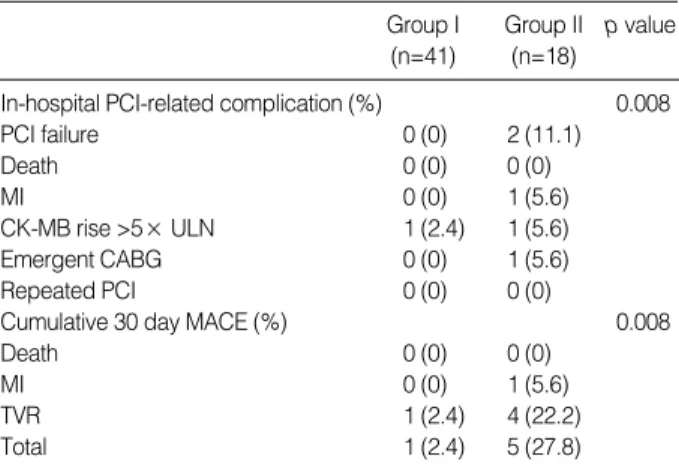
In 59 patients, PCI was performed with an angiographic success rate of 94.9% (Table 3). The procedural failure due to crossing failure of guide wire or balloon catheter occurred in no patient (0.0%) of Group I and 2 patients (11.1%) of Group II. Procedure-related complications included one case of myocardial infarction (2.4%) in Group I; and two cases of myocardial infarction (11.1%), one case of emergent CABG (5.6%) in Group II. The procedure-related complications were more prevalent in Group II than Group I (p=0.008). The MACE at 30-day follow-up was 1 case (2.4%) of TVR in Group I and 4 cases (22.2%) of TVR in Group II, and the incidence of MACE was significantly higher in Group I than Group II (p=0.008).
PCI, percutaneous coronary intervention; MI, myocardial infarction; BMI, body mass index.
Group I (n=71) Group II (n=27) p value
Age (yr) 58±12.1 61±10.7 0.708
Male (%) 44 (62.0) 19 (70.4) 0.488
Hypertension (%) 23 (32.4) 11 (40.7) 0.481
Dyslipidemia (%) 23 (32.4) 11 (40.7) 0.348
Smoker (%) 20 (28.2) 5 (18.5) 0.239
Family history (%) 8 (11.3) 3 (11.1) 0.645
Previous MI (%) 4 (5.6) 0 (0.0) 0.269
Previous PCI (%) 4 (5.6) 3 (11.1) 0.294
Ejection fraction (%) 64.4±10.39 63.7±10.18 0.079 BMI (kg/m2) 23.9±2.41 26.4±3.09 0.116
Clinical diagnosis on admission (%) 0.187
Stable angina 22 (31.0) 7 (25.9)
Unstable angina 49 (69.0) 21 (74.1)
Table 1.Baseline clinical characteristics of Group I (the value of HOMA-IR [homeostasis model assessment of insulin resistance]
<2.6) and Group II (the value of HOMA-IR ≥2.6)
Hb, hemoglobin; HOMA-IR, homeostasis model assessment of insulin resistance; hsCRP, high-sensitivity C-reactive protein; HDL, high densi- ty lipoprotein; LDL, low density lipoprotein.
Group I (n=71) Group II (n=27) p value Laboratory findings
Fasting glucose (mg/dL) 101.2±13.89 108.4±9.79 0.072 Fasting insulin ( U/mL) 11.9±4.99 31.8±14.23 <0.001
HbA1c (%) 5.6±0.38 5.8±0.34 0.404
HOMA-IR 1.4±0.51 4.4±1.82 <0.001
Monocyte (×103) 443.8±223.75 558.1±305.54 0.364
hsCRP (mg/dL) 1.0±3.11 0.8±1.84 0.431
Total cholesterol (mg/dL) 188.9±34.00 189.9±35.33 0.643 Triglyceride (mg/dL) 96.5±62.86 151.4±192.43 0.028 HDL-cholesterol (mg/dL) 50.3±13.19 48.3±14.67 0.755 LDL-cholesterol (mg/dL) 124.9±33.68 121.7±34.31 0.872 Lipoprotein (a) (mg/dL) 27.3±19.53 29.7±21.12 0.415 Apolipoprotein AI (mg/dL) 125.1±27.29 121.8±25.68 0.805 Apolipoprotein B (mg/dL) 98.6±22.70 98.2±23.23 0.621 Homocysteine (mg/dL) 9.6±3.95 9.9±4.45 0.945 Fibrinogen (mg/dL) 264.8±72.01 253.7±71.78 0.671
Coronary angiographic findings 0.164
No significant stenosis (%) 28 (39.4) 6 (22.2) Single vessel disease (%) 28 (39.4) 11 (40.7) Multivessel disease (%) 15 (21.1) 10 (37.0) Table 2.Laboratory and coronary angiographic findings
PTCA, percutaneous transluminal coronary angioplasty; MI, myocardial infarction; ULN, upper limit of normal; CABG, coronary artery bypass graft; TVR, target vessel revascularization.
Group I Group II p value (n=41) (n=18) In-hospital PCI-related complication (%) 0.008
PCI failure 0 (0) 2 (11.1)
Death 0 (0) 0 (0)
MI 0 (0) 1 (5.6)
CK-MB rise >5× ULN 1 (2.4) 1 (5.6)
Emergent CABG 0 (0) 1 (5.6)
Repeated PCI 0 (0) 0 (0)
Cumulative 30 day MACE (%) 0.008
Death 0 (0) 0 (0)
MI 0 (0) 1 (5.6)
TVR 1 (2.4) 4 (22.2)
Total 1 (2.4) 5 (27.8)
Table 3.The incidences of in-hospital and 30-day major adverse cardiac events
HOMA-IR, homeostasis model assessment of insulin resistance; TIMI, Thrombolysis in myocardial infarction; ACC/AHA, American College of Cardiology/American Heart Association.
Variables Odds ratio 95% CI p value
HOMA ≥2.6 49.27 1.05-231.13 0.048
Fasting glucose ≥110 mg/dL 4.58 0.28-83.98 0.277 Fasting insulin ≥20 U/mL 78.54 0.25-251-32 0.138 Preprocedural TIMI flow <3 5.35 0.08-34.69 0.431 ACC/AHA lesion type B2, C 1.29 0.02-66.03 0.899
Age > 65 yr 3.25 0.07-140.38 0.539
Multivessel disease 3.17 1.00-104.88 0.518
Dyslipidemia 5.89 0.17-204.55 0.327
Table 4.Independent predictors for 30-day major adverse car- diac events after percutaneous coronary intervention
Multivariate analysis revealed that the value of HOMA-IR
≥2.6 was the independent prognostic indicators for MACE (p=0.048) (Table 4).
DISCUSSION
Our clinical study indicated that high HOMA-IR value is related with the high incidence of complications and short- term major adverse cardiac events (MACE) following PCI even in non-diabetic patients.
Insulin resistance plays a crucial role in the development of metabolic disorder accompanied by obesity, dyslipidemia, dia- betes mellitus, impaired glucose tolerance and hypertension;
and is also closely related to the incidence of cardiovascular disease (14-22). Hyperinsulinemia is an only indirect indica- tor for insulin sensitivity. Several large-scale studies have re- vealed that hyperinsulinemia is closely associated with the mortality due to cardiovascular disease (14, 21, 23). To date, however, few large-scale studies have been conducted to exam- ine the relationship between insulin resistance and coronary heart disease (24, 25). Recently, IRAS (insulin resistance atheroscleorsis study) group has conducted large-scale epi- demiologic studies, and has reported that insulin resistance rather than insulin concentration is an independent, power- ful risk factor for coronary heart disease (26).
The mechanism by which insulin resistance provokes car- diovascular disease is mainly associated with the development of metabolic syndrome. It is well established that non-diabet- ic patients with insulin resistance exhibits high concentration of serum triglyceride, hypertension and low concentration of HDL-cholesterol (1, 4, 23, 24). In recent years, among several types of apolipoprotein that determines the conformational stability and the metabolic direction of lipoprotein, apolipop- rotein B has been elevated particularly in patients with insulin resistance (27, 28). This has indicated that apolipoprotein B is associated with the development of cardiovascular disease.
In addition, insulin resistance has been reported to reduce flow-mediated vasodilation (FMD) of brachial artery, and to thereby trigger the endothelial dysfunction (20). The present study has shown that the value of HOMA-IR was correlated with levels of triglyceride and inversely correlated with that of HDL-cholesterol, which is similar to the results of previous studies (1, 23, 24).
To date, only few studies based on coronary angiography have examined the relationship between insulin resistance and coronary atherosclerosis (8, 24, 25). According to Takeza- ko et al., the profile of insulin resistance based on HOMA-IR model was correlated with severity of coronary atherosclero- sis based on Gensini’s score (8). Other studies have reported that the incidence of MACE following PCI was higher in non- diabetic patients with high concentration of HbA1c than those with low concentration of HbA1c, although these stud- ies did not evaluate using insulin resistance (29). Here, the
concentration of HbA1c was lower in non-diabetic patients than in diabetic patients although it was ‘high’. On the other hand, some studies have reported that hyperinsulinemia and insulin resistance measured by HOMA are closely associated with restenosis following stenting in non-diabetic patients (9, 30). In our series, we have predicted that since insulin re- sistance is the risk factor for coronary atherosclerosis and is associated with the mortality due to cardiovascular disease, it will affect the prognosis following PCI. As predicted, the re- sults were that the higher value of HOMA-IR was associated with the higher incidence of MACE, since more TVR was performed in cases with higher value of HOMA-IR. Although our results were based on non-diabetic patients, these findings suggested that patients with high insulin resistance exhibited high incidences of complex and heavy calcified lesions, com- monly noted in coronary artery of patients with diabetes melli- tus. Henceforth, comparative studies will be conducted regard- ing this matter between diabetic and non-diabetic patients.
The limitation of present study has disclosed that it exam- ined the small number of patients within a short period of time. To date, however, few studies have been conducted to examine whether insulin resistance is correlated with the prognosis following PCI. For this reason, the present study has its own value that it is a preliminary study for further large-scale studies. Moreover, the present study failed to tes- tify the reproducibility since it did not measure the value of HOMA-IR in a repetitive manner during the period of ad- mission. The concentrations of serum glucose and insulin can be altered at each different measuring time, although the pre- sent study measured them only once in the morning that diagnostic coronary angiogram was performed. However, HOMA-IR index is well reflected in euglycemic hyperinsu- linemic clamp test, a standard test, and the clinical useful- ness has been well established.
In conclusion, insulin resistance is associated with poor prognosis in non-diabetic patients after PCI. However, long- term large clinical follow-up studies should be conducted in diabetic and non-diabetic patients.
REFERENCES
1. Laws A, Reaven GM. Evidence for an independent relationship between insulin resistance and fasting plasma HDL-cholesterol, tri- glyceride and insulin concentrations. J Intern Med 1992; 231: 25-30.
2. Reaven GM. Role of insulin resistance in human disease. Diabetes 1988; 37: 1595-607.
3. Isomaa B, Almgren P, Tuomi T, Forsen B, Lahti K, Nissen M, Task- inen MR, Groop L. Cardiovascular morbidity and mortality associ- ated with the metabolic syndrome. Diabetes Care 2001; 24: 683-9.
4. Mykkanen L, Zaccaro DJ, Wagenknecht LE, Robbins DC, Gabriel M, Haffner SM. Microalbuminuria is associated with insulin resis- tance in nondiabetic subjects. Diabetes 1998; 47: 793-800.
5. DeFronzo RA, Tobin JD, Andres R. Glucose clamp technique: a
method for quantifying insulin secretion and resistance. Am J Physiol 1979; 237: E214-23.
6. Matthews DR, Hosker JP, Rudenski AS, Naylor BA, Treacher DF, Turner RC. Homeostasis model assessment: insulin resistance and -cell function from fasting plasma glucose and insulin concentra- tions in man. Diabetologia 1985; 28: 412-9.
7. Haffner SM, Miettinen H, Stern MP. The homeostasis model in the San Antonio heart study. Diabetes Care 1997; 20: 1087-92.
8. Takezako T, Saku K, Zhang B, Shirai K, Arakawa K. Insulin resis- tance and angiographical characteristics of coronary atherosclero- sis. Jpn Circ J 1999; 63: 666-76.
9. Radke PW, Voswinkel M, Reith M, Kaiser A, Haager PK, Hanrath P, Hoffmann R. Relation of fasting insulin plasma levels to resteno- sis after elective coronary stent implantation in patients without dia- betes mellitus. Am J Cardiol 2004; 93: 639-41.
10. Austen WG, Edwards JE, Frye RL, Gensini GG, Gott VL, Griffith LS, McGoon DC, Murphy ML, Roe BB. A reporting system on pa- tients evaluated for coronary artery disease. Circulation 1975; 51:
5-40.
11. Ellis SG, Vandormael MG, Cowley MJ, DiSciascio G, Deligonul U, Topol EJ, Bulle TM. Coronary morphologic and clinical determinants of procedural outcome with angioplasty for multivessel coronary dis- ease. Implications for patient selection. Multivessel Angioplasty Prog- nosis Study Group. Circulation 1990; 82: 1193-202.
12. Moussa I, Di Mario C, Reimers B, Akiyama T, Tobis J, Colombo A.
Subacute stent thrombosis in the era of intravascular ultrasound-guid- ed coronary stenting without anticoagulation: frequency, predictors and clinical outcome. J Am Coll Cardiol 1997; 29: 6-12.
13. Gibson CM, Cannon CP, Daley WL, Dodge JT Jr, Alexander B Jr, Marble SJ, McCabe CH, Raymond L, Fortin T, Poole WK, Braun- wald E. TIMI frame count: a quantitative method of assessing coro- nary artery flow. Circulation 1996; 93: 879-88.
14. Pyorala M, Miettinen H, Laakso M, Pyorala K. Hyperinsulinemia and the risk of stroke in healthy middle-aged men: The 22-year fol- low-up results of the Helsinki Policemen Study. Stroke 1998; 29:
1860-6.
15. Sung KC, Kim BJ, Kim BS, Kang JH, Lee MH, Park JR, Rhee EJ, Lee WY, Kim SW, Kim H, Lee KB, Ryu SH. In normoglycemic Koreans, insulin resistance and adipocity are independently corre- lated with high blood pressure. Circ J 2004; 68: 898-902.
16. Folsom AR, Eckfeldt JH, Weitzman S, Ma J, Chambless LE, Barnes RW, Cram KB, Hutchinson RG. Relation of carotid artery wall thick- ness to diabetes mellitus, fasting glucose and insulin, body size, and physical activity. Atherosclerosis Risk in Communities (ARIC) Study Investigators. Stroke 1994; 25: 66-73.
17. Howard G, O’Leary DH, Zaccaro D, Haffner S, Rewers M, Hamman R, Selby JV, Saad MF, Savage P, Bergman R. Insulin sensitivity and atherosclerosis. The Insulin Resistance Atherosclerosis Study (IRAS) Investigators. Circulation 1996; 93: 1809-17.
18. Haffner SM, D’Agostino R, Mykkanen L, Hales CN, Savage PJ, Bergman RN, O'Leary D, Rewers M, Selby J, Tracy R, Saad MF.
Proinsulin and insulin concentrations in relation to carotid wall
thickness: Insulin Resistance Atherosclerosis Study. Stroke 1998;
29: 1498-503.
19. Bavenholm P, Proudler A, Tornvall P, Godsland I, Landou C, de Faire U, Hamsten A. Insulin, intact and split proinsulin, and coronary artery disease in young men. Circulation 1995; 92: 1422-9.
20. Mizuno T, Matsui H, Imamura A, Numaguchi Y, Sakai K, Murohara T, Okumura K. Insulin resistance increases circulating malondialde- hyde-modified LDL and impairs endothelial function in healthy young men. Int J Cardiol 2004; 97: 455-61.
21. Despres JP, Lamarche B, Mauriege P, Cantin B, Dagenais GR, Moor- jani S, Lupien PJ. Hyperinsulinemia as an independent risk factor for ischemic heart disease. N Engl J Med 1996; 334: 952-7.
22. Yanase M, Takatsu F, Tagawa T, Kato T, Arai K, Koyasu M, Horibe H, Nomoto S, Takemoto K, Shimizu S, Watarai M. Insulin resistance and fasting hyperinsulinemia are risk factors for new cardiovascular events in patients with prior coronary artery disease and normal glu- cose tolerance. Circ J 2004; 68: 47-52.
23. Yarnell JW, Sweetnam PM, Marks V, Teale JD, Bolton CH. Insulin in ischemic heart disease: are associations explained by triglyceride concentrations: the Caerpholly prospective study. Br Heart J 1994;
171: 293-6.
24. Young MH, Jeng CY, Sheu WH, Shieh SM, Fuh MM, Chen YD, Reaven GM. Insulin resistance, glucose intolerance, hyperinsuline- mia and dyslipidemia in patients with angiographycally demonstrat- ed coronary artery disease. Am J Cardiol 1993; 72: 458-60.
25. Shinozaki K, Suzuki M, Ikebuchi M, Hara Y, Harano Y. Demonstra- tion of insulin resistance in coronary artery disease documented with angiography. Diabetes Care 1996; 19: 1-7.
26. Rewers M, Zaccaro D, D’Agostino R, Haffner S, Saad MF, Selby JV, Bergman R, Savage P; Insulin Resistance Atherosclerosis Study Investigators. Insulin sensitivity, insulinemia, and coronary artery disease: the Insulin Resistance Atherosclerosis Study. Diabetes Care 2004; 27: 781-7.
27. Taghibiglou C, Carpentier A, Van Iderstine SC, Chen B, Rudy D, Aiton A, Lewis GF, Adeli K. Mechanisms of hepatic artery very low density lipoprotein overproduction in insulin resistance: evidence for enhanced lipoprotein assembly, reduced intracellular ApoB degra- dation, and increased microsomal triglyceride transfer protein in a fructose-fed hamster model. J Biol Chem 2000; 275: 8416-25.
28. Hwang ST, Sung KC, Kim BJ, Kim BS, Kang JH, Lee MH, Park JR, Rhee EJ, Lee WY, Kim SW, Jeon WK, Lee SJ. Insulin resistance and apolipoprotein B as a metabolic syndrome risk factor in normal glucose tolerance. Korean J Med 2004; 66: 156-66.
29. Corpus RA, O’Neill WW, Dixon SR, Timmis GC, Devlin WH. Rela- tion of hemoglobin A1c to rate of major adverse cardiac events in non- diabetic patients undergoing percutaneous coronary revasculariza- tion. Am J Cardiol 2003; 92: 1282-6.
30. Sekiguchi M, Kurabayashi M, Adachi H, Hoshizaki H, Oshima S, Taniguchi K. Usefulness of insulin resistance measured by home- ostasis model assessment in predicting restenosis after coronary stent placement in nondiabetic patients. Am J Cardiol 2004; 93: 920-2.