Fatal Cases of 2009 Pandemic Influenza A (H1N1) in Korea
The aim of this study was to describe the features of deaths associated with the 2009 pandemic influenza A (H1N1) by 26 November 2009 in Korea. We collected standardized case reports on 115 confirmed deaths through a nationwide enhanced influenza
surveillance system. The median age was 61 yr (interquartile range [IQR], 0.2-97 yr) and 58 (50.4%) were females. The case fatality rate was estimated as 16 per 100,000 cases. The age-related mortality rate had a J-shaped curve. Eighty-three patients (72.2%) had at least 1 underlying medical disease. Bacterial co-infections were detected in the blood or sputum specimens from 34 patients. Of the 63 patients who were hospitalized in the intensive care unit (ICU), the median time from symptom onset to hospital admission was 2 days (IQR, 0-22 days), and the median time from hospitalization to ICU admission was 1 day (IQR, 0-17 days). Neuraminidase inhibitors were administered to 100 patients (87.0%), 36% of whom began treatment within 2 days. In conclusion, fatal cases from the 2009 influenza A (H1N1) infection in Korea are mainly aged individuals with underlying disease, and associated with pneumonia, bacterial co-infections, and multi-organ failure.
Key Words: Influenza A Virus, H1N1 Subtype; Mortality; Complications; Korea Hyun Su Kim1, Joon Hyung Kim1,
Soo Youn Shin1, Young A Kang1, Ha Gyung Lee1, Jin Seok Kim1, Jong Koo Lee1, and Belong Cho2
1Korea Centers for Disease Control and Prevention (KCDC), Seoul; 2Department of Family Medicine, Seoul National University College of Medicine, Seoul, Korea
Received: 18 March 2010 Accepted: 16 June 2010 Address for Correspondence:
Belong Cho, MD
Department of Family Medicine, Seoul National University Hospital, 101 Daehang-no, Jongno-gu, Seoul 110-744, Korea Tel: +82.2-2072-2195, Fax: +82.2-766-3276
E-mail: [email protected]
DOI: 10.3346/jkms.2011.26.1.22 • J Korean Med Sci 2011; 26: 22-27
INTRODUCTION
During the spring of 2009, the Centers for Disease Control and Prevention (CDC) confirmed the first two cases of human in- fection with the 2009 influenza A (H1N1) virus in the USA (1).
As the virus spread rapidly to other regions of the world, the World Health Organization (WHO) declared the first phase 6 global influenza pandemic of the century on 11 June 2009 (2).
By 23 December 2009, there were > 11,516 deaths worldwide;
however, the reported number of fatal cases is an under-repre- sentation of the actual number as many deaths are never tested or recognized as influenza-related (3).
In Korea, the first confirmed case of H1N1 influenza was re- ported on 1 May 2009 and the first fatal case was documented on 15 August 2009. Until 31 January 2010, 740,835 patients were confirmed with pandemic H1N1 2009 and 225 of them were reported to have died. (4). In spite of various reports of critical illness caused by the 2009 H1N1 influenza in North America, Europe, and Oceania, there have been no reports focusing on deaths in Asia (5-10). The current report describes the clinical and epidemiologic characteristics of the first 115 deaths associ- ated with the 2009 pandemic influenza A (H1N1) virus infection in Korea between 15 August and 26 November 2009.
MATERIALS AND METHODS
Prior to the H1N1 epidemic, Korea had a passive influenza sur-
veillance system. Every week, 820 private medical institutions reported patients with influenza-like illnesses (ILIs) and 105 pri- vate medical institutions updated respiratory viral infections by laboratory tests.
From late June 2009, the Korea Centers for Disease Control and Prevention (KCDC) began active mortality surveillance in inpatients within nationwide community-based hospitals, as determined by the government. A fatal case was defined as a person with a confirmed H1N1 influenza 2009 infection based on ante- or post-mortem specimens who died from a clinically compatible illness or complications attributable to that infection.
There should be no period of complete recovery between illness and death, and no alternative agreed upon cause of death.
With the first fatal case report of the 2009 pandemic influenza A (H1N1) virus on 15 August, we collected several types of data for the fatal cases using a standardized format that included de- mographic features, underlying diseases, initial symptoms, warn- ing signs, chest radiographs, laboratory findings, complications, and treatment course. Data were obtained by physicians (Epi- demic Intelligence Service Officers) based on a review of medi- cal records and discussions with the patient’s attending physi- cian. All laboratory findings were clinically driven and recorded during the hospitalization, regardless of the type of ward the patient was admitted to.
The body mass index (BMI; kg/m2) was calculated for pa- tients in whom the height and weight were available to deter- mine whether or not the patient was obese. Obesity was defined
as a BMI ≥ 25 kg/m2 in adults ≥ 18 yr of age, as based on WHO Asia-Pacific diagnostic criteria (11).
All analyses were carried out with the use of SPSS 12.0K.
Ethics statement
The protocol and standardized clinical format was approved by the KCDC’s institutional review board (2010-01-EXP-03-P). The informed consent was exempted.
RESULTS
The median age of the dead patients was 61 yr (interquartile range [IQR], 0.2-97 yr). Fifty-eight patients (50.4%) were females, 16 patients were < 20 yr of age, and most of the patients (53.9%) were > 60 yr of age. In contrast, the majority of confirmed pa- tients (78.1%) were < 20 yr of age and only 1.2% of the patients were > 60 yr of age (Fig. 1).
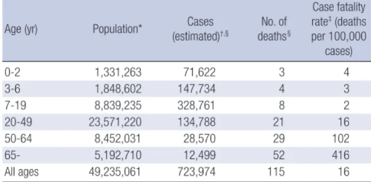
A total of 723,974 people in Korea had ILIs based on positive reverse transcriptase polymerase chain reaction (RT-PCR) dur- ing the study period. With this denominator, the ILI mortality rate was 16 deaths per 100,000 cases. The age-specific mortality rate had a J-shaped curve (Table 1).
Fifty-six patients (48.7%) were at high risk based on age (< 5 yr or ≥ 65 yr), and 83 patients (72.2%) had 1 or more underlying medical conditions. Ninety-six patients (83.5%) had a high-risk condition (age or underlying disease). A high proportion of chil- dren (64.3% of 0-17 yr of age) had no documented underlying disease, while 86% of people > 65 yr of age had an underlying disease. The most frequently identified underlying medical con- dition was chronic lung disease in 30 patients (26.1%). Other underlying conditions included cancers, diabetes, neurologic disorders, cardiac disease, chronic renal disease, chronic liver disease, and immunosuppressed conditions. Height and weight were available for 47 of 115 patients (40.9%) who were > 18 yr of age. Of the 47 patients, 11 (23.4%) were obese (BMI ≥ 25 kg/m2).
There were no deaths in pregnant woman (Table 2).
Table 1. Age-specific mortality from the pandemic A (H1N1) in 2009
Age (yr) Population* Cases
(estimated)†,§ No. of deaths§
Case fatality rate‡ (deaths per 100,000
cases)
0-2 1,331,263 71,622 3 4
3-6 1,848,602 147,734 4 3
7-19 8,839,235 328,761 8 2
20-49 23,571,220 134,788 21 16
50-64 8,452,031 28,570 29 102
65- 5,192,710 12,499 52 416
All ages 49,235,061 723,974 115 16
*From National Statistics Office age-related estimates 2009; †(ILI cases) × (2009 pan- demic H1N1 positivity rates of samples of ILI cases) × (total private medical institutions proportion); ‡No. of deaths × 100,000/Cases (estimated); §The number of estimated cases and deaths was reported until 26 November 2009.
Fig. 1. Age distribution of confirmed cases and 115 fatalities.
Proportion of patients (%)
Age (yr)
0-9 10-19 20-29 30-39 40-49 50-59 60- 60
50 40 30 20 10 0
Fatal cases Confirmed patients
Table 2. Characteristics of fatal cases with confirmed 2009 influenza A (H1N1) in Korea
Characteristics No. (%)
(N = 115)
Age, median (range), yr 61.0 (0.2-97)
Female gender 58 (50.4)
Age of high risk for complications Children (< 5 yr)
Adults (≥ 65 yr)
56 (48.7) 6 (5.2) 50 (43.5) Medical condition
Any of high risk complications* 83 (72.2)
Hypertension 30 (26.1)
Chronic lung disease
Chronic obstructive pulmonary disease Asthma
Other
30 (26.1) 14 (12.2) 9 (7.8) 7 (6.1) Cancer†
Metastatic solid tumor Hematologic malignancy
28 (24.3) 23 (20.0) 5 (4.3) Chronic metabolic disorder
Diabetes (type 1 or 2) Adrenal insufficiency
25 (21.8) 24 (20.9) 1 (0.9) Neurologic disorder
Cerebrovascular disease Epilepsy
Cerebral palsy Mental retardation Other
21 (18.3) 11 (9.6)
4 (3.5) 2 (1.7) 2 (1.7) 6 (5.2) Heart disease
Ischemic heart disease Congestive heart failure Arrhythmia
Valvular heart disease Congenital heart disease
17 (14.8) 9 (7.8) 8 (7.0) 2 (1.7) 1 (0.9) 1 (0.9)
Chronic renal disease 14 (12.2)
Immunosuppression Chemotherapy‡ Immnosuppresive therapy
11 (9.6) 6 (5.2) 5 (4.3)
Obesity§ 11 (23.4)||
Chronic liver disease 7 (6.1)
Autoimmune disease 3 (2.6)
Pregnancy 0 (0.0)
*Exclude hypertension; †Exclude survivors with free symptoms > 5 yr; ‡Patients treated within 2 weeks; §Defined as a BMI > 25 kg/m2; ||Total number was 47 patients who were available for height and weight recording.
Diagnostic findings
Evidence of concurrent bacterial infections was found in spu- tum or blood specimens from 34 patients (29.6%). Of these 34 patients, 28 had confirmed bacterial infections in sputum spec- imens. The most common bacteria isolated in the sputum spec- imens were Staphylococcus aureus and Klebsiella pneumoniae.
These infections were documented in eight patients each. One- half of S. aureus isolates were methicillin-resistant (MRSA). Based on bronchoalveolar lavage (BAL) specimens, five patients had Acinetobacter baumannii, four patients had Candida albicans, three patients had Streptococcus pneumonia, two patients had Pseudomonas aeruginosa, and three patients had Aspergillus fumigatus. In addition, sputum positive for acid-fast bacilli, Esch- erichia coli, and Pneumocystis jiroveci based on BAL specimens were demonstrated in one patient each. Although different from the sputum culture, a 7-yr-old child was seropositive for Myco- plasma IgM and had a 4-fold rise in serial titers for anti-Myco- plasma IgG.
Bacterial organisms were cultured in blood specimens from 10 patients. Three patients had Klebsiella pneumoniae, one pa- tient had C. albicans, and one patient each had vancomycin- resistant Enterococcus, Enterobacter, Pseudomonas aeruginosa, Streptococcus pneumoniae, S. capitis, and yeast. In addition,
Citrobacter braakii and E. coli were isolated from ascites and urine.
Leukocytosis and thrombocytopenia occurred frequently in patients with ILIs. Anemia and abnormal liver function tests was also common. Six patients who had no evidence of hepatitis B or C virus infections were thought to have severe acute hepati- tis. The erythrocyte sedimentation rate (ESR), C-reactive protein (CRP), creatine kinase (CK), and lactate dehydrogenase (LDH) levels were increased in patients with ILIs (Table 3).
Complications
Of the 113 patients who underwent chest radiography on ad- mission, 97 patients (85.8%) had findings that were consistent with pneumonia. Pneumothoraces occurred in two patients af- ter central line insertion and ventilator care. The most common cardiac complication was myocarditis (13 patients [11.3%]). Pul- monary emboli were detected in two patients by CT or echocar- diography and blood testing. Pericarditis was documented in two patients. Myocardial infarctions were noted in 2 patients; 1 patient had angina with 3-vessel disease as an underlying con- dition and the other patient was a smoker without other under- lying diseases. In addition, a ruptured abdominal aortic aneu- rysm was founded.
Seven patients (6.1%) had encephalopathy based on EEG, brain MRI, or brain CT; 2 of the patients were shown to have the 2009 pandemic influenza (H1N1) virus by RT-PCR of cere- brospinial fluid. Except these patients, intracranial hemorrhage and acute cerebral infarction were noted in 1 patient each.
Five patients (4.3%) had gastrointestinal bleeding; 2 of the pa- tients had colon cancer and the others had no definite underly- ing disease to account for the bleeding. Intestinal obstruction occurred in three patients; one of the patients had a history of intestinal surgery. Among other two patients with peritonitis, one patient had pancreatic cancer and one patient had no un- derlying disease. A liver abscess was documented in one patient with diabetes and stroke.
Eighteen patients (15.7%) who had no underlying chronic re- nal disease were suspected to have acute renal failure; 9 of the patients were treated with dialysis, continuous renal replace- ment therapy, or diuretics, and the other patient had elevated creatinine levels (> 2.0 mg/dL). Rhabdomyolysis was noted in 4 patients (3.5%) who had a CK level > 3,000 U/L (10 times the nor- mal upper range in adults); 1 patient had a CK level > 30,000 U/L.
Anti-viral treatment
Of the 115 patients, 100 (87%) received anti-viral drugs. Of these 100 patients, 98 received oseltamivir, 1 received zanamivir, and 1 received combination therapy with oseltamivir plus zanamivir.
All of the patients who were treated with oseltamivir received the recommended dose that is approved by the US Food and Drug Administration, but 15 adult patients were treated with high- Table 3. Laboratory findings of fatal cases with confirmed 2009 influenza A (H1N1)
in Korea
Characteristics No. (%)
Bacterial co-infection Any growth of sputum culture*
Any growth of blood culture 29 (25.2%)
10 (8.7%) Selected Laboratory Abnormalities†
Leukopenia (WBC < 5,000 µL) Neutropenia (ANC < 500 µL) Leucotytosis (WBC > 11,000 µL) Anemia‡
Thrombocytopenia (platelet count < 150,000 µL) Thrombocytosis (platelet count > 350,000 µL) Elevated alanine aminotransferase§ Any elevation
≥ 2 × the upper limit of the normal range Elevated aspartate aminotransferase||
Any elevation
≥ 2 × the upper limit of the normal range Severe hepatitis (AST or ALT > 300 U/L) Elevated total bilirubin (> 1.2 mg/dL)
No./Total No. (%) 23/111 (20.7%) 3/111 (2.7%) 49/111 (44.1%) 59/108 (54.6%) 45/109 (41.3%) 12/109 (11.0%) 50/111 (45.0%) 19/111 (17.1%) 65/111 (58.6%) 36/111 (32.4%) 7/111 (6.3%) 29/106 (27.4%) Other Laboratory Findings
Creatinine, mg/dL (n = 111) Creatinine kinase, U/L (n = 63) CK-mb, U/L (n = 70)
Lactate dehydrogenase, U/L (n = 69) ESR, mm/h (n = 46)
Median (Range) 1.1 (0.4-10.7) 125 (4-10,700) 4.0 (0-175.2) 595 (116-7,159)
31.5 (2-120)
*Including 1 case of positive Mycoplasma IgM Ab; †Laboratory values are based on Custer and Rau (12); ‡The presence of anemia was determined according to age, as follows: ≥ 12 yr of age (Hb < 12 for women and Hb < 13 for men); 2-12 yr of age (Hb
< 11.5); 6 months to 2 yr of age (Hb < 10.5); 2-6 months (Hb < 9.5); §The alanine aminotransferase level was considered to be elevated if it was > 30 U/L in patients ≥ 1 yr of age and > 54 U/L in those < 1 yr of age; ||The aspartate aminotransferase level was considered to be elevated if it was > 35 U/L in patients ≥ 1 yr of age and > 65 U/L in those < 1 yr of age.
dose oseltamivir (300 mg a day). The median time from the on- set of illness to the initiation of anti-viral therapy was 3 days (IQR, 0-21 days); 36% of these patients received anti-viral therapy with- in 48 hr after the onset of symptoms.
Clinical course
The median time from symptom onset to death was 8 days (IQR, 1-33 days). Fifteen patients died within 48 hr since the onset of symptoms and 4 patients died within 24 hr.
We evaluated 63 patients who were admitted to the intensive care unit (ICU), except confirmed cases during hospitalization for other diseases and cases with unknown symptom onset. The median time from symptom onset to hospital admission was 2 days (IQR, 0-22 days), the median time from hospitalization to ICU admission was 0 days (IQR, 0-17 days), and the median time from ICU admission to death was 4 days (IQR, 0-26 days).
DISCUSSION
Our data suggest that age ≥ 65 yr has the lowest incidence and the highest case fatality rate for the 2009 influenza A (H1N1) vi- rus. Our age-specific case fatality rates follow a pattern similar to the J-shaped distribution described in the Mexican, English, and Japanese studies of the current pandemic (13-15). The in- cline of the curve became steeper with older age older. The me- dian age of our analysis was much older than the median age reported in other countries (median age range, 37-53 yr) (7-9, 16). These results differ from the early 2009 pandemic in which there was a high case fatality occurring among young healthy adults (7, 17). The case fatality of our study (16 per 100,000) is lower than that of previous report (30 per 100,000) based on the confirmed patients in Korea (4). This is because not all patients with ILI were confirmed by RT-PCR.
The finding that the majority of deaths occurred in older pa- tients explains that the proportion of patients who had gastro- intestinal symptoms, headaches, and myalgias (systemic symp- toms) at the onset of illness was much lower than previous re- ports (5, 8, 9). Generally, the younger age group is more likely to have systemic symptoms in seasonal influenza infections (18).
There were no pregnant women enrolled in our study.
There are a greater number of cancer patients (24.3%) with underlying medical conditions compared to previous studies (range, 2.6%-7.9%) (7, 14). Interestingly, cancer patients are in- fected more often during hospitalization to manage other med- ical diseases (P = 0.005).
Among critically ill patients with severe 2009 influenza A (H1N1) infections, obesity was more common than in the gen- eral population; however, increased BMI has not emerged as a predictor of mortality (8, 9). Although data regarding height and weight was available for only 47 adult patients (40.1%) in our study, 11 of these patients (23.4%) were obese based on the Asia-
Pacific definition of obesity (11). The prevalence of obesity in our study was much lower than in the general adult Korean pop- ulation (31.7%) (19).
Even though all of the patients were not evaluated with spu- tum cultures, evidence of concurrent bacterial co-infections in sputum specimens was shown in 29.6% of 115 fatal cases. The result was similar to the report in the USA in which a 29% bac- terial co-infection rate was shown in lung tissue specimens from 77 deaths (20). The most common pathogens recovered were Klebsiella pneumoniae (8 patients) and S. aureus (8 patients) in our study, compared to Streptococcus pneumoniae (10/77) in the USA.
The data of our study were derived from the KCDC, which had a detailed description of deaths associated with the 2009 influenza A (H1N1), and differed with respect to race based on a previously published report. These observations of the epide- miologic risk factors and typical clinical features of fatal cases will help in the diagnosis and clinical management of severely ill patients with pandemic influenza A (H1N1).
This study had several limitations. First, we evaluated only fatal cases with confirmed 2009 H1N1 infections, so a compara- tive study of survivors and non-survivors could not be conduct- ed. The group in our study may not be representative of those who were not tested. Second, our data were gathered while the epidemic was ongoing in Korea; the findings after the end of the pandemic may be different owing to the effect of massive vac- cination. Third, the absolute low number of deaths in the very young age group could lead to a different interpretation. Finally, it is impossible to compare our case fatality rate to that of other countries directly because every country has a different calcu- lation method of the estimated cases.
In Korea, from the end of October, anti-viral medications were administered to patients who met the definition of an ILI with or without laboratory confirmation. As of 31 December, approx- imately 9 million people have been vaccinated, including stu- dents in elementary, middle, and high schools since early No- vember, through the national pandemic influenza H1N1 vacci- nation program. As a result, the trend of ILIs and deaths decre- ased rapidly. Considering the second wave of the outbreak in the spring of 2010, we have to make a vaccination plan for the older age group with underlying disease and prevent known complications. If possible, we expect to understand the 2009 H1N1 virus and manage patients appropriately by performing a mathematically well-designed mortality study and a compar- ative study involving survivors versus non-survivors.
ACKNOWLEDGMENTS
We declare that we have no conflicts of interest and financial support. We are grateful to the EIS officers in Korea who supplied a standardized case report of deaths associated with 2009 pan-
demic influenza A, Mina Baek for managing the database, Hoc- hun Choi and Jinsu Song for writing assistance, and members in The Division of KCDC’s Infectious Disease Surveillance for providing estimated case numbers.
REFERENCES
1. Centers for Disease Control and Prevention (CDC). Swine influenza A (H1N1) infection in two children–Southern California, March-April 2009. MMWR Morb Mortal Wkly Rep 2009; 58: 400-2.
2. WHO. World now at the start of Pandemic influenza. Available at http:
//www.who.int/mediacentre/news/statements/2009/h1n1_pandemic_
phase6 _20090611/en/index.html [accessed on 30 Dec 2009].
3. WHO. Pandemic (H1N1) 2009 – update 80. Available at http://www.
who.int/csr/don/2009_12_23/en/index.html [accessed on 30 Dec 2009].
4. Kim JH, Yoo HS, Lee JS, Lee EG, Park HK, Sung YH, Kim S, Kim HS, Shin SY, Lee JK. The spread of pandemic H1N1 2009 by age and region and the comparison among monitoring tools. J Korean Med Sci 2010;
25: 1109-12.
5. Centers for Disease Control and Prevention (CDC). Hospitalized pa- tients with novel influenza A (H1N1) virus infection–California, April- May, 2009. MMWR Morb Mortal Wkly Rep 2009; 58: 536-41.
6. Centers for Disease Control and Prevention (CDC). Intensive-care pa- tients with severe novel influenza A (H1N1) virus infection –Michigan, June 2009. MMWR Morb Mortal Wkly Rep 2009; 58: 749-52.
7. Vaillant L, La Ruche G, Tarantola A, Barboza P; epidemic intelligence team at InVS. Epidemiology of fatal cases associated with pandemic H1N1 influenza 2009. Euro Surveill 2009; 14: pii=19309.
8. Kumar A, Zarychanski R, Pinto R, Cook DJ, Marshall J, Lacroix J, Stelfox T, Bagshaw S, Choong K, Lamontagne F, Turgeon AF, Lapinsky S, Ahern SP, Smith O, Siddiqui F, Jouvet P, Khwaja K, McIntyre L, Menon, K, Hutchison J, Hornstein D, Joffe A, Lauzier F, Singh J, Karachi T, Wiebe K, Olafson K, Ramsey C, Sharma S, Dedek P, Meade M, Hall R, Fowler RA;
Canadian Critical Care Trials Group H1N1 Collaborative. Critically ill patients with 2009 influenza A (H1N1) infection in Canada. JAMA 2009;
302: 1872-9.
9. Domínguez-Cherit G, Lapinsky SE, Macias AE, Pinto R, Espinosa-Perez L, de la Torre A, Poblano-Morales M, Baltazar-Torres JA, Bautista E, Martinez A, Martinez MA, Rivero E, Valdez R, Ruiz-Palacios G, Hernan- dez M, Stewart TE, Fowler RA. Critically ill patients with 2009 influenza
A (H1N1) in Mexico. JAMA 2009; 302: 1880-7.
10. ANZIC Influenza Investigators, Webb SA, Pettilä V, Seppelt I, Bellomo R, Bailey M, Cooper DJ, Cretikos M, Davies AR, Finfer S, Harrigan PW, Hart GK, Howe B, Iredell JR, McArthur C, Mitchell I, Morrison S, Nichol AD, Paterson DL, Peake S, Richards B, Stephens D, Turner A, Yung M.
Critical care services and 2009 H1N1 influenza in Australia and New Zealand. N Engl J Med 2009; 361: 1925-34.
11. WHO regional office for the western pacific. The Asia-pacific perspective;
redefining obesity and its treatment. Manila: 2000. Available at http://
www.wpro.who.int/internet/resources.ashx/NUT/Redefining+obesity.
pdf [accessed on 30 Dec 2009].
12. Custer JW, Rau RE, eds. The Harriet Lane handbook. 18th ed. Philadel- phia: Elsevier Mosby; 2009.
13. Echevarria-Zuno S, Mejia-Arangure JM, Mar-Obeso AJ, Grajales-Muniz C, Robles-Perez E, Gonzalez-Leon M, Ortega-Alvarez MC, Gonzalez- Bonilla C, Rascon-Pacheco RA, Borja-Aburto VH. Infection and death from influenza A H1N1 virus in Mexico: a retrospective analysis. Lancet 2009; 374: 2072-9.
14. Donaldson LJ, Rutter PD, Ellis BM, Greaves FE, Mytton OT, Pebody RG, Yardley IE. Mortality from pandemic A/H1N1 2009 influenza in Eng- land: public health surveillance study. BMJ 2009; 339: b5213.
15. Kamigaki T, Oshitani H. Epidemiological characteristics and low case fatality of pandemic (H1N1) 2009 in Japan. PLoS Curr Influenza 2009;
RRN1139.
16. Bishop JF, Murnane MP, Owen R. Australia’s winter with the 2009 pan- demic influenza A (H1N1) virus. N Engl J Med 2009; 361: 2591-4.
17. Louie JK, Acosta M, Winter K, Jean C, Gavali S, Schechter R, Vugia D, Harriman K, Matyas B, Glaser CA, Samuel MC, Rosenberg J, Talarico J, Hatch D; California Pandemic (H1N1) Working Group. Factors associ- ated with death or hospitalization due to pandemic 2009 influenza A (H1N1) infection in California. JAMA 2009; 302: 1896-902.
18. Treanor JJ. Influenza virus. In: Mandell GL, Bennett JE, Dolin R, eds. Man- dell, Douglas, and Bennett’s Principles and Practice of Infectious disease.
6th ed (2). Philadelphia, Elsevier Churchill Livingstone, 2004; 2069-71.
19. Korea Centers for Disease Control and Prevention. The pattern of obesi- ty prevalence in Korea from KNHANES. Available at http://knhanes.cdc.
go.kr. [accessed on 30 Dec 2009].
20. Centers for Disease Control and Prevention (CDC). Bacterial coinfec- tions in lung tissue specimens from fatal cases of 2009 pandemic influ- enza A (H1N1) – United States, May-August 2009. MMWR Morb Mortal Wkly Rep 2009; 58: 1071-74.
AUTHOR SUMMARY
Fatal Cases of 2009 Pandemic Influenza A (H1N1) in Korea
Hyun Su Kim, Joon Hyung Kim, Soo Youn Shin, Young A Kang, Ha Gyung Lee, Jin Seok Kim, Jong Koo Lee, and Belong Cho The aim of this study was to describe the features of deaths associated with the 2009 pandemic influenza A (H1N1) by 26 November 2009 in Korea. We collected standardized case reports on 115 confirmed deaths through a nationwide enhanced influenza surveillance system. The median age was 61 years (interquartile range, 0.2-97 yr). The case fatality rate was estimated as 16 per 100,000 cases. The age-related mortality rate had a J-shaped curve. Eighty-three patients (72.2%) had at least 1 underlying medical disease. Coinfections were detected in the blood or sputum specimens from 34 patients. Neuraminidase inhibitors were administered to 100 patients (87.0%), 36% of whom began treatment within 2 days. Fatal cases with 2009 influenza A (H1N1) infection in Korea occurred in older individuals with underlying disease, and were associated with pneumonia, bacterial co- infection, and multi-organ failure.