Introduction
In many cases with class III malocclusion, orthognathic surgery is required to improve function and aesthetics with significant changes to hard and soft tissues.
1-15One of the main purposes of orthognathic surgery is to create a sym- metric face. Accurate diagnosis of facial asymmetry is not only essential for presurgical treatment planning but also for postsurgical evaluation. Three-dimensional (3D) com- puted tomography (CT) depicts the human face without
superimposition, magnification, and distortion of anatomic structures, which are inherent in two-dimensional (2D) cephalometric radiographs. Three-dimensional evaluation is valuable because postsurgical changes, including those not evident in that 2D imaging modalities, can be assessed.
Improvements to facial symmetry may be more accurately assessing 3D modalities to evaluate facial changes after surgery than are 2D modalities.
14,15In the 3D analysis of facial asymmetry, the linear dis- tances and angles among structures are commonly used.
14-17A spherical coordinate system can be modified to analyze facial lines. Recently, a new spherical coordinate system for the 3D analysis of facial asymmetry was introduced.
Using this new system, facial lines can be more clearly defined by their length as well as vertical and horizontal
Use of spherical coordinates to evaluate three-dimensional facial changes after orthognathic surgery
Suk-Ja Yoon
1,*, Rui-Feng Wang
2, Sun-Youl Ryu
3, Hyeon-Shik Hwang
4, Byung-Cheol Kang
1, Jae-Seo Lee
1, Juan M. Palomo
51Department of Oral and Maxillofacial Radiology, School of Dentistry, Chonnam National University, Gwangju, Korea
2Department of Biologic and Material Sciences, School of Dentistry, University of Michigan, Ann Arbor, MI, USA
3Department of Oral and Maxillofacial Surgery, School of Dentistry, Chonnam National University, Gwangju, Korea
4Department of Orthodontics, School of Dentistry, Chonnam National University, Gwangju, Korea
5Department of Orthodontics, School of Dental Medicine, Case Western Reserve University, Cleveland, OH, USA
ABSTRACT
Purpose: This study aimed to assess the three-dimensional (3D) facial changes after orthognathic surgery by eval- uating the spherical coordinates of facial lines using 3D computed tomography (CT).
Materials and Methods: A 19-year-old girl was diagnosed with class III malocclusion and facial asymmetry. Orthog- nathic surgery was performed after orthodontic treatment. Facial CT scans were taken before and after orthognathic surgery. The patient had a menton deviation of 12.72 mm before surgery and 0.83 mm after surgery. The spherical coordinates of four bilateral facial lines (ramal height, ramal lateral, ramal posterior and mandibular body) were estimated from CT scans before and after surgery on the deviated and opposite side.
Results: The spherical coordinates of all facial lines changed after orthognathic surgery. Moreover, the bilateral differences of all facial lines changed after surgery, and no bilateral differences were zero.
Conclusion: The spherical coordinate system was useful to compare differences between the presurgical and post- surgical changes to facial lines. (Imaging Sci Dent 2014; 44 : 15-20)
KEY WORDS: Tomography, X-Ray Computed; Facial Asymmetry; Orthognathic Surgery
Received July 26, 2013; Revised September 23, 2013; Accepted September 28, 2013
*Correspondence to : Prof. Suk-Ja Yoon
Department of Oral and Maxillofacial Radiology, School of Dentistry, Chonnam National University, 77 Yongbongro, Bukgu, Gwangju 500-757, Korea
Tel) 82-62-530-5680, Fax) 82-62-530-5689, E-mail) [email protected]
http://dx.doi.org/10.5624/isd.2014.44.1.15
Copyright ⓒ 2014 by Korean Academy of Oral and Maxillofacial Radiology
This is an Open Access article distributed under the terms of the Creative Commons Attribution Non-Commercial License (http://creativecommons.org/licenses/by-nc/3.0) which permits unrestricted non-commercial use, distribution, and reproduction in any medium, provided the original work is properly cited.
Imaging Science in Dentistry∙pISSN 2233-7822 eISSN 2233-7830
angle in the spherical coordinate system.
18-20The spheri- cal coordinate system may be used to indicate definitive changes to the facial area before and after orthognathic surgery.
The purpose of this study was to assess changes to the facial area of one patient with Class III malocclusion and significant facial asymmetry before and after orthognathic surgery by analyzing facial lines (spherical coordinate sys- tem) from 3D CT images.
Materials and Methods
Study subject
A19-year-old female diagnosed with mandibular prog- nathism, maxillary hypoplasia, and lip canting was eval- uated before and after orthognathic surgery via 3D CT.
The orthognathic surgery included a mandibular setback, Le Fort I canting correction, and right mandibular angle reduction. The menton (Me) deviation was reduced from 12.72 mm to 0.83 mm after surgery, which was measured as the distance of Me from the midsagittal reference plane on the reconstructed 3D CT image.
CT scans
CT scans were obtained using a spiral CT scanner (Light Speed QX/I, GE Medical Systems, Milwaukee, WI, USA) with a 512×512 matrix (120 kV, 200 mA, and a gantry angle of zero). The axial image thickness was 2.5 mm, the table speed was 3 mm/sec, and the scanning time was 0.8 sec. Digital imaging and communication in medicine images were created at a slice thickness of 1.0 mm. The acquired data from these images were input into a personal computer, and the CT data were used to construct 3D images with the software Vworks
++Vsurgery (Cybermed, Seoul, Korea).
Landmarks and facial lines
Three orthogonal reference planes, the horizontal (xy plane), midsagittal (yz plane), and coronal (xz plane) planes, were established. The horizontal reference plane was first established using the right Po, right Or, and left Or. The midsagittal reference plane was formed perpendicular to the horizontal reference plane and passing through Na and Op. The coronal reference plane was defined as perpendi- cular to both the midsagittal and the horizontal reference planes passing through Op. Next, the condylar landmarks were identified as the most superior (Cd
sup), lateral (Cd
lat),
and posterior (Cd
post) points of the condylar head. The gonion landmarks were identified as the most inferior (Go
inf), lateral (Go
lat), and posterior (Go
post) points of the gonion area. Additionally, the Me was defined as the most inferior point on the mandibular symphysis. The side of the face with the Me was identified as the deviated side, while the contralateral side of the face was considered the opposite side. The four facial lines that were established included the ramal height (Cd
sup-Go
inf), ramal lateral (Cd
lat- Go
lat), ramal posterior (Cd
post-Go
post), and mandibular body (Go
post-Me) with their respective landmarks. The spherical
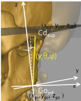
Fig. 1.CT scans from a 19-year-old female with class III malocclu- sion and facial asymmetry. A. Before orthognathic surgery. B. After orthognathic surgery. The menton deviation was reduced from 12.72 mm before surgery to 0.83 mm after surgery.
A B
Fig. 2. A. Spherical coordinates of the analyzed facial lines and ramal height is shown. Length is indicated as v, the midsagittal inclination angle (between the line and the midsagittal reference plane) as θ, and the coronal inclination angle (between the line and the coronal reference plane) as φ.
coordinates (length, midsagittal inclination angle, and coronal inclination angle) of the bilateral facial lines were mathematically calculated. Midsagittal and coronal incli- nation angles were considered the angles between the facial lines and midsagittal and coronal reference planes, respec-
tively. All postsurgical changes to the spherical coordinates were estimated on both the deviated and opposite side.
Additionally, the bilateral differences among facial lines before and after surgery were evaluated (Figs. 1-3).
19,20Fig. 3.CT images of the facial lines before (A-H) and after (I-P) orthognathic surgery. A-D. The facial lines, ramal height and ramal lateral, before surgery. E-H. The facial lines, ramal posterior and mandibular body, before surgery. I-L. The facial lines, ramal height and ramal lateral, after surgery. M-P. The facial lines, ramal posterior and mandibular body, after surgery. The face remained three-dimensionally asymmetrical after surgery.
A B C D
E F G H
I J K L
M N O P
Results
The differences of length of facial lines (v) by surgery were between -0.71 mm and 17.16 mm. The differences of midsagittal inclination angle (θ) were between -6.67�
and 5.08�. The differences of coronal inclination angle (ϕ) were between 1.45�and 12.60�(Table 1). The bilateral differences of the facial lines had changes after surgery. The changes of bilateral differences by surgery were between
-10.75 mm and 2.09 mm in length (dv), between -10.49�
and -0.49�in midsagittal inclination angle (dθ), and bet- ween -1.27�and 9.76�in coronal inclination angle (dϕ) (Table 2).
Discussion
In this study, the spherical coordinate system was used to evaluate 3D facial changes after orthognathic surgery.
Table 1. Spherical coordinates of facial lines before and after orthognathic surgery
Facial line Face side Spherical
coordinate Presurgical Postsurgical Difference
Ramal height Deviated side v 55.25 45.38 9.86
(Cdsup-Goinf) θ -1.96 4.18 -6.14
ϕ 11.16 5.13 6.03
Opposite side v 58.16 42.59 15.57
θ 0.44 3.49 -3.04
ϕ 16.84 9.55 7.3
Ramal lateral Deviated side v 49.47 32.32 17.16
(Cdlat-Golat) θ 4.95 10.5 -5.55
ϕ 10.27 -2.33 12.6
Opposite side v 46.73 31.66 15.07
θ 7.41 12.48 -5.06
ϕ 13.15 10.31 2.84
Ramal posterior Deviated side v 33.73 28.44 5.29
(CdPost-GoPost) θ -5.48 1.19 -6.67
ϕ 8.91 6.38 2.53
Opposite side v 37.44 32.25 5.19
θ -2.16 -2.58 0.42
ϕ 17.83 16.39 1.45
Mandibular body Deviated side v 93.5 94.2 -0.71
(GoPost-Me) θ 20.18 25.59 -5.41
ϕ 56.87 48.44 8.42
Opposite side v 100.06 90.02 10.05
θ 38.55 33.47 5.08
ϕ 52.83 43.26 9.57
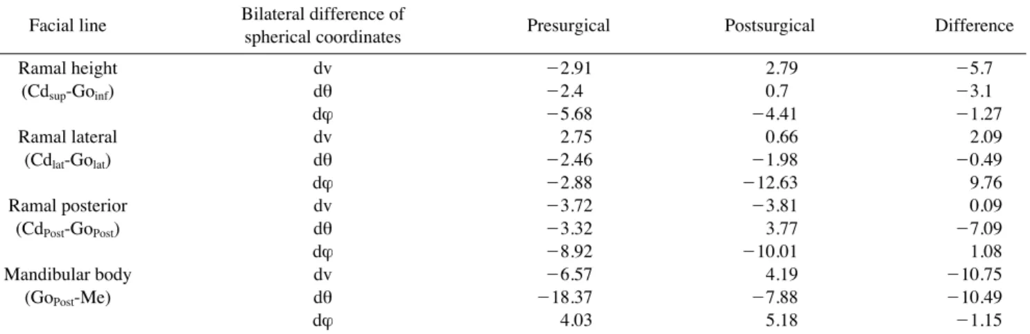
Table 2. Bilateral differences of facial lines before and after orthognathic surgery in the spherical coordinates Facial line Bilateral difference of
Presurgical Postsurgical Difference
spherical coordinates
Ramal height dv -2.91 2.79 -5.7
(Cdsup-Goinf) dθ -2.4 0.7 -3.1
dϕ -5.68 -4.41 -1.27
Ramal lateral dv 2.75 0.66 2.09
(Cdlat-Golat) dθ -2.46 -1.98 -0.49
dϕ -2.88 -12.63 9.76
Ramal posterior dv -3.72 -3.81 0.09
(CdPost-GoPost) dθ -3.32 3.77 -7.09
dϕ -8.92 -10.01 1.08
Mandibular body dv -6.57 4.19 -10.75
(GoPost-Me) dθ -18.37 -7.88 -10.49
dϕ 4.03 5.18 -1.15
(dv, dθ, dϕ)==spherical coordinates of deviated side-spherical cooridnate of opposite side
Spherical coordinate systems are used in various fields such as astronomy and geology. In geology, a spherical coordinate system is used to describe a flying object over the earth according to its distance from the center of the earth and its latitude and longitude. Similarly, the symme- try of human anatomical structures in 3D space can be also evaluated with the spherical coordinate system.
18This system can be modified to define the length, midsagittal, and coronal inclinations of facial lines in 3D images of human structures.
19,20All spherical coordinates on both sides of the patient’s face were three-dimensionally changed after orthognathic surgery (Table 1). In addition, the bilateral differences among all facial lines changed after surgery; however, the patient’s face remained slightly asymmetric. No bilateral differences were zero. Although facial asymmetry was present after surgery, the bilateral differences were not consistent with the findings before surgery (Table 2).
Overall, this patient demonstrated a substantial reduction in facial asymmetry after surgery; the Me deviation, which is commonly used as an index to classify facial asymme- try,
21,22decreased dramatically from 12.72 mm before sur- gery to 0.83 mm after surgery. If only the changes to the Me deviation are considered, facial asymmetry would be considered resolved in this subject. However, 3D analysis of the facial lines by the spherical coordinate system show- ed that the subject still had facial asymmetry after surgery.
Facial asymmetry was resolved in the frontal plane; how- ever, it remained after 3D CT analysis and all facial lines changed after surgery.
Previous reports have indicated that some level of asym- metry can remain after orthognathic surgery, yet surgery tended to result in facial lines that were within the normal range of facial symmetry.
13,15This study applied the spherical coordinate system to comparefacial asymmetry before and after orthognathic surgery. The spherical coordinate substantially changed after surgery. This report suggests that facial asymmetry by surgery might not fully establish a 3D symmetric face in patients with facial asymmetry. Further investigation is required to compare 3D changes to spherical coordinates before and after surgery in a large sample size.
References
1. Aaronson SA. A cephalometric investigation of the surgical correction of mandibular prognathism. Angle Orthod 1967;
37: 251-60.
2. Fromm B, Lundberg M. The soft-tissue facial profile before and after surgical correction of mandibular protrusion. Acta
Odontol Scand 1970; 28: 157-77.
3. Robinson SW, Speidel TM, Isaacson RJ, Worms FW. Soft tissue profile change produced by reduction of mandibular prognathism. Angle Orthod 1972; 42: 227-35.
4. Hershey HG, Smith LH. Soft-tissue profile change associated with surgical correction of the prognathic mandible. Am J Orthod 1974; 65: 483-502.
5. Fanibunda KB. Changes in the facial profile following correc- tion for mandibular prognathism. Br J Oral Maxillofac Surg 1989; 27: 277-86.
6. Gjørup H, Athanasiou AE. Soft-tissue and dentoskeletal pro- file changes associated with mandibular setback osteotomy.
Am J Orthod Dentofacial Orthop 1991; 100: 312-23.
7. McCance AM, Moss JP, Fright WR, James DR, Linney AD.
A three dimensional analysis of soft and hard tissue changes following bimaxillary orthognathic surgery in skeletal III patients. Br J Oral Maxillofac Surg 1992; 30: 305-12.
8. Cho EJ, Yang WS. Soft tissue changes after double jaw sur- gery in skeletal class III malocclusion. Korean J Orthod 1996;
26: 1-16.
9. Lin SS, Kerr WJ. Soft and hard tissue changes in Class III patients treated by bimaxillary surgery. Eur J Orthod 1998;
20: 25-33.
10. Soncul M, Bamber MA. Evaluation of facial soft tissue changes with optical surface scan after surgical correction of Class III deformities. J Oral Maxillofac Surg 2004; 62: 1331-40.
11. Chew MT. Soft and hard tissue changes after bimaxillary sur- gery in Chinese Class III patients. Angle Orthod 2005; 75:
959-63.
12. Park JW, Kim NK, Kim MJ, Chang YI. Three-dimensional analysis of soft and hard tissue changes after mandibular set- back surgery in skeletal Class III patients. Korean J Orthod 2005: 35; 320-9.
13. Sforza C, Peretta R, Grandi G, Ferronato G, Ferrario VF. Three- dimensional facial morphometry in skeletal Class III patients.
A non-invasive study of soft-tissue changes before and after orthognathic surgery. Br J Oral Maxillofac Surg 2007; 45: 138- 44.
14. Baek SH, Cho IS, Chang YI, Kim MJ. Skeletodental factors affecting chin point deviation in female patients with class III malocclusion and facial asymmetry: a three-dimensional analy- sis using computed tomography. Oral Surg Oral Med Oral Pathol Oral Radiol Endod 2007; 104: 628-39.
15. Jung YJ, Kim MJ, Baek SH. Hard and soft tissue changes after correction of mandibular prognathism and facial asymmetry by mandibular setback surgery: three-dimensional using com- puterized tomography. Oral Surg Oral Med Oral Pathol Oral Radiol Endod 2009; 107: 763-71.
16. Hwang HS, Hwang CH, Lee KH, Kang BC. Maxillofacial 3- dimensional image analysis for the diagnosis of facial asym- metry. Am J Orthod Dentofacial Orthop 2006; 130: 779-85.
17. Kim EJ, Palomo JM, Kim SS, Lim HJ, Lee KM, Hwang HS.
Maxillofacial characteristics affecting chin deviation between mandibular retrusion and prognathism patients. Angle Orthod 2011; 81: 988-93
18. Fisher NI, Lewis T, Embleton BJ. Statistical analysis of sphe- rical data. Cambridge: Cambridge University Press; 1987. p.
17-28.
19. Yoon SJ, Wang RF, Hwang HS, Kang BC, Lee JS, Palomo JM. Application of spherical coordinate system to facial asym- metry analysis in mandibular prognathism patients. Imaging Sci Dent 2011; 41: 95-100.
20. Yoon SJ, Wang RF, Na HJ, Palomo JM. Normal range of facial asymmetry in spherical coordinates: a CBCT study. Imaging Sci Dent 2013; 43: 31-6.
21. Masuoka N, Muramatsu A, Ariji Y, Nawa H, Goto S, Ariji E.
Discriminative thresholds of cephalometric indexes in the subjective evaluation of facial asymmetry. Am J Orthod Dento- facial Orthop 2007; 131: 609-13.
22. Haraguchi S, Takada K, Yasuda Y. Facial asymmetry in sub- jects with skeletal Class III deformity. Angle Orthod 2002;
72: 28-35.