Down-regulation of the autophagy gene, ATG7 , protects bone marrow-derived mesenchymal stem cells from stressful conditions
Sedigheh Molaei
1, Mehryar Habibi Roudkenar
1, Fatemeh Amiri
1, Mozhgan Dehghan Harati
1, Marzie Bahadori
1, Fatemeh Jaleh
1, Mohammad Ali Jalili
1, Amaneh Mohammadi Roushandeh
21Blood Transfusion Research Center, High Institute for Research and Education in Transfusion Medicine, Tehran, 2Department of Anatomical Sciences, School of Medicine, Hamadan University of Medical Sciences, Hamadan, Iran
p-ISSN 2287-979X / e-ISSN 2288-0011 http://dx.doi.org/10.5045/br.2015.50.2.80 Blood Res 2015;50:80-6.
Received on January 12, 2015 Revised on February 23, 2015 Accepted on April 2, 2015
Background
Mesenchymal stem cells (MSCs) are valuable for cell-based therapy. However, their appli- cation is limited owing to their low survival rate when exposed to stressful conditions.
Autophagy, the process by which cells recycle the cytoplasm and dispose of defective organelles, is activated by stress stimuli to adapt, tolerate adverse conditions, or trigger the apoptotic machinery. This study aimed to determine whether regulation of autophagy would affect the survival of MSCs under stress conditions.
Methods
Autophagy was induced in bone marrow-derived MSCs (BM-MSCs) by rapamycin, and was inhibited via shRNA-mediated knockdown of the autophagy specific gene, ATG7.
ATG7 expression in BM-MSCs was evaluated by reverse transcription polymerase chain reaction (RT-PCR), western blot, and quantitative PCR (qPCR). Cells were then exposed to harsh microenvironments, and a water-soluble tetrazolium salt (WST)-1 assay was per- formed to determine the cytotoxic effects of the stressful conditions on cells.
Results
Of 4 specific ATG7-inhibitor clones analyzed, only shRNA clone 3 decreased ATG7 expression. Under normal conditions, the induction of autophagy slightly increased the viability of MSCs while autophagy inhibition decreased their viability. However, under stressful conditions such as hypoxia, serum deprivation, and oxidative stress, the in- duction of autophagy resulted in cell death, while its inhibition potentiated MSCs to with- stand the stress conditions. The viability of autophagy-suppressed MSCs was significantly higher than that of relevant controls (P<0.05, P<0.01 and P<0.001).
Conclusion
Autophagy modulation in MSCs can be proposed as a new strategy to improve their surviv- al rate in stressful microenvironments.
Key Words Autophagy, Mesenchymal stem cells, Oxidative stress, Cell survival, shRNA
*This study was supported by the Iran National Science Foundation (INSF).
Correspondence to
Mehryar Habibi Roudkenar, Ph.D.
Blood Transfusion Research Center, High Institute for Research and Education in Transfusion Medicine, Iranian Blood Transfusion Organization (IBTO), Hemmat Exp. Way, 14665-1157, Tehran, Iran
Tel: +982188613423 Fax: +982188601555 E-mail: roudkenar@ibto.ir
Ⓒ 2015 Korean Society of Hematology
INTRODUCTION
Mesenchymal stem cells (MSCs) are valuable cells in cell- based therapy and have been extensively investigated as a new modality for treatment of several diseases [1]. Different tissue-derived MSCs were characterized and expanded in vitro [2] to be utilized for cell-therapy proposes [3]. However, they demonstrate low proliferative and survival rates during long in vitro expansion or after in vivo transplantation [4].
The microenvironments surrounding MSCs contain several hazardous stressors such as poor nutrition [5] and oxidative stress, mainly mediated by reactive oxygen species (ROS) [6].
Practical strategies should therefore be employed to im- prove the underlying mechanisms of MSC resistance to harsh stress conditions, thus leading to higher survival rates and con- sequently higher engraftment rates after transplantation [7].
Autophagy (self-digestion), also described as the garbage disposal system of the cell, involves lysosomal-mediated ca- tabolism of long-lived proteins and damaged organelles,
which are sequestered by autophagosomes or specific autoph- agy vacuoles [8]. Basal autophagy regulates a wide range of physiological and pathophysiological processes, from star- vation adaptation and homeostasis to anti-aging and cell death [9]. Autophagy was initially described as a survival mechanism that recycles degraded intracellular components to serve as new building blocks for the synthesis of macro- molecules and metabolites, which were then used as the cellular energy source for cell survival [10]. However, au- tophagy seems to play dual roles depending on the cell type and context. In the context of cellular death, the persistent and extensive occurrence of autophagy leads to the catabo- lism of cytoplasmic materials, to an extent that causes meta- bolic and bio-energetic cell collapse. Recent studies have shown that autophagy might act as a double-edged sword, leading to either cell death or survival by inactivation of key autophagy genes, particularly ATG7 [11, 12].
ATG7 is an indispensable gene for starvation-induced au- tophagy [13]. Its function in the autophagy process is well known, and its inactivation leads to a significant decrease in the cellular autophagy phenomenon, regardless of any interference from other cellular pathways [14].
Interestingly, few studies have investigated autophagy in stem cells [15], including MSCs, and its role in this cell type is still controversial. This study therefore aimed to ad- dress whether autophagy protects or exacerbates MSCs fol- lowing exposure to various types of stress conditions.
MATERIALS AND METHODS
Isolation of MSCs from human bone marrow aspirates Human MSCs were isolated from the bone marrow of healthy volunteers in the bone marrow transplantation (BMT) center of Shariati hospital under the institutional protocol and consent procedure. These human bone mar- row-derived-mesenchymal stem cells (hBM-MSCs) were successfully isolated using their adhesive characteristics as previously described [16]. hBM-MSC surface markers were detected by flow cytometry and their differentiation po- tentiality for osteogenesis, chondrogenesis, and adipogenesis was analyzed as previously described [16].
Autophagy suppression by gene silencing of ATG7 using short hairpin RNA
To suppress autophagy in hBM-MSCs, the RNAi (RNA interference) technique was performed, since it is the most specific strategy for knockdown studies. SureSilencing short hairpin RNA (shRNA) plasmids targeting human ATG7 were purchased from Qiagen Inc. (SABioscience Qiagen, Germany).
This product contains a negative control and 4 ATG7 shRNA plasmids (no. 1-4), of which only one caused a significant knockdown of ATG7. ATG7-targeting shRNA plasmids were separately cloned into DH5α E. coli (Invitrogen, USA).
Competent cells were cultured in Luria-Bertani (LB) broth medium, followed by plasmid extraction.
Transfection of hBM-MSCs
Following plasmid extraction, hBM-MSCs were trans- fected with the negative control and each of the 4 shRNA vectors using the transfection reagent FuGENE HD (Roche, Germany) according to the manufacturer’s protocol. The transfected cells were named MSC-shRNA 1, MSC-shRNA 2, MSC-shRNA 3, MSC-shRNA 4 according to the number of the shRNA vector, and MSC-shRNA Cont for the negative control shRNA transfected MSCs. A plasmid containing EGFP conjugated ubiquitous microtubule-associated protein 1A/1B-light chain 3 (pEGFP-LC3m) was obtained from Dr.
Y. Kuwahara, Department of Pathology, Institute of Development, Aging and Cancer, Tohoku University, Sendai, Miyagi, Japan as a gift. hBM-MSCs were also transfected with pEGFP-LC3m, and were named MSC-LC3. After trans- fection for 48 hours, MSC-LC3 cells were observed under a fluorescence microscope. RT-PCR analysis was performed to select proper shRNA vectors, which could effectively knock down ATG7 mRNA.
Autophagy induction using rapamycin
Rapamycin (Rapa), a well-known inducer of autophagy, was used to activate the autophagy pathway. To optimize rapamycin (Invitrogen, USA) concentration, hBM-MSCs were cultured in 96-well plates (Orange Scientific, Belgium), and different rapamycin doses (10, 200, and 500 nM) were added and incubated for 24 hours in a standard cell-culturing incubator. The rapamycin-treated hBM-MSCs were named MSC-10 nM Rapa, MSC-200 nM Rapa, and MSC-500 nM Rapa according to the rapamycin dose applied. MSC-LC3, MSC-shRNA Cont, and MSC-shRNA 3 treated with the opti- mized rapamycin dose were named MSC-LC3-Rapa, MSC- shRNA Cont-Rapa and MSC-shRNA 3-Rapa, respectively.
These rapamycin-treated hBM-MSCs were then subjected to western blot analysis.
Western blot analysis
Ubiquitous microtubule-associated protein 1A/1B-light chain 3 (LC3) is a soluble protein in mammalian cells. During the autophagy process, the cytosolic form of LC3 (LC3-I) is converted to a lipid bound form (LC3-II) [17]. Western blot analysis was performed to evaluate these autophagy markers (LC3-I and LC3-II) at the translational level. Whole cell extract was isolated from different rapamycin-treated cells as well as the untreated control. Samples were run on 16% SDS-polyacrylamide gel, and then, transferred to a PDVF membrane. Samples were then incubated with di- luted rabbit anti-LC3-I and LC3-II (1:1,000) (Cell Signaling Technology, USA) primary antibodies for 3 hrs. After primary antibody incubation, the membrane was washed 3 times, and then incubated with an HRP-conjugated goat anti-rabbit secondary antibody for 1.5 hrs. Finally, enhanced chem- iluminescence (ECL) (Abcam, UK) substrate solutions were used to visualize the proteins on the PVDF membrane using a chemiluminescence imaging system. -actin served as an internal loading control protein.
RT-PCR
Total RNA was extracted from transfected hBM-MSCs using the Tripure reagent (Roche, Germany) according to the manufacturer’s instructions. The first-strand cDNA was generated from 1 g of the isolated total RNA using the cDNA synthesis kit (Bioneer, USA) according to the manu- facturer’s instructions. Appropriate primer sets for ATG7 and
-actin I were designed using the Primer3 software and the primers used are as follows: ATG7 forward: 5’-ACCCAG AAGAAGCTGAACGA-3’ and reverse: 5’-CTCATTTGCTGC TTGTTCCA-3’; -actin forward: 5’-TTCTACAATGAGCTG CGTGTGG-3’ and reverse: 5’-GTGTTGAAGGTCTCAAACA TGAT-3’).
PCR reactions were performed using Taq DNA polymerase (Roche, Germany). -actin expression was used to normalize the gene expression. Electrophoresis of DNA fragments on 2% agarose gel and ethidium bromide staining was performed to detect ATG7 expression.
qPCR
For further confirmation of the RT-PCR results, cDNA from MSC-shRNA 3 and MSC-shRNA Cont was subjected to qPCR analysis to quantify the suppression level of ATG7. qPCR was performed in triplicate using specific primers (described above) and the SYBR Green Real-Time PCR Master Mix (Takara, Japan) on the Rotor gene RG-3000 system (Corbett, Germany). The cycle threshold (Ct) was automatically calculated, and the -actin Ct value was used for normalization.
Induction of different stress conditions
To investigate the effects of autophagy on cellular survival rate, the different groups of hBM-MSCs including autoph- agy-suppressed hBM-MSCs (MSC-shRNA 3), Rapamycin- treated hBM-MSCs (MSC-Rapa), MSC-shRNA Cont, and normal h-BM-MSCs without any treatment (MSC-Cont) were cultured under hypoxic, serum-deprived, and oxidative stress conditions. Cells were seeded at a density of 2×10⁴ cells/well in a 96-well plate. To induce oxidative stress, cells were exposed to different doses of 1, 2, 3, 4, and 5 mM hydrogen peroxide (H2O2) (Sigma, Germany) for 1 hour.
For induction of serum deprivation, cells were washed three times with serum-free medium and maintained in this con- dition for various lengths of time. The hypoxic condition was created by transferring plates to a Galaxy 48 R hypoxic incubator (New Brunswick, Germany). These cells were in- cubated at 37oC in the presence of 5% CO2, 1% O2, and N2 for different lengths of time. Each test was done in tripli- cate and the cytotoxic effects of H2O2, hypoxia, or serum deprivation on cells were determined by a WST-1 cytotoxicity assay. The viability of the four MSC groups: MSC-shRNA3, MSC-Rapa, MSC-shRNA Cont and MSC-Cont, were also stud- ied under normal conditions i.e. in the absence of any stress condition.
WST-1 assay
A WST-1 assay was performed to evaluate the viability
of MSCs. The WST-1 reagent (Sigma, Germany) was added to cells at a concentration of 10 L/well (Sigma, Germany) in 100 L culture medium and the samples were incubated at 37oC for 4 hours. The optical density (OD) of each sample was measured using a microplate reader at 450 nM.
Statistical analysis
The quantitative results were analyzed using the SPSS statistical software (version 19) (SPSS Inc., Chicago, IL, USA).
Statistically significant differences (P<0.05) were de- termined by an analysis of variances (one-way ANOVA) analysis.
RESULTS
Down-regulation of ATG7 resulted in suppression of autophagy in MSCs
Four different ATG7 shRNA vectors containing GFP (no.
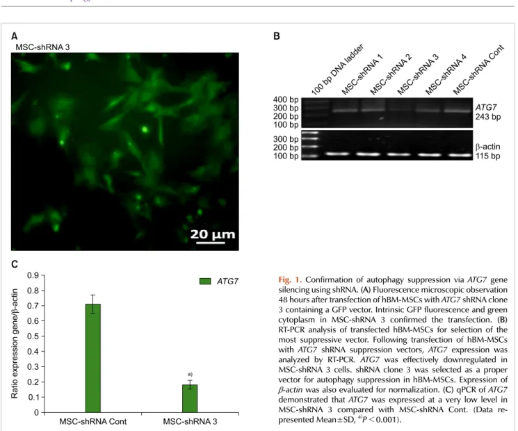
1-4) were transfected into hBM-MSCs, as well as a negative control shRNA vector. After 48 hours, hBM-MSCs that were transfected with the ATG7 shRNA clone 3 (MSC-shRNA 3) were observed under a fluorescence microscope (Fig. 1A).
RT-PCR analysis of the various transfected cells indicated that MSC-shRNA 3 expressed lower levels of ATG7 mRNA than MSC-shRNA Cont. and other shRNA MSC transfected cells (1, 2 and 4) (Fig. 1B).
Next, qPCR was performed to evaluate the level of ATG7 suppression in MSC-shRNA 3 compared to MSC-shRNA Cont. Expression of ATG7 in MSCs-shRNA 3 was consid- erably lower than in MSC-shRNA Cont (P<0.001) (Fig.
1C). Taken together, the shRNA clone 3 was selected as the best ATG7 suppressive vector.
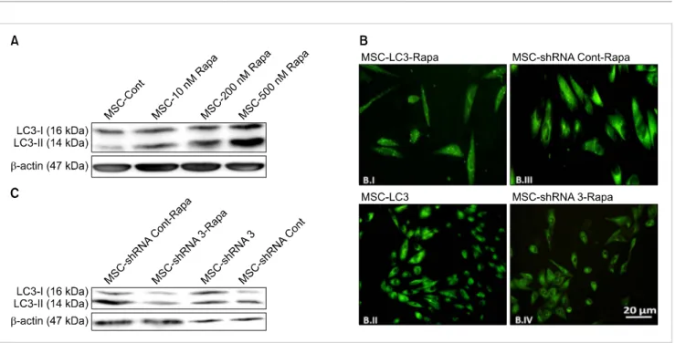
Rapamycin was then added to cells at different concen- trations in order to induce autophagy. Following activation of the autophagy pathway, the conversion of LC3I to LC3II leads to a higher intensity of the LC3II band and an elevated relative ratio of LC3 II to LC3 I. The induction of autophagy was detected after 24-hour-treatment with 500 nM rapamycin (Fig. 2A).
Autophagy was also investigated in hBM-MSCs transfected with GFP-LC3 plasmids (MSC-LC3). MSC-LC3 were exposed to 500 nM rapamycin (the optimized autophagy inducing dose) for 24 hours (MSC-LC3-Rapa), and then observed under a fluorescence microscope. Rapamycin induced stimulation of autophagy resulted in the formation of shiny green dots in MSC-LC3-Rapa (Fig. 2B.I), which were not observed in untreated MSC-LC3 (Fig. 2B.II). MSC-shRNA 3 and MSC-shRNA Cont were also treated with 500 nM rapamycin (MSC-shRNA 3-Rapa and MSC-shRNA Cont-Rapa, re- spectively). Induction of autophagy was observed in MSC-shRNA Cont-Rapa as determined by the presence of shiny green dots (Fig. 2B.III). However this was not observed in MSC-shRNA 3-Rapa, indicating that rapamycin treatment could not induce autophagy under ATG7 suppression.
Western blot analysis of MSC-shRNA Cont-Rapa and MSC-shRNA 3-Rapa also indicated that the down-regulation
Fig. 1.Confirmation of autophagy suppression via ATG7 gene silencing using shRNA. (A) Fluorescence microscopic observation 48 hours after transfection of hBM-MSCs with ATG7 shRNA clone 3 containing a GFP vector. Intrinsic GFP fluorescence and green cytoplasm in MSC-shRNA 3 confirmed the transfection. (B) RT-PCR analysis of transfected hBM-MSCs for selection of the most suppressive vector. Following transfection of hBM-MSCs with ATG7 shRNA suppression vectors, ATG7 expression was analyzed by RT-PCR. ATG7 was effectively downregulated in MSC-shRNA 3 cells. shRNA clone 3 was selected as a proper vector for autophagy suppression in hBM-MSCs. Expression of
-actin was also evaluated for normalization. (C) qPCR of ATG7 demonstrated that ATG7 was expressed at a very low level in MSC-shRNA 3 compared with MSC-shRNA Cont. (Data re- presented Mean±SD, a)P<0.001).
of ATG7 prevented autophagy induction even after rapamycin treatment (Fig. 2C). This was also further confirmed by trans- fection of hBM-MSCs with GFP-LC3 plasmids (data not shown).
Suppression of autophagy protects MSCs from hypoxic, serum-deprived, and oxidative stress conditions
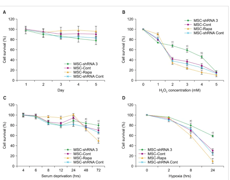
To determine the effects of autophagy induction or in- hibition on the survival rate of MSCs, cells were cultivated under normal conditions for five days, and cell viability was evaluated using the WST-1 assay. Survival of MSCs increased with the induction of autophagy and decreased with autophagy inhibition, however the differences were not significant (P>0.05) (Fig. 3A).
Next, all hBM-MSCs groups including autophagy-sup- pressed MSCs (MSC-shRNA 3), autophagy-induced MSCs (MSC-Rapa) and their relevant controls, MSC-shRNA Cont, and normal MSCs without any treatment (MSC-Cont) were exposed to hypoxic, serum-deprived, and oxidative stress conditions. As shown in Fig. 3B, the cells with ATG7 knock- down demonstrated lower cell death rates at higher H2O2
doses (P<0.01; P<0.001). This observation indicates that autophagy suppression results in cytoprotection against
strong oxidative stress (Fig. 3B). MSC-Rapa cells demon- strated high cell viability compared to MSC-shRNA3 after 24 hours of serum deprivation. This finding demonstrates the protective effects of autophagy on the survival of hBM-MSCs against serum deprivation-induced toxicity 24 hours after starvation. Interestingly, after 48 or 72 hours of serum deprivation, MSC-shRNA 3 demonstrated stronger resistance to cell death compared to other groups (Fig. 3C).
Following 24-hour exposure to hypoxic conditions, the viability of MSC-shRNA 3 was higher than that of MSC-Rapa and the control group. This indicates that ATG7 knockdown protected hBM-MSCs against hypoxia-induced cytotoxicity (Fig. 3D). Overall, these findings suggest that inhibition of ATG7 plays a critical role in the protection of MSCs from hypoxia, serum deprivation, and oxidative stress.
DISCUSSION
The advancement of MSC-based cell therapy is hindered by the low viability of infused MSCs. Harsh microenviron- ments such as those induced by hypoxia, serum deprivation,
Fig. 2. Confirmation of autophagy induction with rapamycin. (A) Western blot analysis of 24 hour-rapamycin-treated hBM-MSCs exposed to 10, 200, and 500 nM rapamycin. LC3-I induction and conversion of LC3-I to LC3-II were determined by western blot analysis. Expression of LC3-II and its significantly increased level compared to LC3-I in MSCs-500 nM Rapa indicated induction of autophagy in these cells. -actin served as the loading control. (B) Microscopic observation of transfected hBM-MSCs before and after rapamycin treatment. hBM-MSCs were transfected with GFP-LC3 containing a GFP vector (MSC-LC3), and 48 hours later, MSC-LC3 was treated with 500 nM rapamycin for 24 hours (MSC-LC3-Rapa).
Fluorescence microscopic observations indicated that MSC-LC3-Rapa manifested green dots (punctate signal) (B.I), which confirmed effective autophagy induction in MSC-LC3-Rapa compared to MSC-LC3 (B.II). Autophagy was observed in MSC-shRNA Cont-Rapa cells, which were transfected with a negative control shRNA vector and treated with rapamycin (shiny green dots) (B.III). Very few green dots were detected in MSC-shRNA 3-Rapa cells, which were treated with rapamycin after suppression of autophagy (B.IV). (C) Western blot analysis of shRNA 3-transfected hBM-MSCs before and after rapamycin treatment. hBM-MSCs were treated with 500 nM rapamycin for 24 hours following transfection with ATG7-shRNA clone 3 (ATG7 suppressive vector) and the negative control shRNA, and subjected to western blot analysis.
and oxidative stress are well-known causes of MSC death in vivo [18, 19].
Autophagy was one of the most ambiguous concepts, due to its dual role in cellular life and death depending on the cell source and the type of stress the cell is exposed to [20].
Our findings indicate that inhibition of ATG7, one of the key autophagy genes, exerts protective influence on MSCs, by allowing them to withstand harsh microenvironments.
We determined that under mild nutrient deprivation or oxi- dative stress, autophagy acts in favor of MSC survival; how- ever, under severe stress conditions, it induces cell death.
Specifically, under conditions of severe stress, inhibition of autophagy plays an important role in the protection of MSCs.
Our findings are consistent with that of Kuwahara et al. [21], where they observed an increase in cell survival when au- tophagy was inhibited in a cancer cell line following radiation exposure [21]. Also in agreement with our results, Han et al. reported that time-dependent induction of autophagy in cardiac myocytes leads to cell injury, while its inhibition results in cell viability [22]. Yu et al. reported that hippo- campal neural stem cells treated with rapamycin followed by insulin withdrawal increased cell death while siRNA knockdown of ATG7 led to cell survival [23]. In the present study, we applied an autophagy specific ATG7-shRNA knockdown method using bacterial plasmids, which are con- sidered transient and safe vectors. However, some studies
demonstrate results that are inconsistent with our findings.
For example, Hou et al. reported that autophagy acts as a preventive factor against ROS generation and irradiation injury in MSCs [24]. Furthermore, Mortensen et al. reported that hematopoietic stem cells (HSCs) lacking ATG7 are un- able to survive in vivo. In their analyses, ATG7 was elimi- nated in the hematopoietic system via ATG7 gene knockout, which resulted in a decrease in the population of HSCs and progenitors of multiple lineages [25]. However, in our study, we induced autophagy knockdown –not knockout– to sup- press the basal autophagy level of MSCs in order to ensure normal cellular homeostasis.
More recently, Song et al. suggested that autophagy in- duction is a survival response to oxidative stress in murine bone marrow-derived mesenchymal stromal cells [26]. In their study, autophagy was inhibited via non-specific genes (ATG5) and inhibitors such as 3MA and bafilomycin [27-29].
They also applied sub-lethal and prolonged low doses of an oxidative agent, which ensured the induction of sen- escence in MSCs [30]; however, our study suppressed ATG7, the most specific autophagy-related gene, and also utilized higher concentrations of H2O2. It is noteworthy that in the present study, the WST-1 results were similar that of Song et al., although lower concentrations of H2O2 were used in their study.
Here, we provide evidence that the induction of autophagy
Fig. 3. WST-1 assay to detect the effects of autophagy on hBM-MSC survival. (A) Survival rates of MSC-shRNA3 (autophagy-suppressed MSCs), MSC-Rapa (autophagy-induced MSCs) and the relevant control groups (MSC-shRNA Cont and MSC-Cont) were assayed at daily intervals during a five-day treatment. (B) Both autophagy modulated and control hBM-MSCs were exposed to different H2O2 concentrations for 1 hour. The viability of MSC-shRNA 3 was higher than MSC-Rapa and controls (MSC-Cont and MSC-shRNA Cont) at 2-4 mM H2O2. (C) After exposing the experimental groups to serum deprivation for different time durations, we found that the suppression of autophagy in MSC-shRNA 3 renders them more resistant to severe SD stress compared to other groups. (D) Following 8 and 24-hour-duration of hypoxia, ATG7-shRNA knockdown protected the hBM-MSCs from cell death compared to other groups. This demonstrated that autophagy knockdown enhances survival of hBM-MSCs under severe persistent stress conditions (Data represented Mean±SD; a)P<0.05, b)P<0.01 and c)P<0.001).
induces cell death while its inhibition protects MSCs in response to severe stressful microenvironments. Our findings highlight the importance of autophagy regulation in the fate of MSCs, and suggest a possible new strategy to prevent the death of engrafted cells in MSC-based cell therapy. It also emphasizes that management of cellular responses to stress can be harnessed in practical applications.
In conclusion, autophagy modulation can be proposed as a new strategy for improving the efficacy of MSC-based cell therapies as a result of enhanced survival rates. However, further ongoing studies including in vivo assessment of the therapeutic potential of autophagy modulated MSCs are required.
ACKNOWLEDGMENTS
We thank Mahshid Mohammadipour and Mohammad Reza Deyhim for technical assistance.
AuthorsÊ Disclosures of Potential Conflicts of Interest
No potential conflicts of interest relevant to this article were reported.
REFERENCES
1. Wang S, Qu X, Zhao RC. Clinical applications of mesenchymal
stem cells. J Hematol Oncol 2012;5:19.
2. Al-Nbaheen M, Vishnubalaji R, Ali D, et al. Human stromal (mesenchymal) stem cells from bone marrow, adipose tissue and skin exhibit differences in molecular phenotype and differen- tiation potential. Stem Cell Rev Rep 2013;9:32-43.
3. Murphy MB, Moncivais K, Caplan AI. Mesenchymal stem cells:
environmentally responsive therapeutics for regenerative medicine. Exp Mol Med 2013;45:e54.
4. Yoon DS, Kim YH, Jung HS, Paik S, Lee JW. Importance of Sox2 in maintenance of cell proliferation and multipotency of mesen- chymal stem cells in low-density culture. Cell Prolif 2011;44:
428-40.
5. Haider HKh, Ashraf M. Strategies to promote donor cell survival:
combining preconditioning approach with stem cell transpla- ntation. J Mol Cell Cardiol 2008;45:554-66.
6. Wei H, Li Z, Hu S, Chen X, Cong X. Apoptosis of mesenchymal stem cells induced by hydrogen peroxide concerns both endoplasmic reticulum stress and mitochondrial death pathway through regulation of caspases, p38 and JNK. J Cell Biochem 2010;111:967-78.
7. Amiri F, Jahanian-Najafabadi A, Roudkenar MH. In vitro augmentation of mesenchymal stem cells viability in stressful microenvironments : In vitro augmentation of mesenchymal stem cells viability. Cell Stress Chaperones 2015;20:237-51.
8. Mizushima N. Autophagy: process and function. Genes Dev 2007;21:2861-73.
9. Shen HM, Mizushima N. At the end of the autophagic road: an emerging understanding of lysosomal functions in autophagy.
Trends Biochem Sci 2014;39:61-71.
10. Glick D, Barth S, Macleod KF. Autophagy: cellular and molecular mechanisms. J Pathol 2010;221:3-12.
11. Yu L, Lenardo MJ, Baehrecke EH. Autophagy and caspases: a new cell death program. Cell Cycle 2004;3:1124-6.
12. Boya P, González-Polo RA, Casares N, et al. Inhibition of macroau- tophagy triggers apoptosis. Mol Cell Biol 2005;25:1025-40.
13. Lee IH, Cao L, Mostoslavsky R, et al. A role for the NAD-dependent deacetylase Sirt1 in the regulation of autophagy. Proc Natl Acad Sci U S A 2008;105:3374-9.
14. Thorburn J, Andrysik Z, Staskiewicz L, et al. Autophagy controls the kinetics and extent of mitochondrial apoptosis by regulating PUMA levels. Cell Rep 2014;7:45-52.
15. Guan JL, Simon AK, Prescott M, et al. Autophagy in stem cells.
Autophagy 2013;9:830-49.
16. Hamedi-Asl P, Halabian R, Bahmani P, et al. Adenovirus-
mediated expression of the HO-1 protein within MSCs decreased cytotoxicity and inhibited apoptosis induced by oxidative stresses.
Cell Stress Chaperones 2012;17:181-90.
17. Tanida I, Ueno T, Kominami E. LC3 and autophagy. Methods Mol Biol 2008;445:77-88.
18. Zhu W, Chen J, Cong X, Hu S, Chen X. Hypoxia and serum deprivation-induced apoptosis in mesenchymal stem cells. Stem Cells 2006;24:416-25.
19. Park HR, Tomida A, Sato S, et al. Effect on tumor cells of blocking survival response to glucose deprivation. J Natl Cancer Inst 2004;
96:1300-10.
20. Baehrecke EH. Autophagy: dual roles in life and death? Nat Rev Mol Cell Biol 2005;6:505-10.
21. Kuwahara Y, Oikawa T, Ochiai Y, et al. Enhancement of autophagy is a potential modality for tumors refractory to radiotherapy. Cell Death Dis 2011;2:e177.
22. Han X, Liu JX, Li XZ. Salvianolic acid B inhibits autophagy and protects starving cardiac myocytes. Acta Pharmacol Sin 2011;32:
38-44.
23. Yu SW, Baek SH, Brennan RT, et al. Autophagic death of adult hippocampal neural stem cells following insulin withdrawal.
Stem Cells 2008;26:2602-10.
24. Hou J, Han ZP, Jing YY, et al. Autophagy prevents irradiation injury and maintains stemness through decreasing ROS generation in mesenchymal stem cells. Cell Death Dis 2013;4:
e844.
25. Mortensen M, Soilleux EJ, Djordjevic G, et al. The autophagy protein Atg7 is essential for hematopoietic stem cell maintenance. J Exp Med 2011;208:455-67.
26. Song C, Song C, Tong F. Autophagy induction is a survival response against oxidative stress in bone marrow-derived mesenchymal stromal cells. Cytotherapy 2014;16:1361-70.
27. Codogno P, Meijer AJ. Atg5: more than an autophagy factor. Nat Cell Biol 2006;8:1045-7.
28. Hou H, Zhang Y, Huang Y, et al. Inhibitors of phosphatidylinositol 3'-kinases promote mitotic cell death in HeLa cells. PLoS One 2012;7:e35665.
29. Kanematsu S, Uehara N, Miki H, et al. Autophagy inhibition enhances sulforaphane-induced apoptosis in human breast cancer cells. Anticancer Res 2010;30:3381-90.
30. Brandl A, Meyer M, Bechmann V, Nerlich M, Angele P. Oxidative stress induces senescence in human mesenchymal stem cells. Exp Cell Res 2011;317:1541-7.