12 http://www.ecevr.org/
CLINICAL
EXPERIMENTAL VACCINE
RESEARCH
Introduction
Vaccination is a deliberate attempt to protect humans against disease. The modern history of vaccination began in 1796, when Edward Jenner used a cowpox virus prepa- ration from a milkmaid for prevention of smallpox. Since the time of Edward Jenner, vaccination has controlled the 12 major diseases, at least in some parts of the world:
smallpox, diphtheria, tetanus, yellow fever, pertussis, Haemophilus influenzae type b disease, poliomyelitis, measles, mumps, rubella, typhoid and rabies. The global cam- paign for smallpox vaccination was very successful so that this disease has disappeared from natural occurring of smallpox in the world. Cases of poliomyelitis have been re- duced by 99% thanks to vaccination in most parts of the world. Vaccinations against many other diseases including influenza have made major headway. However, much remains to be done.
Isolation of the first human influenza A virus in 1933 contributed to the identifica- tion of the cause of previous epidemics and pandemics of respiratory disease, as well
© Korean Vaccine Society.
This is an Open Access article distributed under the terms of the Creative Commons Attribution Non-Com- mercial License (http://creativecommons.org/licenses/
by-nc/3.0) which permits unrestricted non-commercial use, distribution, and reproduction in any medium, pro- vided the original work is properly cited.
K O R E A N V A C C I N E S O C I E T Y
K O R E A N K O R E A N A C C I N E O C I E T Y V
S
Clin Exp Vaccine Res 2014;3:12-28 http://dx.doi.org/10.7774/cevr.2014.3.1.12 pISSN 2287-3651 • eISSN 2287-366X
Young-Tae Lee1, Ki-Hye Kim1, Eun-Ju Ko1, Yu-Na Lee1,
Min-Chul Kim2, Young-Man Kwon1, Yinghua Tang1, Min-Kyoung Cho1, Youn-Jeong Lee2, Sang-Moo Kang1
1Center for Inflammation, Immunity & Infection, and Department of Biology, Georgia State University, Atlanta, GA, USA; 2Animal and Plant Quarantine Agency, Anyang, Korea
Received: November 3, 2013 Revised: November 15, 2013 Accepted: November 20, 2013
Corresponding author: Sang-Moo Kang, PhD Center for Inflammation, Immunity & Infection, and Department of Biology, Georgia State University, Atlanta, GA 30303, USA Tel: +1-404-413-3588, Fax: +1-404-413-3580 E-mail: [email protected]
No potential conflict of interest relevant to this article was reported.
This work was partially supported by NIH/NIAID grants AI105170 (S.M.K.) and AI093772 (S.M.K.).
Vaccination is one of the most effective and cost-benefit interventions that prevent the mortal- ity and reduce morbidity from infectious pathogens. However, the licensed influenza vaccine induces strain-specific immunity and must be updated annually based on predicted strains that will circulate in the upcoming season. Influenza virus still causes significant health prob- lems worldwide due to the low vaccine efficacy from unexpected outbreaks of next epidemic strains or the emergence of pandemic viruses. Current influenza vaccines are based on im- munity to the hemagglutinin antigen that is highly variable among different influenza viruses circulating in humans and animals. Several scientific advances have been endeavored to develop universal vaccines that will induce broad protection. Universal vaccines have been focused on regions of viral proteins that are highly conserved across different virus subtypes.
The strategies of universal vaccines include the matrix 2 protein, the hemagglutinin HA2 stalk domain, and T cell-based multivalent antigens. Supplemented and/or adjuvanted vaccina- tion in combination with universal target antigenic vaccines would have much promise. This review summarizes encouraging scientific advances in the field with a focus on novel vaccine designs.
Keywords: Universal vaccines, M2 protein, Stalk domain, T cell immunity, Supplemented vac- cination
New vaccines against influenza
virus
as the development of influenza vaccines [1,2]. Influenza vi- rus infections can occur in wild animals and livestock as well as in all age groups of human populations. The resulting ill- ness substantially contributes to work and school time losses, increases in influenza-related hospitalizations, and deaths [3-5].
Influenza virus contains eight segmented negative sense RNA genomes within the lipid-bilayer envelope, which be- longs to the family Orthomyxoviridae. There are three distinct types of influenza virus, designated A, B, and C, with types A and B of influenza viruses being the major pathogens in hu- mans. Influenza A viruses occur in birds, humans, horses and other species, whereas types B and C are primarily found in man. The envelope surface of the influenza virus has viral proteins. The hemagglutinin (HA) surface protein is respon- sible for attachment of the virus to sialic acid-containing re- ceptors and viral entry by membrane fusion. The neuramini- dase (NA) surface protein is a receptor-destroying enzyme which plays important roles in viral release and cell-to-cell spread [6,7]. Influenza A viruses can be further divided into different subtypes of HA and NA. There are 17 HA subtypes of influenza virus whereas 9 subtypes of NA are known to be present [8].
Licensed Influenza Vaccines
Conventional inactivated influenza vaccines
The first vaccines using whole- inactivated influenza virus were approved for use in the United States in 1945 [9,10]. In- activated influenza A and B virus vaccines have been exten- sively used in humans. The vaccines consist of purified virus that has been chemically inactivated with formalin or β-pro- piolactone. Influenza B viruses, the H1 and H3 subtypes of influenza A viruses can cause epidemic infections in the hu- man population. Therefore, current vaccines against influen- za epidemics contain two influenza A subtypes (H1N1 and H3N2) and one or two variants of influenza B virus. The com- position of the trivalent vaccine contain two influenza A sub- types (H1N1 and H3N2) and one variant of influenza B virus, which is based on the strains of virus that are expected to cir- culate in the human population during the winter flu season.
The influenza A subtypes of vaccine strains are adapted to grow in embryonated eggs, or may be reassortant viruses con- taining HA and NA of strains needed for vaccination and oth- er remaining genes (polymerase basic protein [PB] 1, PB2, polymerase acidic protein [PA], nucleoprotein [NP], M1-M2,
NS) which encode the internal proteins from A/Puerto Ri- co/8/34 (PR8) (H1N1) virus which confer high growth capac- ity in eggs [11].
Since the dissolution of the lipid envelope still retain the major antigen HA protein and its immunogenicity with re- duction in reactogenicity, detergent mediated disrupting (splitting) influenza viruses to produce subvirion prepara- tions has been most commonly used in recent vaccines. Al- though whole-virus vaccines are still in use in some countries and are highly effective, most vaccines manufactured since the 1970s have been ‘split’ preparations [12-15].
Live attenuated influenza virus (LAIV) vaccines
Another platform of influenza vaccines was developed to over- come the variable efficacy of the inactivated vaccine, its short duration of protective immunity, and low capacity to induce local or cellular immunity. LAIV vaccines administered via nasal spray (FluMist) have been successfully developed. These LAIV strains have the properties with cold-adapted (ca) (i.e., they replicate efficiently at 25°C, a temperature that is restric- tive for replication of most wild-type viruses); temperature- sensitive (ts) (i.e., they are restricted in replication at 37°C or 39°C); and are attenuated (att) to prevent illness. LAIV is a re- assortant of internal proteins of a master donor virus and sur- face proteins (HA, NA) of a wild-type influenza virus. The strains of A/Ann Arbor/6/60 and B/Ann Arbor/1/66 were de- veloped as master donor viruses which acquired the ca, ts, and att phenotypes as a result of multiple mutations in the gene segments that encode internal viral proteins [16].
The efficacy of LAIV is relatively high in children compared to the inactivated vaccines [17,18]. Intranasal delivery of LAIV is likely to induce both serum IgG and mucosal IgA antibod- ies [19]. Unlike inactivated vaccines, LAIV evokes mucosal and systemic humoral and cellular immunity against native HA and NA glycoproteins similar to those by natural influen- za infection. LAIV is considered to be safe and well tolerated in children aged over 2 year and adults, but some concerns have been raised regarding its safety in younger children and subjects with previous asthma or recurrent wheezing [20,21].
However, LAIV is less effective in adults, and thus it is not ap- proved for use in persons over the age of 50, and inactivated split vaccines are recommended for adult populations [18,22].
Currently, five seasonal LAIV backbone strains reached re- gulatory approval status: A/Len/134/17/57, A/Len/134/47/57, B/USSR/60/69, A/Ann Arbor/6/60, and B/Ann Arbor/1/66.
With the exception of the A/Len/134/47/57 strain, all are pres-
ently used as master donor strains in the production of sea- sonal LAIV vaccines. LAIV is licensed under the trade name FluMist in the United States and Canada, and Fluenz in Eu- rope. Early animal experimental data suggest that a new class of ‘replication-deficient vaccine’ could be developed in the more distant future, with the plausibility of combining the contrasting theoretical advantages of both LAIV and the in- activated vaccines [23].
Quadrivalent influenza vaccines
Circulating influenza viruses are either Yamagata-like or Vic- toria-like strain. Unfortunately, approximately 2% of the type B influenza viruses matched the vaccine strain (called the
“Victoria” strain) during the 2007-2008 influenza season in the United States. The following season, 2008-2009, only 17%
of type B influenza detected by surveillance matched the vac- cine strain which was the “Yamagata” strain. Type B viruses of a strain different than the vaccine can circulate, causing disease due to a mismatch on the type B strain. Vaccine man- ufacturers have been working on a “quadrivalent” vaccine that contains four strains of influenza to address this type B mismatching, which contains two subtype A strains (H1N1, H3N2) and two type B strains (Victoria, Yamagata). The first quadrivalent LAIV vaccine was MedImmune’s nasal spray vaccine, FluMist quadrivalent and licensed by Food and Drug Administration (FDA). Sanofi Pasteur also announced the re- sults of its Phase II and Phase III clinical trials of their inject- able quadrivalent influenza vaccine. New quadrivalent vac- cines in addition to the trivalent influenza vaccines are on the market.
Experimental Universal Vaccines
Influenza viruses are continuously evolving, introducing vari- ous mutations in particular to the surface major glycoprotein HA. Most commonly, these changes result from point muta- tions in the viral genome RNA encoding HA, and are respon- sible for emergence of new strains responsible for seasonal epidemics. Influenza A viruses sometimes emerge with novel surface proteins that are completely unrelated to pre-existing human strains, as a result of introduction of new HA and/or NA genes from other species. These “major antigenic shifts”
result in novel antigenic subtypes of the HA and/or NA glyco- proteins that had not previously infected most of the human population, and therefore can spread rapidly causing global disease pandemics. Three global pandemics of influenza oc-
curred during the 20th century, which were caused by H1N1 subtype viruses in 1918, H2N2 viruses in 1957, and H3N2 vi- ruses in 1968. In addition to the human influenza subtypes, avian origin influenza viruses including H5N1, H7N2, H7N3, H7N7 and H9N2 subtypes were reported to overcome the species barrier and to infect humans [24-29]. There was a re- cent outbreak and report on significant mortality as a result of H7N9 avian influenza virus transmission to humans [30].
The continuous emergence of highly pathogenic avian influ- enza H5N1 viruses in domestic poultry and the repeated cas- es of direct transmission of avian viruses to humans under- score a persistent threat to public health [31-35]. The 2009 outbreak of a new swine-origin H1N1 pandemic virus repre- sents a good example of how fast a new pandemic virus can spread in the human population once it acquires the ability to transmit among humans [36,37]. The presently used vacci- nation programs showed a significant delay in controlling the spread of new pandemics. Indeed, the recent experience with the 2009 H1N1 virus demonstrates the high priority to devel- op novel vaccines to overcome the limited efficacy of current strain-specific vaccines based on HA as well as improved me- thods of immunization.
A truly universal vaccine should be able to protect against all subtypes of influenza A viruses and both lineages of influ- enza B viruses. However, it would be extremely difficult to de- velop such a universal vaccine that protects against both types of influenza A and B viruses. Due to more variants of influen- za A types in both humans and animals, developing universal vaccines have been focused on influenza A viruses. It would be highly promising to develop a vaccine that is broadly cross- protective compared to currently licensed influenza vaccines.
The concept behind developing universal vaccines is to uti- lize the highly conserved antigenic target and to make it im- munogenic sufficient enough for inducing protective immu- nity (Table 1). The M2 ion channel protein and the stalk do- main (HA2) of HA of influenza A virus have been extensively investigated as a promising antigenic target for developing a universal vaccine.
Universal vaccines based on influenza virus M2 Influenza virus M2
M2 is a pH-dependent proton-selective ion channel protein and plays a role during virus entry [38-40]. M2 is a specific target of anti-influenza drugs such as amantadine and riman- tadine [41,42]. The amino acid sequence in the extracellular domain of M2 (M2e) is highly conserved among human in-
fluenza A viruses. For example, the N (amino)-terminal epit- ope SLLTEVET (residues 2-9) in M2e was found to be con- served at a rate of 100% among human influenza A virus iso- lates and approximately over 99% among all influenza A sub- types [43,44].
M2e conjugate vaccines
The extracellular 23-amino acid residue of the M2 protein (M2e) is a poor immunogen in its own. Even mice infected with influenza virus do not induce high levels of antibodies recognizing M2 (data not shown). Nonetheless, M2e specific monoclonal antibodies were shown to reduce the plaque size or the growth of some influenza A virus strains in vitro [45- 47]. Passive transfer of M2 monoclonal antibodies was shown to protect mice by lowering lung virus titers upon subsequent infection with influenza A virus [48,49]. Therefore, induction of adaptive anti-M2 immunity would be a cost effective and practical strategy for controlling influenza epidemics or pan- demics.
Because of low immunogenic property of M2, different ap- proaches to link M2e peptide to carriers and/or use of potent adjuvants were explored. The first study on protection was reported with a full-length M2 vaccine expressed in insect cells after immunization of mice in the presence of incom- plete Freund’s adjuvant [50]. This recombinant M2 vaccine providedsurvival protection [50]. Since then, many studies have reported recombinant M2e fusion constructs using a variety of carrier molecules or systems: hepatitis B virus core (HBVc) particles [51-53], human papillomavirus L protein vi- rus-like particles (VLPs)[54], phage Qβ-derived VLPs [55], keyhole limpet hemocyanin [56], bacterial outer membrane complex [52,57], liposomes [58], and flagellin [59]. Using vari- ous adjuvants inappropriate for human use, recombinant M2e-carrier vaccines were demonstrated to provide protec-
tion against lethal challenge with H1N1, H3N2, and H5N1 in- fluenza A viruses. In particular, different forms of M2e vac- cines fused to the HBVc VLPs were shown to induce high lev- els of anit-M2e antibody responses [43,51,60-62]. However, M2e-mediated protection by conjugate vaccines was rela- tively much weaker than HA-mediated protection [63]. Also, M2e antibodies induced by conjugate vaccines were not ef- fective in binding to the virus [63]. Probably, chemical or ge- netic fusion of M2e would not be effective in inducing anti- bodies recognizing M2 in a native conformation on virus [63].
A novel approach was pursued in an attempt to facilitate the formation of tetrameric structure of M2e. Genetically linking M2e to the tetramer-forming leucine zipper domain of the yeast transcription factor general control nondepressible-4 (GCN4) was demonstrated to form recombinant tetrameric M2e vaccines [64]. The recombinant M2e-GCN4 conjugate vaccine induced M2e-specific antibody responses conferring 100% survival protection to vaccinated mice [64].
M2e VLP vaccines
Enveloped VLPs expressing HA and/or NA were demonstrat- ed to be immunogenic and induce protective immunity [65- 68]. Therefore, we explored an alternative approach to ex- press M2 proteins in a membrane-anchored form mimicking the native conformation of M2. A full-length M2 was present- ed on influenza M1-derived VLPs containing M2 alone (M2 VLPs) without HA and NA to avoid the shielding effect by large size glycoproteins [69]. Mice vaccinated with M2 VLP vaccines induced higher levels of M2e specific antibodies than those by whole inactivated influenza virus vaccination [70]. In addition, M2e specific antibodies induced by M2 VLP vaccination via the intranasal route were cross reactive to and protective against diverse subtypes H1N1, H3N2, and H5N1 influenza viruses [69,70]. However, the wild type M2 protein Table 1. Viral antigenic targets of influenza vaccines
Viral protein Targeted site Proposed mechanism(s) of protection
Hemagglutinin (HA1) Receptor binding globular head domain
Strain specific neutralizing antibodies block the virus entry; weak cellular immunity
Hemagglutinin (HA2) Stalk domain with fusion
activity (Non-neutralizing) antibodies, inhibition of fusion, maturation of the HA, and antibody dependent cell-mediated cytotoxicity
Matrix 2 ion channel
(M2) Ectodomain of M2 (M2e) Non-neutralizing antibodies, alveolar macrophages, Fc receptor, antibody dependent natural killer cell activity, complement mediated lysis, antibody dependent cell-mediated cytotoxicity, CD4 and CD8 T cell mediated protection Nucleoprotein (NP) T-cell epitopes Cell lysis by CD8+ cytotoxic T-lymphocytes (CTL), CD4+ T lymphocyte mediated cytolysis
Neuraminidase (NA) Conserved sialidase active site Non-neutralizing antibodies, Inhibition of virus release, virus spread Matrix (M1) T-cell epitopes Cell lysis by CD8+ CTL, CD4+ T lymphocyte mediated cytolysis
was incorporated into VLPs at low levels similar to those in influenza viruses [69]. The levels of M2 specific antibodies were relatively very low and substantial weight loss was ob- served in M2 VLP alone immunized mice after lethal chal- lenge [69].
There is another caveat in developing truly universal vac- cines based on M2e. There are few amino acid variations de- pending on the host species (human, avian, swine, equine, and other hosts) where influenza viruses were isolated [43,44].
The five amino acids within the residues 10-20 of M2e (under- lined residues) were observed to be host restricted: PIRNEW- GCRCN (aa 10-20, human isolates), PTRNGWECKCS (aa 10- 20, avian isolates), LTRNGWGCRCS (aa 10-20, avian origin human isolates), and PIRNGWECRCN (aa 10-20, swine iso- lates) [44,71]. Therefore, to improve the protective efficacy of M2e vaccines, we genetically engineered a tandem repeat of M2e epitope sequences (M2e5x) of human, swine, and avian origin influenza A viruses, which was expressed in a mem- brane-anchored form and incorporated in VLPs [71]. In addi- tion, the chimeric M2e5x construct with the transmembrane domain being replaced by that of HA was found to incorpo- rate heterologous M2e epitopes into VLPs at a several hun- dred-fold higher level than that on influenza virions or the wild type M2 VLPs [71,72]. Intramuscular immunization with M2e5x VLP vaccines was highly effective in inducing M2e specific antibodies reactive to different influenza viruses, mu- cosal and systemic immune responses, and cross-protection regardless of influenza virus subtypes in the absence of adju- vant. Importantly, immune sera were found to be sufficient for conferring protection in naïve mice, which was long-lived and cross protective. Anti-M2e antibodies induced by heter- ologous M2e5x VLP immunization conferred a wider range of cross reactivity to influenza virus at higher levels than those by live virus infection, homologous M2e VLPs, or M2e mono- clonal antibody 14C2 [72]. As a protective mechanism via M2e- mediated immunity, Fc receptors were found to be important for mediating protection by immune sera from M2e5x VLP vaccination [72]. Thus, molecular designing and presenting M2e immunogens on the surfaces of VLPs provide a promis- ing platform for developing universal influenza vaccines with- out using adjuvants, which can be more effective in inducing protective M2e immunity than natural virus infection and further supports an approach for developing an effective uni- versal influenza vaccine.
M2e antibodies have virus non-neutralizing property. M2e based immunity alone is infection-permissive and is difficult
to eliminate disease symptoms so far even in animal models.
A desirable universal influenza vaccine should be able to di- minish both morbidity and mortality. A novel approach is to use such vaccines of conserved antigenic targets as adjunct to current vaccine formulations with a variable antigenic tar- get HA. In this regard, use of M2e-based VLP vaccines as a supplement could significantly improve the efficacy of cross protection by HA-based current vaccines. Wild type M2 pre- sented on VLPs in a membrane-anchored form was found to significantly improve cross protection when used in combi- nation with inactivated whole viral vaccine in mice [70]. Mice that were intranasally immunized with a mixture of an inacti- vated virus and wild type M2 VLP were protected against a low dose of challenge virus from both mortality and morbidi- ty. In addition, M2 VLP-supplemented inactivated influenza virus vaccine (A/PR/8/34, H1N1) conferred broad cross pro- tection to the immunized mice against lethal challenge with 2009 H1N1 pandemic virus, heterosubtypic H3N2, or H5N1 influenza viruses [70]. The concept of M2 VLP supplementa- tion to improve cross protection was extended to the seasonal influenza split vaccines as well as to the more commonly used route of intramuscular immunization (unpublished data).
Intramuscular immunization with H1N1 split vaccine (A/
California/07/2009) supplemented with heterologous tan- dem repeat M2e5x VLPs induced M2e specific humoral and cellular immune responses contributing to improved cross protection of licensed split vaccines. More importantly, sup- plementation of M2e5x VLPs was able to shape the host re- sponses to the split vaccine in the direction of T helper type 1 responses inducing dominant IgG2a isotype antibodies as well as interferon gamma (IFN-γ) producing cells in systemic and mucosal sites (unpublished data). These recent new ap- proaches support evidence that supplementation of split vac- cines with M2e5x VLPs is a promising new vaccination for overcoming the limitation of strain-specific protection by cur- rently marketed influenza split vaccines.Ultimately, clinical trials should be carried out to validate this supplementation method as a potential universal vaccine for human use.
Influenza M2-mediated immunity
The protective immune mechanisms of the immune respons- es induced by M2 vaccination remain to be fully elucidated and further studies are needed for a better understanding (Table 1). Antibody-dependent, natural killer cell mediated cytotoxicity was found to be important for conferring protec- tion by vaccination with M2e-hepatitis B core vaccine [63].
Another M2e-carrier vaccine using Mycobacterium tubercu- losis heat shock protein 70 (M2e-HSP70359-610) was shown to provide protection via alveolar macrophages and Fc receptor- dependent elimination of influenza A virus-infected cells [73].
We demonstrated that dendritic and macrophage cells were needed for protection by immune sera of M2 VLP vaccina- tion in a mouse model since depletion of these cells using clodronate-liposomes abrogated the protection [69,70]. Oth- er studies suggest that M2 antibodies can restrict the growth of influenza viruses as shown by inhibiting the plaque size or replication of in vitro cultured viruses [45,74,75]. Our recent studies also found that Fc receptors played an important role in conferring protection by M2e immunity of M2e5x VLP im- munization [72]. Multiple mechanisms are likely to be involv- ed in conferring protection against influenza after immuniza- tion with M2 based vaccines.
Clinical trials of influenza M2 vaccines
The efficacy of some early recombinant M2e vaccines has been tested in human clinical trials. Sanofi Pasteur Biologics Co., formerly Acambis (Cambridge, MA, USA) reported the safety and immunogenicity of a recombinant vaccine candi- date, M2e-HBc fusion protein (ACAM-FLU-A) in its Phase I trial study [76]. The ACAM-FLU-A vaccine was demonstrated to be immunogenic and well-tolerated with no significant side-effects. M2e-HBc carrier protein vaccine also conferred partial protection of 70% survival from infection by the highly lethal avian H5N1 influenza strain in ferrets a more relevant animal model. The flagellin-adjuvanted HA fusion protein vaccine was tested in a clinical trial and shown to be immu- nogenic in elderly vaccinees [77]. An M2e-flagellin fusion vac- cine (STF2.4xM2e) was also tested in healthy young volun- teers, aged 18-49 as reported in the first Phase I clinical trial study by VaxInnate Corp. (Cranbury, NJ, USA) [76,78]. Low doses of flagellin-M2 vaccines (0.3 and 1.0 µg doses) were found to be safe and tolerated in subjects tested. Also, these M2e-flagellin conjugate vaccines were immunogenic in 75%
vaccinees after the first dose and 96% after the second dose [76,78]. However, two high doses (3 and 10 µg doses) of fla- gellin-M2 vaccines were associated with the appearance of adverse effects of illness symptoms in some of the subjects.
Therefore, toxicity of flagellin adjuvant might be an issue at higher doses of vaccines. Development of a safer vaccine based on M2 will be needed and VLP-based vaccines can be an attractive candidate without using additional adjuvants.
These clinical trials merit further studies for developing safe
and effective universal influenza vaccines.
Cross protective immunity by hemagglutinin stalk domain (HA2) vaccines
Functionally, influenza virus HA is composed of a receptor binding globular head domain of HA1 and a membrane-fu- sion inducing stalk domain of HA2 for virus entry [79]. Cur- rent influenza vaccination is based on immunity to the glob- ular head domain of HA1 surrounded by variable antigenic sites contributing to the generation of numerous escape mu- tants [80,81]. The failure or low efficacy of influenza vaccina- tion is primarily due to mutations occurring in the HA1 glob- ular head domains of predicted influenza virus pathogens [82,83].
In an effort to identify conserved epitopes, recent studies have provided evidence that the HA stalk domain can be a potential target for developing universal vaccines. The se- quence homology of the HA2 subunit among different sub- types is in a range of 51-80%, and the sequence conservation of HA2 is relatively higher than that of the HA1 subunit do- main which is in a wider range between 34% and 59% [84].
The sequence conservation of the HA2 subunit among influ- enza virus strains within the same subtype is even higher [84].
In particular, specific regions in the HA stalk domains were identified to be highly conserved among different subtypes of influenza viruses, which can be divided into two main phylo- genetic groups [8]. The group 1 includes the subtypes of H1, H2, H5, H6, H8, H9, H11, H12, H13, H16, and H17. The group 2 contains the subtypes of H3, H4, H7, H10, H14, and H15. A long α-helix domain in the HA2 subunit (the 76-130 amino acid region) was shown to have this conservation among dif- ferent HA subtypes [85,86].
Recent studies demonstrate the feasibility of stalk domain- based vaccines. The chemically synthesized fusion peptide (amino acids 1-38 of HA2) was conjugated to the keyhole lim- pet hemocyanin and tested in mice as a vaccine [87,88]. Simi- larly, this vaccine provided survival protection against a low dose challenge with homologous and heterologous virus in immunized mice but showed severe weight loss, indicating its incapability of preventing illness [87,88]. A long α-helix HA2 vaccine consisting of the amino acid 76-130 polypeptide was coupled to the carrier protein keyhole limpet hemocya- nin and its vaccine efficacy was tested I a mouse model [85,86].
Sera from immunized mice with an α-helix HA2 conjugate vaccine showed substantial binding antibodies reactive to heterosubtypic viruses [86]. This α-helix HA2 vaccine could
confer partial survival protection against heterosubtypic chal- lenge viruses (A/PR8 H1N1, A/Vietnam/04 H5N1 virus) and the homologous virus H3 subtype. Although the HA2 vaccine immune sera showed broader cross-reactivity with different HA molecules, the breadth was limited to the group 2 HAs (subtypes H3, H4, H7, H10, H14, and H15).
Using recombinant genetic engineering techniques, head- less HA constructs that were composed of membrane-proxi- mal portions of both the HA1 signal peptide region and HA2 subunits were stably expressed on the cell surfaces [89]. As well, these headless HA constructs were intended to avoid the highly immunogenic head domain. Co-expression of the human immunodeficiency virus (HIV) Gag core protein and headless HA protein by transient DNA co-transfections pro- duced chimeric Gag VLPs containing headless HA molecules [89]. Two times vaccinations with DNA constructs (Gag+HA) were followed by boost immunization with chimeric head- less HA VLP vaccines in the presence of Freund’s complete adjuvant [89]. Immunization of mice with Headless HA VLP vaccines provided protection against homologous challenge with moderate body weight loss. The neutralizing activity in immune sera of mice with headless HA VLP vaccines was not conclusively confirmed [89]. Nonetheless, headless HA (A/
PR8) VLP immune sera were likely to show greater reactivity to heterologous strains than the full-length HA vaccine [89].
A/Hong Kong/68 headless HA VLP vaccines did not induce antibodies cross reactive to different group 1 HAs (subtypes H1, H2, H5, H6, H8, H9, H11, H12, H13, and H16). Vaccines of influenza virions stripped of HA1 by treatment with acid or dithiothreitol were found not to be effective in inducing cross reactive antibodies and cross protection compared to the un- treated control vaccines [90,91]. Possible limitations of these approaches include a low yield of headless HA VLPs produced by transient DNA transfection, use of Freund’s adjuvant, and limited breadth due to the specificity of reactivity to certain HA subtypes.
Heterologous prime-boost immunization strategies may elicit cross-reactive anti-HA antibodies. A recent study from the Palese group demonstrated that prime-boost immuniza- tions with chimeric HA influenza vaccine constructs could elicit broadly cross reactive stalk-specific antibodies [92]. Mice were repeatedly immunized with serial chimeric HA protein constructs of H7 HA head domain plus the H3 stalk domain, H5 HA head domain plus the H3 stalk domain, and H4 HA head domain plus the H3 stalk domain. After challenge of mice that were multiple times immunized with chimeric HA
proteins, the immune sera were found to contain cross pro- tective antibodies likely targeted to the stalk domain [92]. In- terestingly, the stalk domain responsive antibodies were in- duced by H3N2 influenza virus infection but not by immuni- zation with inactivated influenza virus vaccines [93]. Interest- ingly, when human subjects who were first primed with an H5 HA DNA vaccine were then boosted with an inactivated H5 vaccine, substantial levels of anti-HA stalk domain anti- bodies were elicited, which showed cross neutralizing activity against the group 1 viruses [94]. However, no promising and effective vaccines capable of inducing antibodies targeted to the stalk domain, which are protective regardless of HA sub- types, have been developed yet. Further development of these vaccines based on the stalk domain of HA would likely con- tribute toward universal influenza vaccines.
T Cell-Based Vaccines for Heterosubtypic Protection
Seasonal influenza viruses undergo continuous mutation in the antigenic sites of HA and NA molecules, which allow vari- ants to escape from the neutralizing antibody responses. Since the majority of cytotoxic T lymphocytes (CTLs) can recognize relatively conserved viral proteins such as the NP, the PA and the matrix 1 protein (M1), they are able to contribute to het- erosubtypic immunity between different subtypes [95,96].
It has been recognized that CTLs play a key role in elimi- nating influenza virus infected cells by inducing apoptosis through contact-dependent interactions [97]. There is evi- dence for the role of influenza virus-specific CTLs, which are elicited by vaccination or virus infection, in protection against infection with influenza A virus of different subtypes (Table 2). Previous studies have demonstrated that influenza virus- specific CTLs provide protective heterosubtypic immunity, which are confirmed by depletion in influenza virus-infected mice or adoptive transfer of influenza virus-specific CTLs into naïve mice [95,98,99].
The majority of T cell-mediated vaccines target highly con- served internal proteins such as NP, PA and M1 proteins. The different approaches for T cell-based vaccine development have been used, which include the LAIVs, plasmid DNAs, vi- ral vectors (modified vaccinia Ankara strain), and VLPs (Table 2). These T cell-based vaccines can also induce humoral an- tibody responses. One of popular trial is the use of LAIV which can be generated by adaptation at low temperatures. In hu- mans, LAIV vaccination induced strong IFN-γ-producing
CTLs than inactivated influenza vaccine [100]. Of interest, conserved NP-, PA-, and PB1-specific CD4 and CD8 T cells generated after attenuated cold-adapted H3N2 or a 2009 pandemic H1N1 vaccine conferred protection against het- erosubtypic lethal challenges in mice [101,102].
In addition to LAIV, DNA vaccination can be another can- didate. A previous study has shown that NP DNA vaccination increased CTL precursor frequency and provided better pro- tection in NP DNA-immunized mice compared to mice vac- cinated with HA DNA in response to challenges with heterol- ogous influenza viruses [103,104]. Furthermore, other vaccine types have been described to induce influenza virus specific- CTL responses. Vaccination with complex adenovirus vector encoding H5, N1 and M1 genes induced antigen specific CTLs that were able to producing IFN-γ [105]. Recently, mice that were immunized with VLPs containing HA protein derived from PR8 were protected against homologous and hetero- subtypic influenza virus infections and this protection was dependent on HA-specific CTL responses [106].
There is evidence that virus-specific CTLs can afford pro- tection against a variety of influenza virus subtypes, but it is also important to consider generating antigen-specific anti- body responses directed to external proteins HA and NA as well as internal proteins. Interestingly, CTLs specific for NP protein cooperatively synergize protective immunity with non-neutralizing antibodies [107,108]. Thus, it suggests that generation of both antigen-specific CTLs and antibodies is prerequisite for robust protective immunity against hetero-
subtypic influenza virus infections in human.
The replication defective vaccinia Ankara strain (MVA) ex- pressing M1 and NP from A/Panama/2007/1999 virus (H3N2) (MVA-NP+M1) was tested in recent clinical trials [109]. Indi- viduals were immunized with MVA-NA+M1 or unimmunized prior to challenge with A/Wisconsin/67/2005 H3N2 virus.
Higher levels of influenza-specific CTL responses were ob- served compared to unvaccinated controls. Also, those who were vaccinated showed fewer days of virus shedding than unvaccinated control subjects. However, fewer numbers of subjects were enrolled and further validation with a larger number of subjects is warranted.
Roles of Innate Immunity in Vaccination
Recognition of microbial pathogens and vaccine antigens is an essential element for initiating adaptive immune respons- es as well as determining the quality of resulting host immune responses. Dendritic cells (DCs) and macrophages play a crit- ical role in inducing protective adaptive immune responses of both T and B cells. We found that presenting HA antigen on VLPs was significantly much more effective in inducing protective T cell and B cell responses than the soluble HA pro- tein antigen [110]. Effective induction of protective immunity by antigens in a VLP form is probably because particulate an- tigens are more likely to activate innate immune cells such as DCs.
Antigen-presenting cells (APCs) such as DCs and macro- Table 2. The evidence of the role of T cells in heterosubtypic immunity
Infection or vaccination Challenge Approach Comments
H1N1 [95] H3N2 Depletion of T cells CTLs reduced viral infection and conferred protective heterosubtypic immunity in mice
NP Vaccina virus [98] H3N2, H1N1 Adoptive transfer of T cells NP specific-CD8 T cells reduced virus replication and protected mice against a lethal influenza virus infection in mice
H3N2 [99] 2009 H1N1 Depletion of CD8 T cells CTLs conferred protection against 2009 H1N1 LAIV H1N1 [100] Intracellular cytokine staining of CD4
and CD8 T cells IFN-γ-producing T cells in blood were increased 10 days post vaccination in humans
LAIV H3N2 [102] H1N1 Tetramer staining and depletion of CD4
or CD8 T cells Depletion of CD8 T cells abrogated protection against a lethal heterosubtypic influenza infection in mice
NP DNA (H1N1) [103] H3N2 Adoptive transfer of NP specific T cells NP-specific CTLs conferred protection in naïve mice
NP DNA plasmid [104] H1N1, H5N1 Depletion of CD4 or CD8 T cells Depletion of both CD4 and CD8 T cells reduced survival rate after mice challenged with influenza viruses in mice
HA-VLPs [106] H1N1, H2N2 Depletion of CD8 T cells Survival rate was diminished after administration of anti-CD8 depletion antibody in mice
MVA-NP+M1 (H3N2, A/
Panama/2007/1999) [109]H3N2 (A/Wisconsin/
67/2005) CTLs Influenza virus specific CTL and virus shedding were determined in humans CTL, cytotoxic T-lymphocyte; NP, nucleoprotein; LAIV, live attenuated influenza virus; IFN-γ, interferon gamma; VLP, virus-like particle.
phages prominently express toll-like receptors (TLRs) which recognize pathogen, vaccine antigen, or adjuvant components.
APCs play critical roles in activating innate immunity by rec- ognizing and capturing pathogens or vaccine antigens at the site of infection or vaccination via receptors such as TLRs.
Antigen-loaded APCs initiate the events of inducing the acti- vation of adaptive T and B cells [111]. In cases where vaccine antigens (such as subunit vaccines) are not effective in stimu- lating APCs, adjuvants should be included in the vaccine for- mulation to activate APCs. Some compounds enhancing an- tigen depot or activating innate immunity by interacting with pathogen recognizing receptors (PRRs) such as TLRs have shown some promise. For example, alum, MF59, or AS04 (TLR agonist) adjuvants are often added to the subunit vaccine for- mulations. But live attenuated (cold-adapted) influenza vac- cines are likely to activate TLR3 and 7 during viral replication intra-cellularly leading to the up-regulation of inflammatory cytokines [112], and thus adjuvants are not needed. Also, the live attenuated yellow fever vaccine, one of the most effective vaccines, was demonstrated to activate multiple DC subsets via TLRs 2, 7, 8, and 9 [113]. Activated DCs up-regulate co- stimulatory molecules through receptor-mediated recogni- tion of a pathogen or vaccine antigen and can elicit the differ- entiation of naïve T cells into different effector T cells leading to the generation of cytotoxic CD8 T cells and activated B cells [111].
B cells are one of the important adaptive immune cells.
Previous studies suggest that follicular B cells express TLR 1, 2, 4, 7, and 9. These B cells also showed proliferative respons- es and isotype switching upon the in vitro stimulation with TLR2 (Pam3Cys), TLR3 [poly(I:C)], TLR4 (lipopolysaccharide [LPS]), TLR7, and TLR9 agonists [114]. There have been con- troversies concerning whether TLR signaling is essential for B cell responses after vaccination. The addition of TLR9 ligand CpGs to B cells in vitro cultures induced the production of Ig- G2a, IgG2b and IgG3 antibodies [115]. An innate adaptor sig- naling molecule, myeloid differentiation factor-88 (MyD88), is known to be a key adaptor componentfor activation from the most TLRs except the TLR3 [116]. MyD88-deficient mice have been shown to have a defect in inducing T helper type 1 immune responses to ovalbumin plus complete Freund ad- juvant [117]. Importantly, we found that MyD88-deficient mice showed a significant defect in generating T helper type 1 isotype switched antibodies, and interferon (IFN-γ) secret- ing T cell responses as well as for eliciting long-lived antibody secreting plasma cells and protective immunity after vaccina-
tion with influenza VLP vaccines [118]. Better understanding of stimulating innate immune components by vaccine anti- gens will provide informative insight into developing effective and safe vaccines.
Initially, TLRs were recognized as a family of PRR host cell proteins that recognize a wide range of pathogen-associated molecular patterns (PAMPs) in the microbial pathogens or vaccine antigens [116,119]. To date, 10 and 13 functional TLRs have been identified in humans and mice, respectively. TLR1, TLR2, TLR4, TLR5, TLR6, and TLR11 are expressed on the host cell surface. TLR3, TLR7, TLR8, TLR9, and orphan recep- tor TLR13 are expressed within intracellular compartments such as endosome and recognize nucleic acids [116,120]. TLR1, TLR2, and TLR6 recognize pathogen-derived molecules such as lipoproteins. TLR3 binds to double-stranded (ds) RNAs that are present in many RNA viruses including influenza vi- rus. TLR4 recognizes bacterial LPS, fusion protein of respira- tory syncytial virus, fibronectin, and heat-shock proteins.
TLR5 is known to recognize bacterial flagellin molecules.
TLR7 and TLR8 are activated by single-stranded (ss) RNA molecules that are present in RNA viruses such as influenza virus whereas TLR9 recognizes bacterial unmethylated DNA molecules. Once TLRs recognize various components of vac- cine antigens or microorganisms including viruses, inflam- matory cytokines are produced, which initiate the activation of signaling cascade leading to the induction of adaptive im- mune responses to pathogens or vaccine antigens.
Another important family of PRRs includes the C-type lec- tin receptors (CLRs), cytosolic proteins such as NOD-like re- ceptors (NLRs) and cytoplasmic RNA helicase retinoic acid inducible gene I (RIG-I) or melanoma differentiation associ- ated gene 5 (named RIG-I like receptors, RLRs) [121]. The representative CLRs are DC-specific ICAM-3 grabbing non- integrin and DC-associated C-type lectin-1 (dectin-1), both of which play key roles in inducing immune responses in re- sponse to fungal, bacterial or virus pathogen molecules. NLRs are expressed intracellulary and respond to various PAMPs to trigger inflammatory responses [121].
TLRs are expressed on various immune cells including ma- crophages, DCs, B cells, specific T cells and even expressed on non-immune cells such as fibroblasts and epithelial cells [114, 122,123]. All TLRs except TLR3 use the downstream adap tor molecule MyD88, whereas TLR3 and TLR4 in part use Toll receptor-associated activator of interferon (TRIF). After TLRs- ligand interaction, TLR activation results in triggering of down- stream signaling cascades through the engagement of signal-
ing intermediates, MyD88, Toll-interleukin (IL)-1 receptor- associated-protein (also known as MAL), TRIF, Toll receptor- associated molecule, IL-1 receptor associated kinase, and tu- mor necrosis factor receptor-associated factor 6. TLR ligation can result in activation of transcription factors (IRF3, IRF7, activator protein-1, nuclear factor-κB) and consequently pro- duce the proinflammatory cytokines and chemokines. Fur- thermore some TLRs (TLR3, 4, 7, 8, 9) are capable of inducing type 1 interferons (IFN-α/β) to elicit antiviral responses [116].
Vaccine Adjuvants
Adjuvants are used as vaccine additives to lower antigen loads in a vaccine or to increase vaccine efficacy [124]. Adjuvants modulate host immune responses and magnify immunoge- nicity of the vaccine. In general, influenza vaccine such as split vaccine or live attenuated vaccine can elicit host immune re- sponses in primed healthy individuals without adjuvants.
However, in the elderly or immune-compromised popula- tions, adjuvants are needed to increase responsiveness to the vaccine [125]. Adjuvants have a critical role in inducing in- nate, adaptive and memory immune responses to some vac- cine antigens.
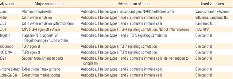
Representative adjuvants and their possible action mecha- nisms are listed in the Table 3. Aluminium adjuvant (Alum) is the most commonly used vaccine adjuvants. It has been used for more than 70 years because of its safety and capacity of making antigen depot [126]. Alum adsorbs the vaccine anti- gen and makes the antigen stay longer in the injection site, so that immunogenicity of the antigen is increased. In an im- munological mechanism study, alum was shown to activate
NALP3 inflammasome and induce IL-1β production to stim- ulate the innate immune system. Alum is likely to induce T helper type 2 immune responses to co-administered antigens in humans [127].
MF59 is an oil-in-water emulsion type of adjuvants, and composed of squalene, polysorbate 80, sorbitan trioleate, tri- sodium citrate dehydrate, citric acid monohydrate and water for injection [128]. MF59 adjvuant is used in influenza vac- cine (Fluad) and pandemic flu vaccine. Use of MF59 adjvuant resulted in stronger antibody responses and vaccine antigen dose sparing effects [129]. MF59 stimulates cells in the sites of injection to express chemokines and cytokines. These che- mokines and cytokines recruit innate immune cells and APCs.
Some antigen presenting cells subsequently uptake antigen- MF59 complex and migrate to draining lymph nodes for the induction of adaptive immune responses [130,131]. AS03 is a modified form of MF59 and is an oil-in-water emulsion with a tocopherol. This form of adjuvant was used in pandemic influenza vaccine (Pandemrix) [132]. TLR agonists are con- sidered as potential adjuvants, so many different kinds of TLR agonists have been tried in experimental animal models and clinical trials. AS04, which contains Alum and monophos- phoryl lipid A (MPL), is a licensed adjuvant in hepatitis B vi- rus (HBV) vaccine (Fendrix) [133] and human papillomavi- rus vaccine (Cervarix) [134]. MPL is a detoxified form of LPS, which is a TLR4 agonist. It can activate immune cells such as monocytes and myeloid dendritic cells directly to induce im- mune responses to the vaccine antigen [124]. Flagellin, a TLR5 agonist and a motor apparatus of bacteria, can induce mixed T helper types 1 and 2 immune responses. Flagellin-antigen fusion proteins were developed to magnify the vaccine and
Table 3. Various adjuvants used in vaccines
Adjuvants Major components Mechanism of action Used vaccines
Alum Aluminium hydroxide Antibodies, T helper type 2, adsorb antigen, NARP3 inflammasome Various human vaccines MF59 Oil in water emulsion Antibodies, T helper types 1 and 2, stimulate immune cells Influenza, pandemic flu AS03 Oil in water emulsion with tocopherol Antibodies, T helper types 1 and 2, stimulate immune cells Pandemic flu AS04 MPL (TLR4 agonist) + Alum Antibodies, T helper type 1, TLR4 signaling stimulation, NLRP3 inflammasome HBV, HPV Flagellin Flagellin (TLR5 agonist) or
Flagellin-antigen fusion protein Antibodies, T helper types 1 and 2, TLR5 signaling stimulation Clinical trial Imiquimod TLR7 agonist Antibodies, T helper type 1, TLR7 signaling stimulation Clinical trial
CpG DNA TLR9 agonist Antibodies, T helper type 1, TLR9 signaling stimulation Clinical trial
QS21 Saponin from American barks Antibodies, T helper types 1 and 2, stimulate immune cells, deliver antigen to cytoplasm
Clinical trial
Ginseng extract Extract from Panax ginseng Antibodies, T helper types 1 and 2, stimulate immune cells Clinical trial Alpha-GalCer Extract from marine sponge Antibodies, T helper types 1 and 2, stimulate immune cells Clinical trial MPL, monophos phoryl lipid A; TLR, toll-like receptor; HBV, hepatitis B virus; HPV, human papillomavirus.
adjuvant effects [135]. Imiquimod, a TLR7 agonist, and CpG DNA, a TLR9 agonist, stimulate plasmacytoid dendritic cells which can produce type 1 interferons to induce immune re- sponses to intracellular pathogens such as virus [124].
Recently, natural substances have received attention as a potential adjuvant compounds. QS21, derived from Ameri- can bark, is a saponin-based adjuvant. QS21 can stimulate dendritic cells directly and also destabilize the membrane of the endosome to deliver antigen to the cytoplasm, so that it can elicit cellular immune responses [136]. Another example of the natural adjuvants is an extract from Panax ginseng. It induced a balanced T helper types 1 and 2 immune response [137]. Many marine organisms, for example, sponge and al- gae extracts, are also investigated actively in experimental and clinical level. Alpha-galactosylceramide (alpha-GalCer), an extract from marine sponge, is known to stimulate natural killer T cells and induce antibody production, and balanced T helper types 1 and 2 immune responses [138].
Conclusion
Until now, most licensed influenza vaccines are manufac- tured by methods that were established more than 50 years ago despite recent scientific advances in vaccinology. New vaccines against influenza viruses to improve the breadth of protection would be feasible but some technical, regulatory, and logistical challenges remain to be resolved.
Many studies reporting universal influenza vaccines have focused on conserved single peptides or proteins as target antigens (M2e, HA2 stalk domain, NP, M1) in the presence of adjuvants in animal models. These conserved antigenic tar- gets have relatively weak immunogenicity (Table 1). Experi- mental vaccines based on the HA2 stalk domain or M2e ex- ternal domain of ion channel protein are not capable of in- ducing neutralizing antibodies although some monoclonal antibodies binding to the HA2 stalk domain are known to have cross neutralizing activity. Immunization with T-cell vaccines could provide survival protection but not prevent infection. Therefore, the protective efficacy of these candi- date universal vaccines would be lower than strain-specific HA based vaccines of inducing neutralizing antibodies.
It is needed to develop new qualitative and quantitative methods to define the potency of the vaccine as well as to iden- tify immune correlates of cross protection. The path to licen- sure of novel influenza vaccines will require demonstration of efficacy in humans. Vaccines that do not prevent infection
but ameliorate disease will need a larger scale of clinical tri- als. Public health authorities will evaluate how truly universal novel influenza vaccines are. Challenging decisions include whom to vaccinate and how often the universal vaccine may require updating.
As alternative approaches to developing truly universal vaccines, supplementation concept will be proven to be fea- sible by complementing and broadening the efficacy of cur- rent vaccines based on the strain-specific immunity. We have shown that supplementing the whole inactivated virus with the conserved M2 VLP vaccine was found to significantly im- prove the heterosubtypic cross protection [70]. With the emer- gence of drafting epidemic strains or an outbreak of pandem- ic, the supplemented vaccination would significantly prevent the mortality and ameliorate morbidity.
It will provide highly informative insight into developing novel new vaccines and adjuvants if we better understand the immunological mechanisms how cross protective vac- cines work and by which certain adjuvants enhance the im- munogenicity of vaccines. Also, deciphering the roles of in- nate immune components in contributing to long term pro- tective immunity will be an important area to be explored for designing and developing effective vaccines. In addition, cur- rent vaccines are mostly given by intramuscular injection us- ing syringe-needles and administered by medical personnel.
It is also an important area in vaccine field to develop new vaccine technologies and alternative routes of immunization such as needle-free skin delivery, oral, nasal, and sublingual immunization.
References
1. Stokes J, Chenoweth AD, Waltz AD, Gladen RG, Shaw D.
Results of immunization by means of active virus of hu- man influenza. J Clin Invest 1937;16:237-43.
2. Davenport FM. Current knowledge of influenza vaccine.
JAMA 1962;182:11-3.
3. Glaser CA, Gilliam S, Thompson WW, et al. Medical care capacity for influenza outbreaks, Los Angeles. Emerg In- fect Dis 2002;8:569-74.
4. Poehling KA, Edwards KM, Weinberg GA, et al. The un- derrecognized burden of influenza in young children. N Engl J Med 2006;355:31-40.
5. Thompson WW, Shay DK, Weintraub E, et al. Influenza- associated hospitalizations in the United States. JAMA 2004;292:1333-40.
6. Palese P, Compans RW. Inhibition of influenza virus re- plication in tissue culture by 2-deoxy-2,3-dehydro-N-tri- fluoroacetylneuraminic acid (FANA): mechanism of ac- tion. J Gen Virol 1976;33:159-63.
7. Matrosovich MN, Matrosovich TY, Gray T, Roberts NA, Klenk HD. Neuraminidase is important for the initiation of influenza virus infection in human airway epithelium.
J Virol 2004;78:12665-7.
8. Pica N, Palese P. Toward a universal influenza virus vac- cine: prospects and challenges. Annu Rev Med 2013;64:
189-202.
9. Francis T Jr, Salk JE, Brace WM. The protective effect of vaccination against epidemic influenza B. J Am Med As- soc 1946;131:275-8.
10. Salk JE, Pearson HE, Brown PN, Francis T. Protective ef- fect of vaccination against induced influenza B. J Clin Invest 1945;24:547-53.
11. Robertson JS, Nicolson C, Newman R, Major D, Dun- leavy U, Wood JM. High growth reassortant influenza vaccine viruses: new approaches to their control. Bio- logicals 1992;20:213-20.
12. al-Mazrou A, Scheifele DW, Soong T, Bjornson G. Com- parison of adverse reactions to whole-virion and split- virion influenza vaccines in hospital personnel. CMAJ 1991;145:213-8.
13. Cate TR, Couch RB, Kasel JA, Six HR. Clinical trials of monovalent influenza A/New Jersey/76 virus vaccines in adults: reactogenicity, antibody response, and anti- body persistence. J Infect Dis 1977;136 Suppl:S450-5.
14. Wright PF, Cherry JD, Foy HM, et al. Antigenicity and re- actogenicity of influenza A/USSR/77 virus vaccine in chil- dren: a multicentered evaluation of dosage and safety.
Rev Infect Dis 1983;5:758-64.
15. Quinnan GV, Schooley R, Dolin R, Ennis FA, Gross P, Gwa- ltney JM. Serologic responses and systemic reactions in adults after vaccination with monovalent A/USSR/77 and trivalent A/USSR/77, A/Texas/77, B/Hong Kong/72 influenza vaccines. Rev Infect Dis 1983;5:748-57.
16. Murphy BR, Coelingh K. Principles underlying the de- velopment and use of live attenuated cold-adapted in- fluenza A and B virus vaccines. Viral Immunol 2002;15:
295-323.
17. Harper SA, Fukuda K, Uyeki TM, et al. Prevention and control of influenza: recommendations of the Advisory Committee on Immunization Practices (ACIP). MMWR Recomm Rep 2004;53:1-40.
18. Belshe RB, Ambrose CS, Yi T. Safety and efficacy of live attenuated influenza vaccine in children 2-7 years of age.
Vaccine 2008;26 Suppl 4:D10-6.
19. Treanor J, Keitel W, Belshe R, et al. Evaluation of a single dose of half strength inactivated influenza vaccine in healthy adults. Vaccine 2002;20:1099-105.
20. Belshe RB, Newman FK, Wilkins K, et al. Comparative immunogenicity of trivalent influenza vaccine adminis- tered by intradermal or intramuscular route in healthy adults. Vaccine 2007;25:6755-63.
21. Bergen R, Black S, Shinefield H, et al. Safety of cold-adapt- ed live attenuated influenza vaccine in a large cohort of children and adolescents. Pediatr Infect Dis J 2004;23:
138-44.
22. Block SL, Yogev R, Hayden FG, Ambrose CS, Zeng W, Walker RE. Shedding and immunogenicity of live atten- uated influenza vaccine virus in subjects 5-49 years of age. Vaccine 2008;26:4940-6.
23. Wacheck V, Egorov A, Groiss F, et al. A novel type of in- fluenza vaccine: safety and immunogenicity of replica- tion-deficient influenza virus created by deletion of the interferon antagonist NS1. J Infect Dis 2010;201:354-62.
24. Peiris M, Yuen KY, Leung CW, et al. Human infection with influenza H9N2. Lancet 1999;354:916-7.
25. Fouchier RA, Schneeberger PM, Rozendaal FW, et al.
Avian influenza A virus (H7N7) associated with human conjunctivitis and a fatal case of acute respiratory dis- tress syndrome. Proc Natl Acad Sci U S A 2004;101:1356- 61.
26. Wong SS, Yuen KY. Avian influenza virus infections in humans. Chest 2006;129:156-68.
27. Cheung CL, Rayner JM, Smith GJ, et al. Distribution of amantadine-resistant H5N1 avian influenza variants in Asia. J Infect Dis 2006;193:1626-9.
28. de Jong MD, Tran TT, Truong HK, et al. Oseltamivir re- sistance during treatment of influenza A (H5N1) infec- tion. N Engl J Med 2005;353:2667-72.
29. Le QM, Kiso M, Someya K, et al. Avian flu: isolation of drug-resistant H5N1 virus. Nature 2005;437:1108.
30. Gao R, Cao B, Hu Y, et al. Human infection with a novel avian-origin influenza A (H7N9) virus. N Engl J Med 2013;
368:1888-97
31. Subbarao K, Klimov A, Katz J, et al. Characterization of an avian influenza A (H5N1) virus isolated from a child with a fatal respiratory illness. Science 1998;279:393-6.
32. Claas EC, Osterhaus AD, van Beek R, et al. Human influ-
enza A H5N1 virus related to a highly pathogenic avian influenza virus. Lancet 1998;351:472-7.
33. Buranathai C, Amonsin A, Chaisigh A, Theamboonlers A, Pariyothorn N, Poovorawan Y. Surveillance activities and molecular analysis of H5N1 highly pathogenic avian influenza viruses from Thailand, 2004-2005. Avian Dis 2007;51(1 Suppl):194-200.
34. Van Borm S, Thomas I, Hanquet G, et al. Highly patho- genic H5N1 influenza virus in smuggled Thai eagles, Bel- gium. Emerg Infect Dis 2005;11:702-5.
35. Wan XF, Nguyen T, Davis CT, et al. Evolution of highly pathogenic H5N1 avian influenza viruses in Vietnam between 2001 and 2007. PLoS One 2008;3:e3462.
36. Nava GM, Attene-Ramos MS, Ang JK, Escorcia M. Origins of the new influenza A(H1N1) virus: time to take action.
Euro Surveill 2009;14:19228.
37. Solovyov A, Palacios G, Briese T, Lipkin WI, Rabadan R.
Cluster analysis of the origins of the new influenza A(H1N1) virus. Euro Surveill 2009;14:19224.
38. Pinto LH, Holsinger LJ, Lamb RA. Influenza virus M2 protein has ion channel activity. Cell 1992;69:517-28.
39. Chizhmakov IV, Geraghty FM, Ogden DC, Hayhurst A, Antoniou M, Hay AJ. Selective proton permeability and pH regulation of the influenza virus M2 channel expressed in mouse erythroleukaemia cells. J Physiol 1996;494(Pt 2):329-36.
40. Mould JA, Drury JE, Frings SM, et al. Permeation and activation of the M2 ion channel of influenza A virus. J Biol Chem 2000;275:31038-50.
41. Leonov H, Astrahan P, Krugliak M, Arkin IT. How do amino- adamantanes block the influenza M2 channel, and how does resistance develop? J Am Chem Soc 2011;133:9903- 11.
42. Lin TI, Heider H, Schroeder C. Different modes of inhi- bition by adamantane amine derivatives and natural polyamines of the functionally reconstituted influenza virus M2 proton channel protein. J Gen Virol 1997;78(Pt 4):767-74.
43. Fiers W, De Filette M, Birkett A, Neirynck S, Min Jou W.
A “universal” human influenza A vaccine. Virus Res 2004;
103:173-6.
44. Liu W, Zou P, Ding J, Lu Y, Chen YH. Sequence compari- son between the extracellular domain of M2 protein hu- man and avian influenza A virus provides new informa- tion for bivalent influenza vaccine design. Microbes In- fect 2005;7:171-7.
45. Zebedee SL, Lamb RA. Influenza A virus M2 protein: mo- noclonal antibody restriction of virus growth and detec- tion of M2 in virions. J Virol 1988;62:2762-72.
46. Hughey PG, Roberts PC, Holsinger LJ, Zebedee SL, Lamb RA, Compans RW. Effects of antibody to the influenza A virus M2 protein on M2 surface expression and virus as- sembly. Virology 1995;212:411-21.
47. Roberts PC, Lamb RA, Compans RW. The M1 and M2 proteins of influenza A virus are important determinants in filamentous particle formation. Virology 1998;240:127- 37.
48. Treanor JJ, Tierney EL, Zebedee SL, Lamb RA, Murphy BR. Passively transferred monoclonal antibody to the M2 protein inhibits influenza A virus replication in mice.
J Virol 1990;64:1375-7.
49. Wang R, Song A, Levin J, et al. Therapeutic potential of a fully human monoclonal antibody against influenza A virus M2 protein. Antiviral Res 2008;80:168-77.
50. Slepushkin VA, Katz JM, Black RA, Gamble WC, Rota PA, Cox NJ. Protection of mice against influenza A virus chal- lenge by vaccination with baculovirus-expressed M2 pro- tein. Vaccine 1995;13:1399-402.
51. Neirynck S, Deroo T, Saelens X, Vanlandschoot P, Jou WM, Fiers W. A universal influenza A vaccine based on the extracellular domain of the M2 protein. Nat Med 1999;5:
1157-63.
52. Fan J, Liang X, Horton MS, et al. Preclinical study of in- fluenza virus A M2 peptide conjugate vaccines in mice, ferrets, and rhesus monkeys. Vaccine 2004;22:2993-3003.
53. De Filette M, Fiers W, Martens W, et al. Improved design and intranasal delivery of an M2e-based human influ- enza A vaccine. Vaccine 2006;24:6597-601.
54. Ionescu RM, Przysiecki CT, Liang X, et al. Pharmaceuti- cal and immunological evaluation of human papilloma- virus viruslike particle as an antigen carrier. J Pharm Sci 2006;95:70-9.
55. Bessa J, Schmitz N, Hinton HJ, Schwarz K, Jegerlehner A, Bachmann MF. Efficient induction of mucosal and sys- temic immune responses by virus-like particles admin- istered intranasally: implications for vaccine design. Eur J Immunol 2008;38:114-26.
56. Tompkins SM, Zhao ZS, Lo CY, et al. Matrix protein 2 vac- cination and protection against influenza viruses, inclu- ding subtype H5N1. Emerg Infect Dis 2007;13:426-35.
57. Fu TM, Grimm KM, Citron MP, et al. Comparative im- munogenicity evaluations of influenza A virus M2 pep-
tide as recombinant virus like particle or conjugate vac- cines in mice and monkeys. Vaccine 2009;27:1440-7.
58. Ernst WA, Kim HJ, Tumpey TM, et al. Protection against H1, H5, H6 and H9 influenza A infection with liposomal matrix 2 epitope vaccines. Vaccine 2006;24:5158-68.
59. Huleatt JW, Nakaar V, Desai P, et al. Potent immunoge- nicity and efficacy of a universal influenza vaccine can- didate comprising a recombinant fusion protein linking influenza M2e to the TLR5 ligand flagellin. Vaccine 2008;
26:201-14.
60. De Filette M, Ramne A, Birkett A, et al. The universal in- fluenza vaccine M2e-HBc administered intranasally in combination with the adjuvant CTA1-DD provides com- plete protection. Vaccine 2006;24:544-51.
61. De Filette M, Min Jou W, Birkett A, et al. Universal influ- enza A vaccine: optimization of M2-based constructs.
Virology 2005;337:149-61.
62. Heinen PP, Rijsewijk FA, de Boer-Luijtze EA, Bianchi AT.
Vaccination of pigs with a DNA construct expressing an influenza virus M2-nucleoprotein fusion protein exacer- bates disease after challenge with influenza A virus. J Gen Virol 2002;83(Pt 8):1851-9.
63. Jegerlehner A, Schmitz N, Storni T, Bachmann MF. Influ- enza A vaccine based on the extracellular domain of M2:
weak protection mediated via antibody-dependent NK cell activity. J Immunol 2004;172:5598-605.
64. De Filette M, Martens W, Roose K, et al. An influenza A vaccine based on tetrameric ectodomain of matrix pro- tein 2. J Biol Chem 2008;283:11382-7.
65. Kang SM, Kim MC, Compans RW. Virus-like particles as universal influenza vaccines. Expert Rev Vaccines 2012;
11:995-1007.
66 Kang SM, Pushko P, Bright RA, Smith G, Compans RW.
Influenza virus-like particles as pandemic vaccines. Curr Top Microbiol Immunol 2009;333:269-89.
67. Rodriguez-Limas WA, Sekar K, Tyo KE. Virus-like parti- cles: the future of microbial factories and cell-free sys- tems as platforms for vaccine development. Curr Opin Biotechnol 2013;24:1089-93.
68. Roldao A, Mellado MC, Castilho LR, Carrondo MJ, Alves PM. Virus-like particles in vaccine development. Expert Rev Vaccines 2010;9:1149-76.
69. Song JM, Wang BZ, Park KM, et al. Influenza virus-like particles containing M2 induce broadly cross protective immunity. PLoS One 2011;6:e14538.
70. Song JM, Van Rooijen N, Bozja J, Compans RW, Kang
SM. Vaccination inducing broad and improved cross protection against multiple subtypes of influenza A vi- rus. Proc Natl Acad Sci U S A 2011;108:757-61.
71. Kim MC, Song JM, O E, et al. Virus-like particles contain- ing multiple M2 extracellular domains confer improved cross-protection against various subtypes of influenza virus. Mol Ther 2013;21:485-92.
72. Kim MC, Lee JS, Kwon YM, et al. Multiple heterologous M2 extracellular domains presented on virus-like parti- cles confer broader and stronger M2 immunity than live influenza A virus infection. Antiviral Res 2013;99:328-35.
73. El Bakkouri K, Descamps F, De Filette M, et al. Universal vaccine based on ectodomain of matrix protein 2 of in- fluenza A: Fc receptors and alveolar macrophages medi- ate protection. J Immunol 2011;186:1022-31.
74. Pei S, Xiong N, Zhang Y, Chen S. Increasing M2 epitope density enhances systemic and mucosal immune respon- ses to influenza A virus. Biotechnol Lett 2009;31:1851-6.
75. Wei G, Meng W, Guo H, et al. Potent neutralization of in- fluenza A virus by a single-domain antibody blocking M2 ion channel protein. PLoS One 2011;6:e28309.
76. Vaxinnate. VaxInnate’s universal flu vaccine candidate shown safe and immunogenic in Phase I clinical study [Internet]. Cranbury: Vaxinnate; 2013 [cited 2013 Sep 1].
Available from: http://www.vaxinnate.com/pages/press- releases/20081026_001.html.
77. Taylor DN, Treanor JJ, Strout C, et al. Induction of a po- tent immune response in the elderly using the TLR-5 ag- onist, flagellin, with a recombinant hemagglutinin influ- enza-flagellin fusion vaccine (VAX125, STF2.HA1 SI). Vac- cine 2011;29:4897-902.
78. Turley CB, Rupp RE, Johnson C, et al. Safety and immu- nogenicity of a recombinant M2e-flagellin influenza vac- cine (STF2.4xM2e) in healthy adults. Vaccine 2011;29:
5145-52.
79. Wilson IA, Skehel JJ, Wiley DC. Structure of the haemag- glutinin membrane glycoprotein of influenza virus at 3 A resolution. Nature 1981;289:366-73.
80. Laver WG, Gerhard W, Webster RG, Frankel ME, Air GM.
Antigenic drift in type A influenza virus: peptide map- ping and antigenic analysis of A/PR/8/34 (HON1) vari- ants selected with monoclonal antibodies. Proc Natl Acad Sci U S A 1979;76:1425-9.
81. Laver WG, Air GM, Dopheide TA, Ward CW. Amino acid sequence changes in the haemagglutinin of A/Hong Kong (H3N2) influenza virus during the period 1968-77.