Address reprint requests to Hwan-Mo Lee, M.D.
Department of Orthopaedic Surgery, Yonsei University College of Medicine
#134 Shinchon-dong, Sodaemun-ku, Seoul, 120-752, Korea
Tel : 82-2-361-5648, Fax : 82-2-363-1139, E-mail : hwanlee@yumc.yonsei.ac.kr
퇴행성 요추질환의 병태생리
이 환 모
연세대학교 의과대학 정형외과학교실
Pathophysiology of Degenerative Disease of the Lumbar Spine
Hwan-Mo Lee, M.D.
Department of Orthopaedic Surgery, Yonsei University College of Medicine, Seoul, Korea
요통은전체 인구의약 80%가 경험할정도로흔한 질 환이며, 특히활동이 많은 젊은층에서는결근의 가장 큰 원인이되고있다. 요통의원인으로는추간판성동통, 후관 절통, 척추성동통, 방사통, 연관통, 근막성동통등여러가 지가 있으나, 그 기전에대해서는많은 논란이 있어왔다.
본 장에서는주로추간판성동통및 방사통의기전에대한 최신지견을살펴보고자한다.
척추 주위 신경의 분포 및 기능
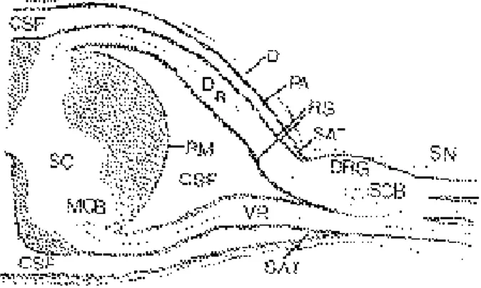
척추 분절에분포하는신경에관한 연구는오래동안이 루어져왔으나, 아직도많은 부분에서논란이거듭되고있 다. 척수에서분지되어나온dorsal root와 ventral root는 dorsal root ganglion(후근절)을 형성하며이후 척수신경 으로이행한다(Fig. 1).
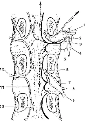
Dorsal primary ramus는 척추분절에분포하는신경으 로 주로내측지가척추골막, 후관절막, 척추주위인대에분 포하며sinuvertebral nerve (척추동신경)은 주로 척추관 내의 구조물에분포한다(Fig. 2). 대부분연구는 D o r s a l primary ramus가 후근절의바로원위부에서나오는척수 신경의분지거나또는 자율 신경 결합지에서분지된다고 보고하고있다30,33,34). 척추동신경의기원에대하여는후근 절의 원위부에서나타난다고알려져왔으나, 현재는척수 신경과 교감신경절을연결하는rami communicantes에 서 분지된다고하며9,26), 이 신경은척수강내에서상하지로
나뉘어최소 2분절 이상의후종인대, 척수경막및 후방 섬 유륜에 분포한다(Fig. 3)2 ). 척추동 신경의 구성성분은 rami communicantes에서 기시되므로spinal nerve와 교 감신경모두 관여하는것으로보이나, 다분절교감신경을 절제하였을때 섬유륜에분포하는신경섬유의숫자가감 소하므로, 교감신경이척추동신경의주된구성 성분인것
Vol. 6, No. 2, pp201~207, 1999
Fig. 1. Microscopic cross section of the spinal cord, nerve roots, and spinal nerve.
SC : Spinal cord VR : Ventral nerve root DR : Dorsal nerve root
MCB : Motor cell body DRG : Dorsal root ganglion SCB : Sensory cell body
SN : Spinal nerve PM : Pia mater RS : Root sheath D : Dura SAT : Subarachnoid triangle
CSF : Cerebrospinal fluid
을 알 수 있다. Nakamura등2 1 )은 이 실험을 통하여 후방 섬유륜에분포하는신경섬유가양측의여러분절에서기시 하는 교감신경에서나오므로추간판성동통이 내장인성 동통 (visceral pain)과 비슷한 성질을 가진다고하였다.
또한 Sekiguchi등32)은 제 2-3 요추의 편측 교감신경절제 후 척수경막에서CGRP에 반응하는nerve ending이 감소 하므로감각신경섬유가교감신경을통해분포한다고하 였다(Fig. 4). 후종인대는척추 분절에서신경섬유가가장 풍부히분포해있으므로, 추간판의이상으로인한 통증의 감지에매우예민하고, 후종인대자체의elevation에 의한 긴장시심한통증을유발할수 있다.
Fig. 2. Nerve innervation of the spinal segment
Fig. 3. Nerve innervation of the posterior aspect of the vertebral body and intervertebral disc.
11. dorsal root ganglion 12. rami communicantes
13. sinuvertebral never and its origin according to Groen and c o l l e a g u e s
14. autonomic ganglion
15. nerve to anterior longitudinal ligament 16. spinal nerve root
17. sinuvertebral nerve arising from distal pole of ganglion (thought to be its most common source before report of Groen and colleagues)
18. dorsal primary ramus of spinal nerve 19. ventral primary ramus of spinal nerve
10. arteries entering basivertebral sinus to supply cancellous bone 11. descending dorsal central branch of vertebromedullary
(spinal) artery
12. ventral branch of vertebromedullary artery
Fig. 4. A schematic illustration of CGRP immunoreactive fibers pathway to the lumbar dura mater.
1. Direct innervation from dorsal root ganglia 2. Fibers descending through the sympathetic trunk
DRG : Dorsal root ganglion SVN : Sinuvertebral nerve ST : Sympathetic trunk Ra : Ramus albus Rg : Ramus griseus
추간판자체는신경섬유및 혈관이없는 구조로알려져 왔으나, 정상 섬유륜의외측에는신경섬유가분포하며39,40) 신생아에서는수핵에도신경섬유가있으나 곧 없어진다
17,41).
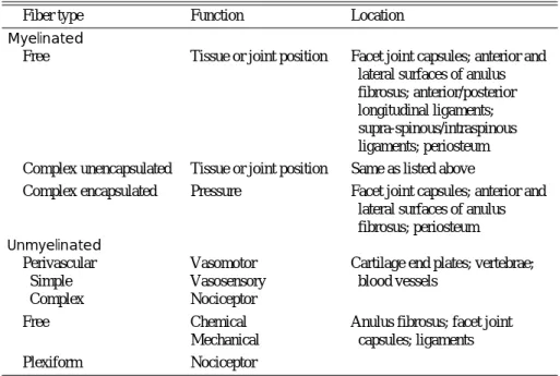
척추에 분포하는신경말단의종류는 다양하게있으며, 그중p l e x i f o r m과 free ending unmyelinated 신경섬유가 화학적또는 기계적인자극에대하여활성화되어 동통을 유발하는n o c i c e p t o r로서 작용한다(Table 1). 말초신경에 서 척수를통하여뇌까지정보를전달하는신경섬유는A - beta, A-delta, C 섬유3가지가있으며3 8 ), A-beta 섬유는굵 기가5 - 1 2㎛, A-delta는1 . 5㎛ C는 1㎛ 미만으로정보의전 달 속도는A-beta 섬유가가장빠르나동통을전달하는작 용은주로A - d e l t a ,와 C -섬유가담당한다(Table 2).
후근절은functional spinal unit에서“brain”에 해당하 는 중요한구조물로서20), 요통의조절과정에서중요한역 할을 한다1 5 ). 요추부에서후근절의위치는 척수신경근의 외측에있으므로척수강내, 추간공내, 추간공외측에위치 할 수 있으나추간공내에위치하는경우가대부분이며원 위부로 갈수록 크기가 커져 제1요추에서는길이와 폭의 평균이4.3㎜와3.7㎜이나제 1천추에서는11.2㎜와 6.2㎜
에 달한다. 후근절의세포는크기에따라 두가지로나뉘며 큰 직경의세포에서는A-beta 섬유가나오고, 작은 직경의 세포에서는주로 동통의전달을담당하는A-delta와 C 섬 유가 나온다. 후근절에서는Substance P, Calcitonin gene-related peptide, Vasoactive intestinal peptide 같 은 신경전달물질들이합성되며이들 모두 동통의매개물 질이고, 후근절은말초신경과척수사이에서동통의 전달 에서필수적인역할을한다.
후근절 자체는 단단한 c a p s u l e로 싸여있고, 신경근에 비해 영 양 공 급 이 저하되어 있 으 므 로 m e c h a n i c a l compre-ssion이 가해지면쉽게 신경내부종을 일으키고 결과적으로후근절내신경세포의혈액공급저하로인한비 정상적신경활동과동통을유발할수 있다31). 후근절의신 경외막에 있는 nervi nervosum 역시 m e c h a n i c a l sensitive nociceptor로 작용하므로후근절의신경외막도 외부 c o m p r e s s i o n이나 mechanical stimulation에 대해 서 직접적으로활성화되어동통을유발할수 있다.
이와같이외부 자극에대하여동통을유발시킬수 있는 신경섬유가분포하는후관절, 추간판인대, 근육, 후근절 및 신경근모두요통을일으킬수 있는원인이된다.
Table 1. Nerve innervation of tissues around the spinal segment
Fiber type Function Location
M y e l i n a t e d
Free Tissue or joint position Facet joint capsules; anterior and lateral surfaces of anulus fibrosus; anterior/posterior longitudinal ligaments;
supra-spinous/intraspinous ligaments; periosteum Complex unencapsulated Tissue or joint position Same as listed above
Complex encapsulated Pressure Facet joint capsules; anterior and lateral surfaces of anulus fibrosus; periosteum U n m y e l i n a t e d
Perivascular Vasomotor Cartilage end plates; vertebrae;
Simple Vasosensory blood vessels
Complex Nociceptor
Free Chemical Anulus fibrosus; facet joint
Mechanical capsules; ligaments
Plexiform Nociceptor
Table 2. Classification and function of nerve endings
Group Function Diameter(㎛) Conduction Velocity(m/s)
Group IA Muscle spindle, primary ending 13-20 180-120
Group IB Golgi tendon organs 12-18 170-110
Group II Muscle spindle, secondary ending 15-12 120-701
Group III A-delta Pricking pain, temperature, crude touch 11-51 12.5-20.0 Group IV C fibers Pain, itch, temperature, crude touch 0.5-2.0 0.5-2.5
추간판성 동통의 기전
성인 요추의추간판은전체 요추의35%를 차지하는큰 구조물이나혈관이 분포하지않으므로대부분의대사가 anaerobic 대사이며d i f f u s i o n에 의해 영양공급을받는 다. 이로 인하여외부의독성 자극에의해 쉽게 변성될수 있다. 추간판은proteoglycan이 풍부한수핵과, collagen 이 대부분을차지하는섬유륜으로나누어지나평균 세포 수는5800/㎣로우리몸의다른조직과비교하여미미하다
18).추간판의변성을촉진시키는요인에는당뇨병, 노화, 진 동같은외부의자극, 흡연, cytokine, 영양부족및 운동부 족 등이있으며, 이와같은변성으로추간판내구성성분의 변화가 나타난다. 즉수분과 p r o t e o g l y c a n은 감소하고, collagen 은 증가하며, keratan sulfate/chondroitin
sulfate의 비도 증가한다. 이렇게퇴행성변화가진행하면 추간판내영양결핍으로인한세포내물질및 세포의파괴 로 인한 추간판구조의변화와불안정성이나타나고이는 후근절에서n e u r o p e p t i d e의 분비를 증가시키며, 이로인 한 prostaglandin E2같은 염증성물질과collagenase 같 은 효소의증가는동통을유발할뿐만 아니라지속적으로 추간판의변성 및 불안정성을악화시킨다4 0 ). 또한 L a c t i c a c i d의 증가로 인한 낮은 p H와 여러 가지 n e u r o g e n i c and non-neurogenic pain mediator는 변성으로인한섬 유륜의파열 부분을통하여nociceptor를 자극하여동통 을 유발할수 있다.
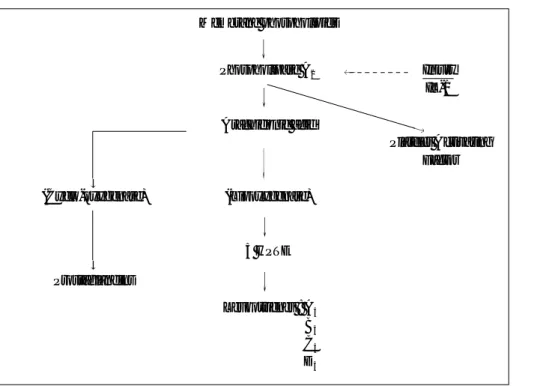
Phospholipase A2 는 arachidonic acid cascade에 관 여하는물질로(Fig. 5) 변성된추간판에서높은농도로발 견된다(Table 3)8). PLA2는 류마토이드관절염, 췌장염, 패 혈증에서도염증을일으키는물질로알려져있으며7,37), in vivo 및 in vitro 실험에서도강력한염증을유발시키는물 질로 보고되었다3,8). PLA2가 추간판내에서외부로노출되 면 직접적으로섬유륜외부의신경섬유를자극하여세포 막의 phospholipid를 분해하여염증을일으킬수도 있으 며, 이로인해형성된염증 매개물질들이신경염을일으켜 동통을 야기시킨다. PLA2에 노출된 신경은 i n t r a n e u r a l edema, demyelination, cellular vacuolization 및 axonal injury가 생기며, 이렇게손상된신경은외부의자 극에민감하여작용하거나자체적으로action potential을 일으킨다. 또한변성된 추간판의 세포는 자체적으로 Matrix Metalloproteinase, Nitric oxide, Interleukin-6, Membrane phospholipids
Phospholipase A2
Arachidonic acid
(Lipoxygenase)
5 HPTE (Cyclo-oxygenase)
Prostaglandins
Leukotrienes : A4
B4
C4
D4
Platelet Activating Factor Injury IL-1
Fig. 5. Inflammatory cascade of PLA2
Table 3. PLA2activity in human tissues
Human Source material PLA2Specific Activity (nmol/min/mg)
PMN 23.2
Platelet 21.4
Sperm 28.0
Plasma 2220.006
Inflammatory synovial fluid 12.1
Mucoid ear effusion 20.3
Herniated lumbar disc 1212222 PLA2= phospholipase A2.
PMN = polymorphonuclear leukocytes.
PG E2를 생산하며12)이들 cytokine은 proteoglycan의 생 성을 억제하여추간판matrix의 파괴와퇴행성변화를촉 진하여 요통을 일으킬 수 있으며, 이들모두 직접적으로 섬유륜과후종인대에분포되어있는 신경 말단을자극하 여 요통의원인이된다.
신경근 병변의 기전
아직까지신경근압박과그로 인한 방사통의발생에관 한 병태 생리학에대해서는정확하게밝혀지지않은 상태 이다. 실험적으로정상신경근에기계적변화를가해도방 사통이나감각 및 운동장애를나타내지않으며, 말초신경 에서도유사한결과를보인다.
손상받지않거나염증이없는 신경에갑자기압박을가 하면 저림, 감각이상, 운동약화등이생길 수 있지만동통 은 발생하지않는 반면, 만성적으로손상받은신경근이나 후근 신경절은기계적압박에민감하게통증을유발하게
된다2 9 ). 정상 신경근과만성적으로손상받았던신경근이
기계적압박에대하여나타내는반응의차이는신경근내
의 혈류 차 이 와 축 삭 형 질 운반 ( a x o n o p l a s m i c transport), 염증, 부종, 탈수초화등과 같은 요소들이관 계가있는것으로알려져있다39).
척수 신경근은말초신경보다압박에쉽게 손상을 받을 수 있는 여러 해부학적특징을갖고 있다. 척수 신경근을 싸고 있는 척수경막, 지주막및 척수연막은말초신경에서 는 신경외막(epineurium), 신경주막(perineurium) 및신 경내막(endoneurium)으로이행되어신경을보호하는기 능을 갖게 된다. 따라서신경근은말초신경계의신경내막 에 해당되는얇은 척수연막에의해서만싸여 있기 때문에 쉽게 압박에의한 손상을받을 수 있다(Fig. 6). 또한신경 근 및 신경근을싸고있는척수경막을후종인대및 척추체 에 고정시켜주는 Hofmann 인대가 추간판탈출증시에는 신경근의뒷부분의압박 없이도신경근의앞부분만을압 박할수 있는해부학적특징을갖고있다33).
신경근조직의기계적 변형은신경내에서다양한 조직 반응을유발시켜신경학적이상과신경근병증등의증상을 나타낸다. 임상적으로신경성파행을지닌 환자에서산소 공급이 감소되면 방사통을 일으키는 점에 착안하여 Evans6)는 신경근의만성적압박은신경근에허혈을일으 킨다고주장하였다. 그러나최근의연구들에의하면협소 한 척추관의공간적제한이내부 정맥혈관계에더 영향을 주는 것으로알려져있다27). Olmarker등24)은 돼지의요천 추부 신경근에낮은 압력 (5-10. ㎜Hg)를 가하면정맥혈 의 울혈로신경내미세 순환에급격한변화를초래한다고 하였다. 이들은평균동맥혈압인130㎜Hg가되면완전허 혈을 볼 수 있고 50㎜Hg로 10분을 압박한후에는신경내 부종이 발생하였으며, 압력이천천히 가해지는경우에는 신경내부종형성또한 현저하지않았다고하였으며, 100
㎜Hg로 두 시간 동안 압박을가하면, 감각신경근의충동 전도가75%에서 장애를 보이고운동신경은55%의 장애 를 보이며, 압박이풀린후운동신경의회복이감각신경보 다 더 빠르고 더 완전하게일어난다고하였다. 신경근에 대한 압박은 혈액순환장애와뇌척수액순환장애로인한 신경내부종을일으키고이로인한신경내압력의증가 및 대사장애는동통 및 신경전도의변화를일으켜신경섬유 의 반흔 및 유착을일으킨다. 그러나실제로인체에서영 상진단방법에서나타난추간판탈출증의정도와방사통의 관 계 가 일 치 하 지 않 으 며1 1 , 3 6 ), 경 막 외 로 주 입 된 corticosteroid는 방사통을효과적으로감소시켜준다4). 그 러므로동통의원인으로신경근의압박 뿐만 아닌 추간판 내 화학물질에의한 chemical neuritis 및 a u t o i m m u n e theory가 있으며, 이를 입증하기위한 많은 임상 및 기초 연구가진행되고있다10,13,14,22,23).
Olmarker등은 돼지 척수강에자가 수핵을놓아서신경 전도 속도가지연되는것을 관찰했으며, 신경기능장애의 Fig. 6. Histology of peripheral nerve and intrathecal nerve
root. Peripheral nerve is surrounded by endoneurium, perineurium, and epineurium, but intrathecal nerve roots are covered only by endoneurium and root sheath.
Intrathecal nerve roots are more susceptible to external compression.
원인으로는Demyelination과 신경세포내미세구조의손 상이라고하였고, 이들은또한 수핵내에서신경근에손상 을 입히는주된 성분은수핵내세포성분인것을 입증하였
다22,23). Kawakami 등13)은 쥐를 이용한실험에서수핵내의
PLA2와 Nitric oxide가 쥐에서나타나는신경근병변에의 한 행동장애의원인이라고하였고, Takahashi등35)은 추간 판 탈출에의해 신경근에가해지는압력이동통의정도와 는 관계가없으나근력감소나감각능력저하와는연관관 계가있다고하였다.
수핵은avascular tissue 이므로 태생기이후 면역체계 에 노출이 되지 않고, 일단 수핵 조직이 c i r c u l a t o r y system에 노출이되면자가면역반응을일으킨다. 이에 대 한 근거로 수핵에 대한 항체를 추간판 탈출증 환자의 serum에서 추출할수 있으며1,19), 추간판탈출증환자에서 cellular migration도 저하되어 있고5 ), 추간판내에서 Immunoglobulin도 발견된다28).
또한 동물실험에서척추강내에삽입된 자가 수핵은 면 역반응에의한 염증반응을일으킬수 있다16). 척수강내로 돌출된수핵이나섬유륜이시간이지남에따라 자연 흡수 되는 것을 많은 연구자들이보고하였고14) contained disc 보다는non-contained disc가 빠른흡수과정을보이는것 은 또 다른면역반응의증거가될 수 있다.
이와같이추간판 탈출증에서신경근 병변을 일으키는 기전으로서 ① compression theory ② c h e m i c a l neuritis theory ③ autoimmune theory 등이 있으며, 이 모든기전이복합적으로작용하는것으로생각된다.
그러나 퇴행성추간판질환에서요통의기전에 대하여 는 우리가 알지 못하는 부분이 아직도 많으며, 앞으로도 많은연구가필요하다.
REFERENCES
11) Bobechko WP and Hirsch C : Autoimmune response to nucleus pulposus in the rabbit. J Bone Joint Surg, 47B:
574-580, 1965.
12) Bogduk N and Twomey LT : Clinical anatomy of the lumbar spine. Melbourne ; Churchill Livingstone, 1987.
13) Chen C, Cavanaugh JM, Ozaktay AC, Kallakuri S a n d King AI : Effects of phospholipase A2 on lumbar nerve root structure and function. Spine, 22:1057-64, 1997.
14) Dilke TFW, Burry JC and Grahame R : E x t r a d u r a l corticoid injection in management of lumbar nerve root compression. BMJ, 2:635-637, 1973.
15) Elves MW, Bucknill T and Sullivan MF : In vitro inhibition leukocyte migration in patients with intervertebral disc lesions. Orthop Clin North Am, 6:59-65, 1975.
16) Evans JG : Neurogenic intermittent claudication. Br Med J, 2:985-987, 1965.
17) Famaey JP : Phospholipases, eicosanoid production and inflammation. Clin Rheumatol, 1:84-94, 1982.
18) Franson RC, Saal JS and Saal JA : Human disc phospholipase A2 is inflammatory. Spine, 175:129-132, 1992.
19) Groen GJ, Baljet B and Drukker J : The innervation of the spinal dura mater : anatomy and clinical implications.
Acta Neurochir, 92:39-46, 1988.
10) Ito T, Yamada M and Ikuta F : Histologic evidence of absorption of sequestration-type herniated disc. Spine, 21:230-234, 1996.
11) Jaffrey D and O’Brien JA : Isolated intervertebral disc resorption : A source of mechanical and inflammatory back pain? Spine, 11:397-401, 1986.
12) Kang JD, Georgescu HI and MacIntyre-Larkin L : Herniated lumbar intervertebral discs spontaneously produce matrix metalloproteinase, nitric oxide, interleukin- 6, and prostaglandin E2. Spine, 21:271-277, 1996.
13) Kawakami M, Tamaki T and Hashizume H : The role of phospholipase A2 and nitric oxide in pain-related behavior produced by an allograft of intervertebral disc material to the sciatic nerve of the rat. Spine, 22:1074-1079, 1997.
14) Kimori H, Shinomiya K and Nakai O : The natural history of herniated nucleus pulposus with radiculopathy. Spine, 21:225-229, 1996.
15) Lindbloom K and Rexed B : Spinal nerve injury in dorso- lateral protrusions of lumbar disks. J Neurosurg, 5:413- 432, 1948.
16) McCarron RF, Wimpee MW, Hudkin RG and Laros GS : The inflammatony effect of nucleus pulposus. A possible element in the pathogenesis of low back pain. Spine, 12:760-764, 1987.
17) Malinsky J : The ontogenetic development of nerve terminations in the intervertebral discs of man. Acta Anat, 38:96-113, 1959.
18) Maroudas A, Stockwell RA and Nachemson A : Factors involved in the nutrition of the human lumbar vertebral disc : cellularity and diffusion of glucose in vitro. J Anat, 120:113-130, 1975.
19) Marshall LL, Trethewie ER and Curtain CC : Chemical radiculitis: A clinical, psychological and immunological study. Clin Orthop, 129:61-67, 1987.
20) Melzack R and Wall PD : Pain mechanisms : A new theory. Science, 150:971-979, 1965.
21) Nakamura S, Takahashi K and Takahashi Y : Origin of nerves supplying the posterior portion of lumbar intervertebral discs in rats. Spine, 21:917-924, 1996.
22) Olmarker K, Blomquist J and Stromberg J : Inflammatogenic properties of nucleus pulposus. Spine, 20:665-669, 1995.
23) Olmarker K, Brisby H and Yabuki S : The effects of normal, frozen, and hyaluronidase-digested nucleus pulposus on nerve root structure and function. Spine, 22:471-476, 1997.
24) Olmarker K, Rydevik B and Holm S : Effects of experimental graded compression on blood flow in spinal nerve root. J Orthop Res, 7:817-823, 1989.
25) Ozaktay AC, Cavanaugh JM, Blagoev DC and King AI : Phospholipase A2-induced electrophysiologic and histologic changes in rabbit dorsal lumbar spine tissues.
Spine, 20:2659-2668, 1995.
26) Paris SB : Functional anatomy of the lumbar spine. PhD Thesis. Union Graduate School, Atlanta, 1983.
27) Parke WW and Watanabe R : The intrinsic vasculature of the lumbosacral spinal nerve roots. Spine, 10:508-515, 1985.
28) Pennington JB, McCarron RF and Laros GS : Identifi- cation of IgG in the canine intervertebral disc. Spine, 13:909-912, 1988.
29) Rydevik B, Brown MD and Lundborg G : P a t h o a n a t o m y and pathophysiology of nerve root compression. Spine, 9:7- 15, 1984.
30) Rydevik B, McLean WG and Sjöstrand J : Blockage of axonal transport induced by acute, graded compression of the rabbit vagus nerve, J Neurol Neurosurg Psychiatry, 43:690-698, 1980.
31) Rydevik B, Myers RR and Powell HC : Pressure increase in the dorsal root ganglion following mechanical compression. Spine, 14:574-576, 1989.
32) Sekiguchi Y, Konnai Y, Kikuchi S and Sugiura Y : An anatomic study of neuropeptide immunoreactivities in the lumbar dura mater after lumbar sympathectomy. Spine, 21:925-930, 1996.
33) Spencer DL, Irvin GS and Miller JA : Anatomy and significance of fixation of the lumbosacral nerve roots in sciatica. Spine, 8:672-679, 1983.
34) Sunderland S : Nerve and nerve injury, In : Peripheral sensory mechanism, 2nd Ed. New York:Churchill Livingstone.
35) Takahashi K, Shima I and Porter RW : The nerve root pressure in lumbar disc herniation. Presented ISSLS meeting, Singapore, June, 2-6, 1997.
36) Thelander U, Fagerlund M and Friberg S : Straight leg raising test vs. radiologic size, shape and position of lumbar disc herniations. Spine, 17:395-399, 1992.
37) Vadas P, Stehanski E and Pruzanski W : C h a r a c t e r i z a t i o n extracellular PLA1 in rheumatoid synovial fluid. Life Sci, 1136:579-587, 1986.
38) Wall PD and Melzack R : Textbook of pain. 1st ed.
Churchill-Livingston, New York, 1984.
39) Weinstein JN, Claverie W and Gibson S : The pain of discography. Spine, 13:1344-1348, 1988.
40) Weinstein JN : Anatomy and neurophysiologic mechanisms of spinal pain. The adult spine 1st ed. Raven Press, New York, 1991.
41) Wyke BD : The neurology of low back pain. The lumbar spine and back pain 2nd ed. Kent:Pitman Medical, 1980.