A COMPARATIVE STUDY ON THE DIMENSIONAL CHANGE OF THE DIFFERENT DENTURE BASES
Myung-Joo Kim, D.D.S., M.S. Ph.D., Chang-Whe Kim, D.D.S., M.S.D., Ph.D.
Department of Prosthodontics, Graduate School, Seoul National University
Statement of problem.Acrylic resin is most commonly used for denture bases. However, acrylic resin has week points of volumetric shrinkage during polymerization that reduces den- ture fit. The expandability of POSS (Polyhedral Oligomeric Silsesquioxane) containing poly- mer could be expected to reduce the polymerization shrinkage of denture bases and would increase the adaptability of the denture to the tissue.
Purpose.The purpose of this study was to compare the dimensional stability in the conventional acrylic resin base, POSS-containing acrylic resin base, and metal bases.
Materials and methods.Thirty six maxillary edentulous casts and dentures of different base were fabricated. Tooth movement and tissue contour change of denture after processing (resin curing, deflasking, decasting and finishing without polishing) and immersion in artificial sali- va at 37℃ for 1 week and 4 weeks were measured using digital measuring microscope and three- dimensional laser scanner.
Results.The results were as follows:
1. The conventional resin group showed significant (p<0.01) dimensional change through- out the procedure (processing and immersion in artificial saliva).
2. After processing, the metal group and POSS resin group showed lower linear and 3-dimen- sional change than conventional resin group (p<0.01).
3. There was no statistically significant linear and 3-dimensional change after immersion for 1 week and 4 weeks in metal and POSS resin group.
4. In all groups, the midline and alveolar ridge crest area presented smaller 3-dimensional change compared with vestibule and posterior palatal seal area after processing and soaking in artificial saliva for 1 week and 4 weeks (p<0.01).
Conclusion. In this study, a reinforced acrylic-based resin with POSS showed good dimen- sional stability.
Key Words
Dimensional change, Acrylic denture base resin, Metal denture base, Polyhedraloli- gosilsesquioxane (POSS), Tooth movement, Laser scanning
J Korean Acad Prosthodont : Volume 44, Number 6, 2006
S
ince the late 1930’s, acrylic resin (PMMA, polymethylmethacrylate) have been the mate- rials of choice for the fabrication of complete denture bases. It has excellent esthetic properties, adequate strength, low water sorption, and low solubility. In addition, it is non-toxic, easy to repair, and can be used by simple molding and processing technique.1However, there are several problems with this material compared with ceramics and metal. One of those is dimensional shrinkage after curing at high temperature and high pressure. For example, dimensional shrinkage occurred in the range of 6~20% after curing (in var- ious commercialized resins) as result of poly- merization contraction, thermal contraction, internal stress release, water absorption, drying, and incomplete polymerization.2This shrinkage results in inaccurate adaptation of the denture to the tissue, reduction in denture stability and retention and changes in the positions of the artificial teeth.3In the past few years, acrylic resin monomer and polymers have also been modified to improve not only physical and mechanical properties, but also the dimensional shrinkage and working
properties that facilitate laboratory techniques of denture fabrication.4These new laboratory tech- niques of denture include microwave- and visi- ble light activated polymerization and vacuum- plus pressure adaptation during low temperature polymerization of resin system.5In spite of all of these advances, no combination of resin materi- al and processing technique has been able to reduce linear distortion to less than 0.2 %.6 Alternative materials such as polyamides, epoxy resin, polystyrene, vinyl acrylic, rubber graft polymers, polycarbonate and nylon have been attempted but no satisfactory material is available to date.4,7 Glass fiber, metal inserts and ultra- high molecular weight polyethylene (UHMPE) fibers have been scrutinized in many ways to
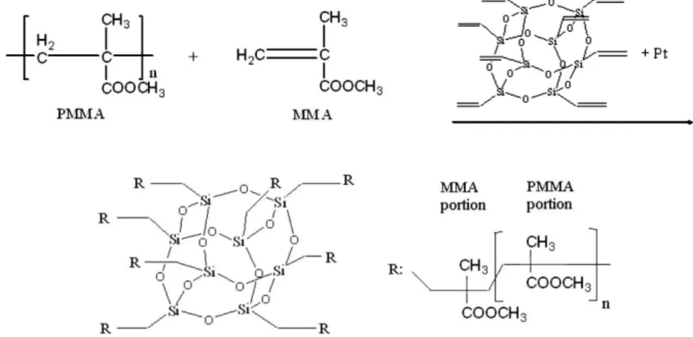
Fig. 1. The basic scheme of the POSS.
Fig. 2. The proposed chemical reactions in copolymerization of the PMMA/MMA with POSS and the structure of network obtained.
improve strength and increase the dimensional sta- bility.8-10However, most of them are unacceptable to the dental technicians because of their poor pro- cessing characteristics.
While, Nam et al.11 attempted to design and synthesized novel organic- inorganic hybrid den- ture materials based acrylic polymeric materials and monomeric vinyloligosilsesquoxane (Polyhedral Oligomeric Silsesquioxane, POSS). The hybrid composite obtained had all the advantage of polymeric materials. In addition it retained the superior characteristics of inorganic materials originated from silicone derivatives.12POSS (Fig.
1), which is one of the expandable monomers, has 8 vinyl functional groups in its own molecular structure, so in polymerization those double bonds in vinyl groups act as crosslinker to make network structure between PMMA and MMA matrices (Fig. 2).
As the results, expandability of POSS contain- ing polymer could be expected to reduce the polymerization shrinkage of denture bases and would increase the adaptability of the denture to the tissue. While the formation of POSS-con- taining denture composite resin successfully resulted in improvements in the mechanical properties of the composites13and solved the
good biocompatibility,14it is not clear whether the novel polymeric denture base resin is shrink- age reducible in comparison with metal denture base.
The purpose of this study was to compare the dimensional stability of the conventional acrylic resin base, prepared resin base of PMMA-POSS hybrid systems (POSS was explored as a par- tial substitute for MMA in the preparation of novel composites), and metal bases after pro- cessing (resin curing, deflasking, decasting and fin- ishing without polishing) and immersion in arti- ficial saliva at 37℃ for 1 week and 4 weeks.
Three-dimensional laser scanning and digital measuring microscope were used as an evaluation method to determine and compare the dimensional change.
MATERIAL AND METHODS 1. Stone cast fabrication
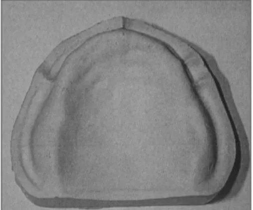
A rubber mold (Fig. 3) was made of non-under- cut edentulous maxillary cast (Fig. 4) using mold- ing silicone (Shimetshu, Japan). This mold was used to prepare 36 identical stone casts. Type V dental stone (Die-Keen, Heraus Kulzer Inc, USA) was
Fig. 3. Silicone mold for duplication of stone cast. Fig. 4. The master cast stone.
measured and mixed with the recommended amount of water (W/P ratio=21 ㎖/100 g) using automatic mixer (MixQueen, Oscotec, Korea).
2. Denture fabrication
A maxillary complete denture (Fig. 5) with palatal thickness of 2 ㎜ was waxed up with acrylic resin denture teeth (Trubyte Bioform 264 and 32M, Dentsply). On 12 stone casts, metal base was fabricated (Ticonium Premium 100, Ticonium Co, USA). Metal bases covered all palate and were extended to residual alveolar ridge with retentive mesh. To ensure uniformity and per- mit comparisons, a mold from silicone rubber was made from the master denture (Fig. 6). With the same mold of teeth the denture wax-up were duplicate by melting and pouring wax into the mold that held the acrylic resin teeth and stone casts in correct relationship to each other. Thirty- six denture wax-ups (12 for conventional resin base, 12 for POSS resin base, and 12 for metal base) were identical in thickness, height, and contour because they were prepared from the same mold. Each group processed into acrylic resin denture using the polymer (Vertex RS, Dentimex Zeist, Holland) and the monomer prepared as follows.
Group 1 ; Acrylic resin base (Control, conventional monomer)-PMMA:MMA system
Group 2 ; Acrylic resin base with POSS monomer- PMMA:MMA:POSS system POSS (Aldrich Chemical, USA) of 1% wt/vol was dissolved in monomer with 5 drops of Pt (Gelset, USA) and stirred for 10 min under vacuum
Group 3 ; Metal base (conventional monomer) A mix of resin was made using 8㎖ of the monomer and 20g of the polymer. A conven- tional heat curing cycle of 70℃ for 30 min and 100
℃ for 2hr was performed with denture curing unit (Curing Unit, Teledyne Hanau, USA).
3. The methods of measurement of dimen- sional change
In this study, laser scanner (Surveyor, Laser design, USA) and microscope (Mitsutoyo, Japan) as the measurement tool of dimensional accura- cy were used. We attempted to evaluate and compare the surface contour change and linear change between the cast and denture base.
(1) linear dimensional changes in the prepared denture base
The movement of teeth after processing of complete denture can be in any direction and may occur as a result of dimensional changes of acrylic resin.15,16Fine crosses were marked Fig. 5. Wax-up for resin base and metal base. Fig. 6. Silicone mold for duplication of
wax denture.
with a surgical scalpel on incisal edges of right cen- tral incisor (point A) and distobuccal cusps of both second molars (point B and C) to be used as ref- erence points of tooth movement measurement (Fig. 7). Distances (AB and BC) on artificial teeth
were measured at the wax-up stage (starting point), after processing, after the dentures had been stored in artificial saliva prepared using magnetic stirrer according to the composition suggested by Indiana University as shown in tableⅠ- in at 37
℃ water bath for 1 week and 4 weeks using dig- ital measuring microscope to the nearest 0.001 ㎜.
Measurements were made by the same operator and repeated 3 times to yield comparable results.
(2) three dimensional changes in the prepared denture base
Thirty-six stone casts were laser-scanned with scanning distance of 0.1㎜, accordingly scanning was repeated over 700 times for each cast. The 3- dimensional tissue surface data were processed through SURFACER 9.0 software program. After the dentures were processed, they were laser scanned within 24 hrs and the 3-dimensional tissue surface data were filed and overlapped onto those of the stone cast to evaluate the absolute average dimensional shrinkage. And then, the dentures were soaked in artificial sali- va for 1week and 4weeks at room temperature, they were again laser scanned. The tissue surface data were again filed and overlapped onto those of the stone cast to compare the dimensional change.
Fig. 7. Positions of reference points.
Table I. Composition and ratio of artificial saliva (g/l)
NaCl 0.4 KCl 0.4 CaCl2∙H2O 0.8 Na2S∙5H2O 0.01 CO(NH2)2Urea 1.0
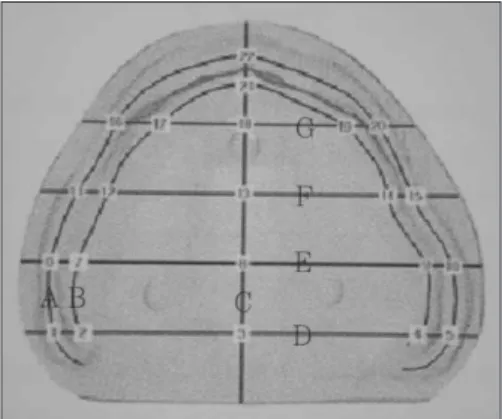
Fig. 8. Sectioning of the cast and denture on the computer screen (22 points).
Line A: middle of vestibule Line B: alveolar ridge crest Line C: midpalatal line Line D: posterior palatal seal
Line E, F, G: equally sectioned line between point 22 & 3 Vestibule area: point 1,5,6,10,11,15,16,20,22
Ridge crest area: point 2,4,7,9,12,14,17,19,21 Midline area: point 3,8,13,18
PPS area: point 1,2,3,4,5
To evaluate the local dimensional accuracy after denture processing, 7 lines were drawn on the overlapped images of the stone cast and den- tures on the computer screen (Fig. 8). The distance between the cast and denture was measured at 22 points.
4. Statistical analysis
The statistical evaluation of the data was per- formed using the software package SPSS/PC+
Statistics™ 10.1 (SPSS Inc., Chicago, IL, USA).
Data are reported as mean ± standard deviations (SD) at a significance level of P < 0.01. Statistics were performed by analysis of variance (ANOVA), followed by Tukey’s test for a post hoc com- parison.
RESULTS
1. Linear dimensional changes in the experi- mental group and procedure
Figure 9 shows linear dimensional changes as difference of AB and BC distances measured
after the processing and immersion of wax den- ture. The shrinkage and expansion are presented as negative and positive values. The metal group showed slight expansion (increase of AB dis- tance) result in comparison with wax denture step specially. Significant dimensional changes were found (p<0.01) among the experimental groups.
The POSS resin and metal group showed sig- nificantly smaller linear dimensional changes in all 3 procedures (after processing and immer- sion for 1week or 4 weeks). The conventional resin group showed significant difference through- out the procedure, while the POSS resin and metal group presented no significant difference.
In other words, some recovery of shrinkage after immersion in artificial saliva was observed in only conventional resin group. There was no statistical difference between immersion for 1 week and 4 weeks.
2. Three dimensional changes in the experi- mental group and procedure
After overlapping the data of stone cast with those of denture, 3 types of value were made.
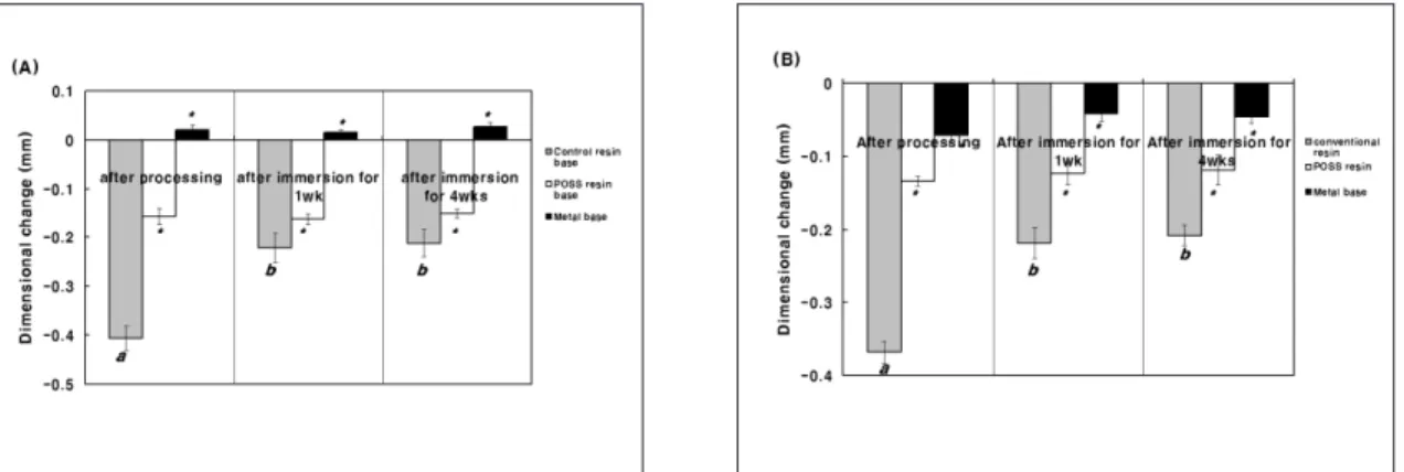
Fig. 9.Linear dimensional changes in AB distance (A) and BC distance (B) measured at the each group and procedure.
Data were expressed as mean values and standard deviations (n=36). Data were analyzed by ANOVA. * denotes a significant difference from the control (conventional resin) group (p<0.01). The different matching letters indicates the significant difference (p< 0.01) between procedures (after processing and immersion) in each group based on Tukey’
s multiple comparison test.
The positive and negative value of the distance between the stone cast and the tissue surface of denture were caused by shrinkage, expansion and deformation. The absolute mean values were measured and used as comparison factor.
The metal group showed the lowest 3-dimen- sional shrinkage and POSS resin group present- ed significantly lower (p<0.01) 3-dimensional change than control (conventional resin) group in all procedures (Fig. 10) in the same manner with linear dimensional change. POSS group and metal group showed non-significant (p>0.01) volumetric change after immersion (Fig. 10).
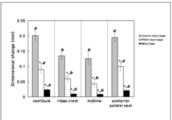
After sectioning the overlapped image of a stone cast and a denture to evaluate the local dimensional shrinkage, the distances were mea- sured 3 times at vestibular area 9 points, ridge crest area 9 points, midline area 4 points, and the pos- terior palatal seal area 5 points. In all groups, the midline and alveolar ridge crest area showed significantly lower dimensional change than the vestibular and posterior palatal seal area (Fig. 11).
DISCUSSION
In this study, digital microscopic measuring and 3-dimensional laser scanning were used to determine the dimensional change. Zissis et al16 reported that 60% of articles which had been published about dimensional accuracy of denture bases during the past 30 years used microscopy, and 25 % used simple hand-held caliper instru- ments. These methods could not evaluate the surface contour changes between the cast and den- ture than simple linear contraction measure- ment. Recently, a 3- dimensional digitization and computer graphic system has been developed to study and reproduce anatomic surfaces. The laser scanner reads an object and obtains numer- ous point data. These point data are converted into surface data through the computer program and reproduction of the original object can be possi- ble on the screen. We attempted to evaluate and compare the linear and three dimensional changes according to denture base materials. Our results Fig. 10. The absolute mean three dimensional shrink-
ages after procedure were measured. Data were ana- lyzed by ANOVA. * denotes a significant difference from the control (conventional resin) group (p<0.01). The dif- ferent matching letters indicates the significant difference (p<0.01) between procedures (after processing and immersion) in each group based on Tukey’s multiple comparison test.
Fig. 11. The local dimensional shrinkage after den- ture processing was measured. Data were analyzed by ANOVA. The different matching letters indicates the significant difference (p<0.01) between denture areas in each group based on Tukey’s multiple comparison test.
showed similar trends in linear volumetric change and tissue surface contour change. In other words, 3-dimensional volumetric shrinkage reflected the linear dimensional change. Therefore, simple linear dimensional change is regarded as a comparable tool in the measurement of dimensional accuracy or stability as well as three- dimensional change.
The first unavoidable dimensional change in all acrylic resin prosthesis is shrinkage that occurs dur- ing processing and finishing. The second change, expansion, occurs when the dentures are either stored in water bath or inserted in the mouth and then absorb oral fluids.17-19Traditionally, PMMA dentures are processed in brass denture flasks by compression molding of the acrylic resin with stone while it is in the doughy stage. The flasks are placed in a temperature-controlled water bath for a specified time to permit resin poly- merization. There has always been a problem with shrinkage of the acrylic resin during poly- merization process. The resin’s coefficient of linear expansion is 8.1×10⁻⁷. The gypsum prod- ucts which form the mold have a coefficient of lin- ear expansion of 1/8 that of acrylic resin. This dif- ference contributes to the dimensional change and induced strain.20 PMMA resin monomer can shrink up to 21 %vol during the polymerization process. In this study, the amount of process- ing shrinkage is larger than those reported by ot- her authors.3,6,15The fast heat-cured resin(boil- able resin) was used in this experiment reported produce significantly less distortion in a den- ture base than conventional acrylic resin by some author20,21Others suggested opposite results.
These differences are thought to be the different test specimen and conditions. In experimental stud- ies on dimensional stability of maxillary complete denture, it is difficult to standardize test condition, for example, palatal shape, arch shape, height of residual ridge, denture thickness. These factors
would have led to different results. Theoretically, water sorption can help compensate for pro- cessing shrinkage by expanding the dentures.
A dimensional shrinkage of conventional resin base group showed recovery after 1week immersion but, no more compensated in 4 weeks immersion.
We confirmed that the greatest dimensional recovery occurs during the first week storage in water and that no significant expansion occurs afterward. The dimensional shrinkage by pro- cessing is actually greater than dimensional expansion by water sorption. Campbell sug- gested that water sorption reflects increased retention of dentures.22
In this experiment, metal base group showed greater dimensional stability after processing and immersion. It can be assumed that all inter- nal stress could not be released due to the rein- forcing effect of metal base - the metal frame had retentive beads and was extended beyond residual ridge- and metal base group has less volume and less surface area of PMMA resin than resin denture groups. The metal base group showed increase (expansion) for distance of ante- rior-posterior direction (AB distance), especially.
This result thought to be due to the U-shaped por- tion of PMMA resin. The contraction of U-shaped portion might occur to the outside direction in con- trast to total resin base contract in a direction to the center.
POSS-containing acrylic resin group showed good dimensional stability (lower dimensional shrinkage) than the conventional heat cured resin after processing immersion for 1week and 4 weeks in the current study. POSS is one of non-shrinkable monomers containing a cage structure consisting of Si-O bonds. POSS can be hybridized with copolymerization of various monomers, such as stryl, acrylics, polyamides and light curing polymers.23It is assumed that pt catalyst act as a trigger for opening the vinyl
groups in POSS, subsequently all of the 8 vinyl par- ticipated in connection reaction with the MMA vinyl groups. Finally a 3-dimensionally cross- linked network structure, which is volumetric sta- ble, was made. As a result, it is thought be the dimensional change after processing and soaking in artificial saliva is relatively small in POSS resin group.
The adaptation of a denture base depends of many factors which include the method and the material used for its construction. It is self-evident that the more dimensionally accurate and stable a material is the more retentive and adaptive will be the denture. Therefore, the properties of the denture base material used are considerable importance in denture fabrication. Additionally, high thermal stability and anti-abrasion ability are also expectable in an anti-shrinkable POSS com- posite because of its network structure. It has been reported that this new resin has better properties in terms of mechanical intensity and sta- bility against heat.11,24,25We hope that these results might help to develop a reinforced acrylic-based denture resin.
CONCLUSION
The movement of artificial teeth and the dif- ference between laser scanned data of cast and den- ture in maxillary complete denture to evaluate the dimensional change of three denture bases: heat- cured conventional acrylic resin base, POSS-con- taining acrylic resin base, and metal base: were measured in this study.
The results were as follows:
1. The conventional resin group showed signif- icant difference (p<0.01) throughout the pro- cedure (after processing and immersion in artificial saliva).
2. After processing, the metal group and POSS resin group showed lower linear and 3-dimen-
sional change than conventional resin group (p<0.01).
3. There was no stastistically significant linear and 3-dimensional change after immersion for 1 week and 4 weeks in metal and POSS resin group.
4. In all groups, the midline and alveolar ridge crest area presented smaller 3-dimensional change compared with vestibule and posteri- or palatal seal area after processing and soak- ing in artificial saliva for 1 week and 4 weeks (p<0.01).
REFERENCES
1. Winkler S. Denture base resins. Dent Clin North Am 1984;28:287-297.
2. de Gee AJ, ten Hakel EC, Davison CL. Measuring procedure for the determination of the three-di- mensional shape of dentures. J Prosth Dent 1979;4:149-153.
3. Woefel JB. Processing complete dentures. Dent Clin North Am 1977,21:329-338.
4. Jagger DC, Harrison A, Jandt KD. Review -The re- inforcement of dentures. J Oral Rehabil 1999;26:185- 94.
5. Takamata T, Setocos JC. Resin denture bases:
Review of accuracy and methods of polymeriza- tion. Int J Prosthodont 1989;2:555-562.
6. Baemmert RJ, Lang BR, BarconMT, Billy EJ. The ef- fects of denture teeth on the dimensional accura- cy of acrylic resin denture bases. Int J Prosthodont 1990;3:528-537.
7. Chow TW. A study of reinforcement of dental polymers with ultral high molecular polyethylene fibers. PhD thesis, Univ of London. 1996.
8. Vallitu PK. Effect of some properties of metal strengtheners on the fracture resistance of acrylic denture base material. J Oral Rehab 1993;20:241-248.
9. Vallitu PK, Lassila VP, LAppalainen R. Acrylic resin fiber composite. Part 1: The effect of fiber con- centration on fracture resistance. J Prosthet Dent 1994;71:607-612.
10. Kaine T, Fujii K, Arikawa H, Inoue K. Flexural prop- erties and impact strength of denture base polymer reinforced with woven glass fibers. Dent Mater 2000;16:150-8.
11. Nam KW, Chang MW, Chang BS, Han DH, Shim JS, Chang IK, Heo SJ, An JH, Chung DJ.
Development of the reinforced acrylic-based hy- brid denture composite resin with vinyloligo- silsesquioxane(POSS). J Kor Acad Prosthodont
2000;38:782-790.
12. Schwab JJ, Lichtenhan JD. Polyhedral oligomeric silsesquioxane (POSS)-based polymers. Appl Organometal Chem 1998;12:707-713.
13. Lichtenhan JD. Polyhedral oligomeric silsesquiox- anes: building blocks for silsesquioxane-based polymers and hybrid materials. Comment Inorg Chem 1995:17:115-30.
14. Kim SK. Cytotoxicity of denture base resins. PhD thesis, Seoul National Univ. 2001.
15. Philips RW. Skinners science of dental materials.
9TH ed. Philadelphia: WB saunders, 1991:177- 213.
16. Zissis A, Huggett R, Harrison A. Measurement methods used for the determination of dimen- sional accuracy and stability of denture base ma- terial. J Den 1991;19:199-206.
17. Sykora O, Sutow EJ. Improved fit of maxillary complete dentures processed on high expansion stone casts. J Prosthet Dent 1997;77:205-208.
18. Johnson DL, Duncanson JM Jr. The plastic post- palatal denture seal/ Quentessence Int 1987;18:457- 462.
19. Jow J. Mechanical undercuts as a means of de- creasing shrinkage un the postpalatal seal region of the maxillary denture. J Prosthet Dent 1989;62:110- 115.
20. Firtell DN, Green AJ, Elahi JM. Posterior periph- eral seal distortion related to processing temper- ature. J Prosthet Dent 1981;45:598-601.
21. Polyzois GL,Karkazis HC, Zissis AJ, Demetriou PP.
Dimensional stability of dentures processed in boilable acrylic resins: A comparative study. J Prosthet Dent 1989;57:639-647.
22. Haddad TS, Lichtehan JD. Hybrid organic-inorganic thermoplastic styryl base polyhedral oligomeric sid- seqioane (POSS)molecules. Macromolecules.
1996;7302-7305.
23. Campell RL. Effects of water sorption on retention of acrylic resin denture bases. J Am Dent Assoc 1956;13:269-282.
24. Gao F, Tong Y, Schricker SR, Culbertson BM.
Evaluation of neat resins based on methacrylates modified with methacryl-POSS, as potential organic- inorganic hybrids for formulating dental restora- tives. Polym Adv Technol 2001;12:355-60.
25. Fong H, Dickens SH, Flaim GM. Evaluation of dental restorative composites containing polyhe- dral oligomeric silsesquioxane methacrylate. Dent Mater 2005;21:520-9.
Reprint request to:
CHANG-WHEKIM, D.D.S., M.S.D., PH.D.
DEPARTMENT OFPROSTHODONTICS ANDDENTALRESEARCH INSTITUTE, GRADUATESCHOOL, SEOULNATIONALUNIVERSITY 28-1, YEUNGUN-DONG,CHONGNO-GU,110-749, SEOUL, KOREA [email protected].