9 Exploratory biomarkers of Alzheimer's disease (AD) are often expensive and invasive, which limits their clinical application.1 One potential biomarker is visual-system assessment in the early stages of AD. The retina is an extension of the central nervous system and can be accessed easily via imaging techniques, such as optical-coherence tomography (OCT).2,3 Many in vivo studies have investigated the accumulation of Aβ plaques and structural abnormalities in the retina of patients with AD.2,3 Patients with mild cognitive impairment (MCI) have thinner retinal-nerve-fiber layers (RNFLs) than control subjects. A number of studies also investigated the association between RNFL thickness and cognitive function, but the results were inconsistent.2,3 In this study, we retrospectively examined the cognitive function of patients with MCI using domain-specific neuropsychological tests, and investigated the association between RNFL thickness and cognitive function in several subdomains.
Between July 2016 and February 2020, we recruited patients with memory deterioration who visited Veterans Health Service Medical Center. We examined the medical records of 57 MCI patients according to the diagnostic criteria of the National Institute of Neurologic and Communicative Disorders and Stroke-Alzheimer Disease and Related Disorders Association.4 All participants underwent a domain-specific neuropsychological test using the Seoul Neuropsychological-Screening Battery.5 Language function was assessed using the Korean version of the Boston Naming Test. Verbal memory was assessed using the Seoul Verbal-Learning Test. Executive function was assessed using the categorical (animal) and phonemic word-fluency scores of the Controlled-Oral-Word-Association Test. Scores that were <1 standard deviation away from the age, gender, education standard were classified as abnormal. RNFL thickness was measured in the total, superior, inferior, nasal, and temporal quadrants using DRI OCT (Topcon Corp., Tokyo, Japan) according to standard procedure (Supplementary Fig. 1). We performed Pearson's correlation analysis followed by linear- regression analysis to determine the independent parameters correlated with cognitive functioning of subdomains in relation to RNFL thickness, age, education year, and gender.
We performed all the statistical analyses using SPSS 18 (SPSS Inc., Chicago, IL, USA). This study was approved by the Medical-Research-Ethics Committee of Veterans Health Service Medical Center (2020-07-003).
We included 57 participants in our analysis, with median age and years of education of 71 and 12 years, respectively. Ophthalmological data are shown in the Supplementary Table 1.
Pearson's correlation analysis did not reveal any correlation between mean or quadrant RNFL Dement Neurocogn Disord. 2021 Jan;20(1):9-11
https://doi.org/10.12779/dnd.2021.20.1.9 pISSN 1738-1495·eISSN 2384-0757
Letter to the Editor
Received: Oct 4, 2020 Revised: Nov 4, 2020 Accepted: Dec 10, 2020 Correspondence to Min Ju Kang
Department of Neurology, Veterans Medical Research Institute, Veterans Health Service Medical Center, 53 Jinhwangdo-ro 61-gil, Gangdong-gu, Seoul, 05368, Korea.
E-mail: [email protected]
© 2021 Korean Dementia Association This is an Open Access article distributed under the terms of the Creative Commons Attribution Non-Commercial License (https://
creativecommons.org/licenses/by-nc/4.0/) which permits unrestricted non-commercial use, distribution, and reproduction in any medium, provided the original work is properly cited.
ORCID iDs Heewon Bae
https://orcid.org/0000-0002-2266-9288 Tae Gu Kang
https://orcid.org/0000-0003-0665-2061 Min Ju Kang
https://orcid.org/0000-0002-7736-6073 Conflict of Interest
The authors have no financial conflicts of interest.
Author Contributions
Conceptualization: Bae H, Kang MJ; Data curation: Bae H; Formal analysis: Bae H, Kang TG; Investigation: Bae H, Kang TG; Project administration: Kang MJ; Resources: Kang TG; Supervision: Kang MJ; Validation: Kang MJ; Writing - original draft: Bae H, Kang MJ;
Writing - review & editing: Kang MJ.
Heewon Bae ,1 Tae Gu Kang ,2 Min Ju Kang 1
1 Department of Neurology, Veterans Medical Research Institute, Veterans Health Service Medical Center, Seoul, Korea
2Department of Ophthalmology, Veterans Health Service Medical Center, Seoul, Korea
Relationship between Retinal Nerve Fiber Layer Thickness and
Cognitive Measures in Mild Cognitive Impairment Patients
https://dnd.or.kr
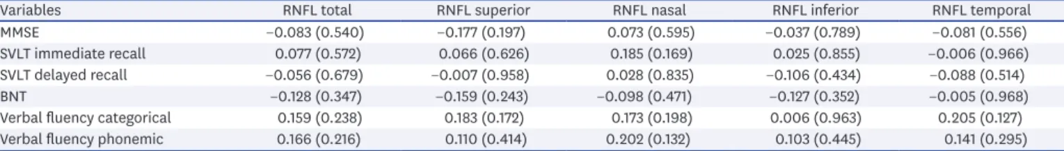
thickness and Mini-Mental State Examination (MMSE) and/or cognitive subdomain scores (Table 1). The univariate-linear-regression analysis did not find a significant correlation after adjusting for age, education years, and sex (data not shown).
The results support previous studies that have also shown the absence of correlation between these parameters. In contrast, many studies have identified a significant correlation between the reduction of RNFL thickness and the severity of cognitive impairment in patients with MCI and AD.2,3 Interestingly, 2 studies reported an inverse relationship between RNFL thickness and cognitive function in patients with MCI.6,7 Knoll et al.7 have suggested that gliotic reactive changes preceded the loss of neurons and resulted in thicker RNFL in MCI patients than in controls.
Previous studies have shown the accumulation of Aβ in the RNFL, the ganglion-cell layer (GCL), the inner plexiform layer, and the inner nuclear layer of the retina. The accumulation of Aβ in the GCL occurs early in the course of neurodegeneration caused by AD8 and leads to microglial activation and disruption of retinal architecture. The timeline of accumulation of Aβ plaques in the retinal structures suggests that RNFL thickness may be observed in the later stages of the disease.9 The pathologic changes associated with Aβ in RNFL may be less pronounced in MCI patients due to the reduced impact of the disease on cognitive impairment. Interestingly, Iseri et al.10 identified a significant decrease in macular volume, which was negatively correlated with the MMSE score but not with RNFL thickness. This suggests that macular volume may be a better predictor of cognitive impairment than RNFL thickness.
This study has several limitations that should be considered. First, the sample size is small, and the study was conducted in a single center with patients of only one ethnicity. Second, the study only evaluated RNFL thickness, and not macular volume or GCL thickness.
In conclusion, this study did not find an association between RNFL thickness and cognitive function in MCI patients. Further studies are required to clarify the relationship of RNFL thickness and disease severity in early-stage of AD.
SUPPLEMENTARY MATERIALS
Supplementary Table 1
Ophthalmological data of the participants Click here to view
10 https://doi.org/10.12779/dnd.2021.20.1.9
RNFL and Cognitive Measures in MCI due to AD
https://dnd.or.kr
Table 1. Correlation matrix between cognitive measures and RNFL thickness
Variables RNFL total RNFL superior RNFL nasal RNFL inferior RNFL temporal
MMSE −0.083 (0.540) −0.177 (0.197) 0.073 (0.595) −0.037 (0.789) −0.081 (0.556)
SVLT immediate recall 0.077 (0.572) 0.066 (0.626) 0.185 (0.169) 0.025 (0.855) −0.006 (0.966)
SVLT delayed recall −0.056 (0.679) −0.007 (0.958) 0.028 (0.835) −0.106 (0.434) −0.088 (0.514)
BNT −0.128 (0.347) −0.159 (0.243) −0.098 (0.471) −0.127 (0.352) −0.005 (0.968)
Verbal fluency categorical 0.159 (0.238) 0.183 (0.172) 0.173 (0.198) 0.006 (0.963) 0.205 (0.127)
Verbal fluency phonemic 0.166 (0.216) 0.110 (0.414) 0.202 (0.132) 0.103 (0.445) 0.141 (0.295)
Data are presented as correlation coefficients (p-values).
BNT: Boston Naming Test, MMSE: Mini-Mental State Examination, RNFL: retinal-nerve-fiber layer, SVLT: Seoul Verbal-Learning test.
Supplementary Fig. 1
OCT. OCT image of fundus (A). The RNFL thickness measured in the study was shown (arrow) (B), and the measured value throughout 360° was analyzed for comparison with the average values (C).
Click here to view
REFERENCES
1. Park KH, Lim JS, Seo SW, Jeong Y, Noh Y, Koh SH, et al. Executive summary of the 2019 international conference of Korean Dementia Association: exploring the novel concept of Alzheimer's disease and other dementia: a report from the Academic Committee of the Korean Dementia Association. Dement Neurocogn Disord 2020;19:39-53.
PUBMED | CROSSREF
2. Coppola G, Di Renzo A, Ziccardi L, Martelli F, Fadda A, Manni G, et al. Optical coherence tomography in Alzheimer's disease: a meta-analysis. PLoS One 2015;10:e0134750.
PUBMED | CROSSREF
3. Doustar J, Torbati T, Black KL, Koronyo Y, Koronyo-Hamaoui M. Optical coherence tomography in Alzheimer's disease and other neurodegenerative diseases. Front Neurol 2017;8:701.
PUBMED | CROSSREF
4. Langa KM, Levine DA. The diagnosis and management of mild cognitive impairment: a clinical review.
JAMA 2014;312:2551-2561.
PUBMED | CROSSREF
5. Kang YW, Na DL. Seoul Neuropsychological Screening Battery (SNSB). Seoul: Human Brain Research &
Consulting Co., 2003.
6. Shen Y, Liu L, Cheng Y, Feng W, Shi Z, Zhu Y, et al. Retinal nerve fiber layer thickness is associated with episodic memory deficit in mild cognitive impairment patients. Curr Alzheimer Res 2014;11:259-266.
PUBMED | CROSSREF
7. Knoll B, Simonett J, Volpe NJ, Farsiu S, Ward M, Rademaker A, et al. Retinal nerve fiber layer thickness in amnestic mild cognitive impairment: case-control study and meta-analysis. Alzheimers Dement (Amst) 2016;4:85-93.
PUBMED | CROSSREF
8. La Morgia C, Ross-Cisneros FN, Hannibal J, Montagna P, Sadun AA, Carelli V. Melanopsin-expressing retinal ganglion cells: implications for human diseases. Vision Res 2011;51:296-302.
PUBMED | CROSSREF
9. Ramirez AI, de Hoz R, Salobrar-Garcia E, Salazar JJ, Rojas B, Ajoy D, et al. The role of microglia in retinal neurodegeneration: Alzheimer's disease, Parkinson, and glaucoma. Front Aging Neurosci 2017;9:214.
PUBMED | CROSSREF
10. Iseri PK, Altinaş O, Tokay T, Yüksel N. Relationship between cognitive impairment and retinal
morphological and visual functional abnormalities in Alzheimer disease. J Neuroophthalmol 2006;26:18-24.
PUBMED | CROSSREF
11 https://doi.org/10.12779/dnd.2021.20.1.9
RNFL and Cognitive Measures in MCI due to AD
https://dnd.or.kr