www.jpis.org
pISSN 2093-2278 eISSN 2093-2286 Copyright © 2011 Korean Academy of PeriodontologyThis is an Open Access article distributed under the terms of the Creative Commons Attribution Non-Commercial License (http://creativecommons.org/licenses/by-nc/3.0/).
The biological effect of cyanoacrylate-combined calcium phosphate in rabbit calvarial defects
Yun-Young Chang1, Surangi Dissanayake1, Jeong-Ho Yun1, Ui-Won Jung1, Chang-Sung Kim1, Kyeong-Jun Park2, Jung-Kiu Chai1, Seong-Ho Choi1,*
1Department of Periodontology, Research Institute for Periodontal Regeneration, Yonsei University College of Dentistry, Seoul, Korea
2 YESBIO Co., Seoul, Korea
Purpose: The purpose of this study was to determine the biological effects of cyanoacrylate-combined calcium phosphate (CCP), in particular its potential to act as a physical barrier - functioning like a membrane - in rabbit calvarial defects.
Methods: In each animal, four circular calvarial defects with a diameter of 8 mm were prepared and then filled with either nothing (control group) or one of three different experimental materials. In the experimental conditions, they were filled with CCP alone (CCP group), filled with biphasic calcium phosphate (BCP) and then covered with an absorbable collagen sponge (ACS; BCP/ACS group), or filled with BCP and then covered by CCP (BCP/CCP group).
Results: After 4 and 8 weeks of healing, new bone formation appeared to be lower in the CCP group than in the control group, but the difference was not statistically significant. In both the CCP and BCP/CCP groups, inflammatory cells could be seen af- ter 4 and 8 weeks of healing.
Conclusions: Within the limits of this study, CCP exhibited limited osteoconductivity in rabbit calvarial defects and was his- tologically associated with the presence of inflammatory cells. However, CCP demonstrated its ability to stabilize graft parti- cles and its potential as an effective defect filler in bone augmentation, if the biocompatibility and osteoconductivity of CCP were improved.
Keywords: Octylcyanoacrylate, Calcium phosphate, Rabbits, Bone regeneration.
INTRODUCTION
Bone-grafting techniques have become essential in the res- toration of both function and esthetics in the treatment of maxillofacial osseous defects. In dentistry, it has been used widely to treat periodontal osseous defects and to regenerate the bone necessary to permit placement of implants, such as sinus grafts and guided bone regeneration [1-3]. Although autogenous bone has excellent osteogenic properties and elicits no immune response, it has several disadvantages and limitations with regard to patient morbidity, harvest quantity, and complications such as paresthesia and infection. Research-
ers have been working to develop the ideal substitute for au- togenous bone. Synthetic materials have been suggested as alternatives and have been investigated for with adequate osteoconductive properties.
Cyanoacrylates have been used successfully as tissue adhe- sives in various surgical applications [4,5]. It has also been re- ported that cyanoacrylate has hemostatic and antibacterial properties [6,7] when used instead of traditional suture mate- rials to close surgical wounds. Many recent investigations have found that cyanoacrylate does not induce tissue toxicity when it is used as a wound dressing for the treatment of open wounds as well as wound closure [8-10]. Kutcher [11] re- Received: Apr. 7, 2011; Accepted: May 3, 2011
*Correspondence: Seong-Ho Choi
Department of Periodontology, Research Institute for Periodontal Regeneration, Yonsei University College of Dentistry, 134 Sinchon-dong, Seodaemun-gu, Seoul 120-752, Korea
E-mail: [email protected], Tel: +82-2-2228-3189, Fax: +82-2-392-0398
fective for pain relief in oral ulcerations. According to Bhas- kar et al. [12], the application of cyanoacrylate spray to the oral mucosa did not induce any adverse effects in the sur- rounding tissues.
It has recently been suggested that synthetic materials pro- duced using cyanoacrylate can be used as a bone substitute for the restoration of osseous defects. Lee et al. [13] reported that rat calvarial defects reconstructed with cyanoacrylate- combined calcium phosphate (CCP) demonstrated new bone formation, and Park et al. [14] revealed that CCP could be a candidate for osseous healing in bone defects. CCP is pre- pared by mixing liquid cyanoacrylate and inorganic bioc- eramic powders. This material has unique properties differ- ent from those of conventional bone substitutes. Traditional granular-type bone substitutes may be lost and are difficult to manipulate when applied to osseous defects that are espe- cially large or that have a limited bony wall for supporting the graft materials. Conversely, because of its plasticity, adhe- siveness, and rapid hardening properties (hardens within 3 to 5 minutes), CCP can prevent such loss of graft particles and is easily manipulated within osseous defects. In addition, when it is used in combination with different kinds of alloplasts or allografts, CCP is able to bind to and immobilize other graft materials within the defect. It is thus possible to consider CCP a physical binder that can prevent loss of graft particles, like a plastic membrane. CCP is also more advantageous than a block-type bone substitute, which is difficult to plasticize in nonstandardized osseous defects. CCP has physical and me- chanical benefits; however, clinical and histologic evaluations of this material as a bone-graft material are lacking. Further- more, although cyanoacrylate has been used effectively and reliably for the closure of superficial wounds and lacerations, the biocompatibility of cyanoacrylate as an implant material in vivo has not yet been established.
The purpose of this study is to histologically determine the biologic effects of a novel CCP material and its potential as a physical binder in surgically prepared rabbit calvarial defects.
MATERIALS AND METHODS
Animals
Twelve adult male New Zealand white rabbits weighing 2.5 to 3.0 kg were used. All animals were given a 1-week period of adaptation before the start of the surgical procedure. The animal selection and management, surgical protocols, and preparation followed the protocols approved by the Institu- tional Animal Care and Use Committee, Yonsei Medical Center, Seoul, Korea (08-167).
CCP was prepared by mixing two pastes: one containing 0.1 g of liquid cyanoacrylate and solid inorganic materials, the other containing 0.22 g of osteoconductive β-tricalcium phosphate (β-TCP) with a particle size of 10 to 50 μm and 0.14 g of glycerin. Inorganic materials in the first paste comprised 0.23 g of monocalcium phosphate (particle size 50 to 100 μm) and 0.03 g of dicalcium phosphate (particle size 10 to 20 μm).
Biphasic calcium phosphate (BCP; Osteon, Genoss Co., Su- won, Korea) with a hydroxyapatite (HA)/β-TCP ratio of 70/30, a porosity of 77%, and a pore size of 300 to 500 μm was used in this study. The HA in this BCP was coated with β-TCP. An absorbable collagen sponge (ACS; Collatape, Zimmer Dental, Carlsbad, CA, USA) was used to cover the bone substitute.
Study design
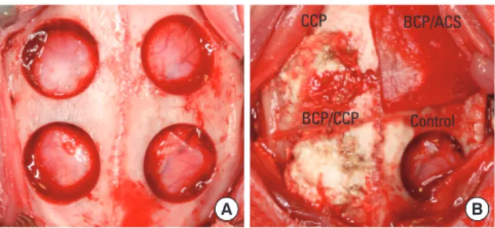
Four circular defects, each with a diameter of 8 mm, were prepared in each rabbit calvarium (Fig. 1). Each of the four defects in each animal was immediately filled with different graft materials according to the experimental condition, as follows (the location of the implant material in each animal was assigned randomly). The animals underwent a healing period of either 4 or 8 weeks (six animals per group for each period).
1. Control group (12 defects): defects were left unfilled, to serve as the surgical control.
2. CCP group (12 defects): defects were filled with only CCP.
3. BCP/ACS group (12 defects): defects were filled with BCP and then covered by ACS.
4. BCP/CCP group (12 defects): defects were filled with BCP and then covered by CCP.
Surgical procedures
All animals were anesthetized using an intramuscular in- jection of a mixture of ketamine hydrochloride (Ketar, Yuhan,
Figure 1. Clinical photographs of defect preparation (A) and appli- cation of the experimental materials (B). CCP: cyanoacrylate-com- bined calcium phosphate, BCP: biphasic calcium phosphate, ACS:
absorbable collagen sponge.
CCP BCP/ACS
BCP/CCP Control
A B
rea). The head of the rabbit was shaved and disinfected with povidone iodine. An incision was made along the midline of the parietal bone from the frontal bone to the occipital bone.
A full-thickness flap was elevated. Under copious saline irri- gation, four standardized round defects, each 8 mm in diam- eter, were created using a trephine bur (Fig. 1). The resected bones were removed carefully to avoid injury to the underly- ing brain tissue. The interdefect distance was more than 4 mm, allowing normal healing and easy harvest of the speci- mens for histologic analysis. The defects were filled with dif- ferent experimental materials, depending on the study group (see above). The flaps were repositioned and sutured with re- sorbable suture material (Polyglactin 910, braided absorbable suture, Ethicon, Johnson & Johnson, Edinburgh, UK). The an- imals were sacrificed at either 4 or 8 weeks postsurgery. The skin flaps were then reflected and the entire calvarium was harvested from each animal using a small sharp fissure bur.
Histological processing
Block sections of the surgical sites were fixed in 10% for- malin for 10 days. The fixed specimens were decalcified in 5%
formic acid for 14 days and then embedded in paraffin. Seri- al, 5-μm-thick sections were cut along the midline of the cal- varial defects. Only sections located at the middle of the de- fect were selected, and these were stained with hematoxylin- eosin for histologic and histometric analysis.
Analysis
Clinical observations; Visual observations were performed after 4 and 8 weeks of healing for clinical evaluation.
Radiologic analysis; After sacrificing the animals, all speci- mens were radiographed using an X-ray machine for de- scriptive radiographic analysis.
Histologic analysis; Histologic slides were examined with the aid of a binocular microscope (Leica DM LB, Leica Mi- crosystems, Wetzlat, Germany) equipped with a camera (Leica DC300F, Leica Microsystems, Heerbrugg, Switzerland). The slides were photographed and the obtained images saved as JPEG files.
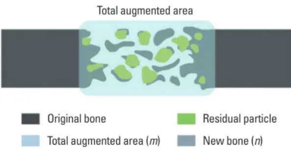
Histometric analysis; Histometric measurement was per- formed using an automated image-analysis computer pro- gram (Image-Pro Plus, Media Cybernetics, Sliver Spring, MD, USA). The augmented area (all tissue within boundaries of the defect: newly formed bone, residual graft material, con- nective tissue, bone marrow) and new bone area (mm2; Fig. 2) were measured.
Statistics
The mean and standard deviation of values for each group
were calculated. The significance of differences between groups was determined using the Kruskal-Wallis test (P<0.05).
The Mann-Whitney test was used to analyze the differences between values at 4 and 8 weeks. The Bonferroni correction was used to analyze differences that were significant at the 5% level (P<0.05).
RESULTS
Clinical observations
Healing was uneventful for all animals during the postop- erative period. None of the animals exhibited any complica- tions such as infection or exposure of the graft material after surgery.
Radiologic observations
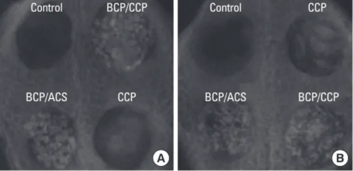
In the control group, a small radiopaque area was observed from the defect border after 4 weeks of healing; this area had increased after 8 weeks of healing. In the CCP group, a large radiopaque area surrounded by a radiolucent rim was ob- served after 4 weeks of healing. There was an overall decrease in the radiopaque area at 8 weeks. In the BCP/ACS group, ra- diopaque graft particles were densely packed and there was no significant difference between the findings after 4 and 8 weeks of healing. In addition, the radiographic appearance of the BCP/CCP-filled defects was very similar to that of the BCP/ACS-filled defects, except that the former was slightly more radiopaque (Fig. 3).
Histologic analysis
Control group; A slight bony ingrowth from the border of the defects that appeared to proceed to the central portion was observed in the control group. Most of the area of the defects was filled with fibrous connective tissue that inter- connected with both defect margins. The space observed within the defect did not persist, collapsing during the heal- ing period. Some specimens exhibited bony islands within
Original bone
Total augmented area (m)
Residual particle New bone (n)
Figure 2. Schematic diagram of a calvarial osteotomy defect show- ing the histometric analysis. New bone area (mm2)=n; residual bio- material, fibrovascular tissue, bone marrow=m; augmented area (mm2)=n+m.
the defects. The amount of new bone observed was greater after 8 weeks than after 4 weeks of healing (Fig. 4).
CCP group; There was a minimal amount of newly formed bone in the CCP group, and an inflammatory response was detected within the defects. When viewed at a higher magni- fication, inflammatory cells such as multinucleated giant cells, neutrophils, and lymphocytes were detected. The inflamma- tory reaction persisted at the 8-week follow-up (Figs. 5, 6).
BCP/ACS group; A bony ingrowth originating from the pe- riphery of the defect was observed in the BCP/ACS group, with immature woven bone in close contact with the graft particles. The regenerated bone contained many osteocytes, and osteoblastic cells surrounded the graft materials. The amount of new bone formation was greater after 8 weeks than after 4 weeks of healing (Figs. 7, 8).
BCP/CCP group; Histologic findings of the BCP/CCP group were very similar to those of the CCP group. Many inflam- matory cells were seen, and there was limited bone forma-
tion, which was seen only at the defect margin. Moreover, immature woven bone was only barely observed around the BCP particles, in contrast to the BCP/ACS group (see above).
Comparison of the specimens after 4 and 8 weeks of healing revealed them to be histologically indistinguishable from each other (Figs. 9, 10).
Histometric analysis
The results of the histometric analysis are presented in Ta- ble 1. The augmented area in the control group was smaller Figure 3. Radiologic presentation of calvarial defects after 4 weeks
(A) and 8 weeks (B) of healing. CCP: cyanoacrylate-combined calci- um phosphate, BCP: biphasic calcium phosphate, ACS: absorbable collagen sponge.
BCP/ACS CCP BCP/ACS BCP/CCP
A B
Figure 5. Histologic presentation of the cyanoacrylate-combined calcium phosphate group after 4 weeks of healing. A limited amount of new bone formation and inflammatory infiltration can be seen (A, H&E, ×10; B, H&E, ×100). arrowheads, defect margin.
A B
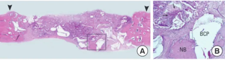
Figure 7. Histologic presentation of the biphasic calcium phos- phate (BCP)/absorbable collagen sponge group after 4 weeks of healing. New bone is seen in close contact with the graft material (A, H&E, ×10; B, H&E, ×100). arrowheads, defect margin; NB: new bone, BCP: biphasic calcium phosphate.
A B
NB
BCP
Figure 8. Histologic presentation of the biphasic calcium phos- phate (BCP)/absorbable collagen sponge group after 8 weeks of healing. Enhanced and mature bone surrounds the graft materials (A, H&E, ×10; B, H&E, ×100). arrowheads, defect margin; NB: new bone, BCP: biphasic calcium phosphate.
A NB B
BCP
Figure 6. Histologic presentation of the cyanoacrylate-combined calcium phosphate group after 8 weeks of healing (A, H&E, ×10; B, H&E, ×100). arrowheads, defect margin; NB: new bone.
A B
NB
Figure 4. Histologic presentation of a specimen from the control group after 4 weeks (A) and 8 weeks (B) of healing. Slight bony in- growth can be seen from the border of the defects, along with col- lapse of the connective tissue (A, H&E, ×10; B, H&E, ×10). arrow- heads, defect margin.
B A
to that of the experimental groups, with a new bone area of 1.04 ±0.69 mm2 after 4 weeks and 2.29 ±0.86 mm2 after 8 weeks of healing. Although the amount of bone increased with healing time (i.e., 4 and 8 weeks), the difference was not statistically significant. The mean bone regeneration in the CCP group was lower than in the control group after both 4 and 8 weeks of healing. After 8 weeks of healing in the CCP group, an increase was observed in the area of new bone, but again this change was minimal and not statistically signifi- cant. Meanwhile, a significant increase in the area of new bone was observed in the BCP/ACS group, from 2.10±0.38 mm2 after 4 weeks of healing to 2.84±1.24 mm2 after 8 weeks.
The amount of new bone found in the BCP/CCP group was 1.26±0.93 mm2 and 0.64±0.31 mm2 after 4 weeks and 8 weeks of healing, respectively. A statistically significant difference in new bone formation was found between the BCP/CCP and BCP/ACS groups after 8 weeks of healing. The mean bone regeneration in the CCP group was significantly lower than in the BCP/ACS group after both 4 and 8 weeks of heal- ing.
DISCUSSION
This study evaluated the biological effects of CCP in the treatment of rabbit calvarial defects. Various homologues of cyanoacrylate compounds exist, such as methylcyanoacrylate, ethylcyanoacrylate, butylcyanoacrylate, and octylcyanoacry- late. Cyanoacrylate with a longer carbon side chain has a slower degradation rate and also a lower toxicity [15]. Only
butylcyanoacrylate and octylcyanoacrylate have been used thus far in medical and dental applications. In particular, 2-octylcyanoacrylate was approved by the Food and Drug Administration in 1998 and was reported to be a well-estab- lished tissue adhesive for surgical wound closure [8]. The CCP used in this study also contained liquid 2-octylcyanoac- rylate and was manufactured by combining 2-octylcyanoac- rylate with several inorganic calcium phosphates. The mono- calcium phosphate in this particular CCP controls the veloci- ty of polymerization. This was necessary, since cyanoacrylate rapidly polymerizes and hardens when it is exposed to mois- ture at room temperature. The addition of monocalcium phosphate extends the working time necessary to manipu- late the CCP in order to fill defects. The polymerization and hardening of CCP takes approximately 3 minutes. Among the several other calcium phosphates in CCP, dicalcium phos- phate serves as a filler, and β-TCP, which is a well-established osteoconductive synthetic biomaterial that has been demon- strated to be absorbed in vivo and replaced with new bone, [16,17] is also added to CCP.
The defects reconstructed by only CCP exhibited a limited amount of new bone formation compared to the control group after both 4 and 8 weeks of healing. A slight increase was observed at 8 weeks but was not statistically significant.
Inflammatory infiltrates with numerous multinucleated gi- ant cells, lymphocytes, and neutrophils were observed in the histologic specimens. Although healing progressed to 8 weeks, the extent of inflammation was constant.
A recent study that investigated the same material in a ca- nine model found similar results to the present study, where- in CCP resulted in slight bone and cementum regeneration in periodontal one-wall intrabony defects [18]. Lee et al. [13]
reported no inflammatory response in a defect filled with a cyanoacrylate-based filling material, and further, new bone formation was observed in rat calvarial defects. These incon- Figure 10. Histologic presentation of the biphasic calcium phos-
phate (BCP)/cyanoacrylate-combined calcium phosphate group af- ter 8 weeks of healing. The connective tissue is infiltrated with in- flammatory cells (A, H&E, ×10; B, H&E, ×100). arrowheads, defect margin.
A B
BCP
Figure 9. Histologic presentation of the biphasic calcium phos- phate (BCP)/cyanoacrylate-combined calcium phosphate group af- ter 4 weeks of healing. Limited bone formation is observed at the defect margin and in contact with the BCP materials (A, H&E, ×10;
B, H&E, ×100). arrowheads, defect margin; NB: new bone.
A NB B
BCP
Parameter Control CCP BCP/ACS BCP/CCP
4 wk (n=6)
Augmented area 5.07±1.25 15.30±5.13a) 11.11±0.96 19.09±3.85a),b) New bone area 1.04±0.69 0.4±0.17b) 2.10±0.38 1.26±0.93 8 wk (n=6)
Augmented area 5.52±1.87 12.24±1.88a) 10.50±1.81a) 14.43±1.89a),c) New bone area 2.29±0.86 0.75±0.58b) 2.84±1.24 0.64±0.31b) Values are presented as mean±SD.
CCP: cyanoacrylate-combined calcium phosphate, BCP: biphasic calcium phosphate, ACS: absorbable collagen sponge.
a) Significant statistical difference compared to control group at each week (P<0.05).
b) Significant statistical difference compared to BCP/ACS at each week (P<0.05).
c) Significant statistical difference compared to 4 weeks (P<0.05).
perimental materials, namely N-butyl-2-cyanoacrylate and β-TCP used in the study of Lee et al. [13]. According to those authors, when this compound was mixed, the temperature generated during the polymerization did not increase signif- icantly relative to that generated during the polymerization of N-butyl-2-cyanoacrylate alone. It has been reported that the polymerization process of cyanoacrylate generates heat that could damage the cells and surrounding tissues, ulti- mately accounting, at least in part, for the cytotoxicity of cya- noacrylate [15]. The exothermic properties of CCP in vivo have not yet been verified. However, it is possible that the heat re- leased during the polymerization of CCP is associated with the inflammatory response observed in our histologic analy- sis. Another possible explanation for this finding is a foreign- body reaction to the 2-octylcyanoacrylate itself, when it is implanted in vivo. Dragu et al. [19] reported a foreign-body reaction when a tissue adhesive composed of 2-octylcyano- acrylate was applied to a wrist laceration wound, and identi- fied inflammatory histopathologic results.
On the other hand, it has been reported that cyanoacrylate has hemostatic properties and could be considered a thera- peutic option for the prevention of microvascular bleeding and postoperative hemorrhage in surgical procedures [20-22].
However, this means that the hemostatic effect of cyanoac- rylate may ironically reduce the osteoconductivity of CCP.
New bone formation is achieved by providing the synthetic material with a sufficient blood supply, and so this hemostat- ic effect may in some way be responsible for the minimal new bone formation found in the CCP group. However, fur- ther investigation is necessary to verify whether the hemo- static effects of cyanoacrylate can adversely influence the os- teoconductivity of CCP.
The defects reconstructed with BCP and ACS exhibited sta- tistically significant new bone formation after both 4 and 8 weeks of healing, compared to the CCP group. There was also statistically significant bone formation at 8 weeks when compared to the BCP/CCP group. Histologically, grafts ex- hibited many osteoblasts surrounding the graft material, and immature woven bone. The maturity and quantity of new bone increased with healing time. These histologic and his- tometric results concur with those of previous studies dem- onstrating the osteoconductive effects of BCP in both clinical and animal experiments [23-25].
The ACS that was applied to cover the BCP was completely biodegraded within 10 to 14 days. During this period it was possible to separate the bone graft material from the cutane- ous flap and prevent graft particles from escaping the defects.
In our previous study, we confirmed that placement of the ACS over the particles was an effective method for accelerat-
bony defects [26]. Moreira-Gonzalez et al. [27] described the importance of maintaining graft particles, finding that the migration of particles into the surrounding tissue resulted in limited bone regeneration. The ability of CCP to act as a me- chanical binder and physical barrier to keep the graft materi- als within the defect was also evaluated by comparing it to ACS with regard to bone regeneration. Histologic observa- tions of the BCP/CCP groups showed that the BCP granules located under CCP were stably maintained without migrat- ing out of defect, and there were no exceptions in all speci- mens of the BCP/CCP group. This indicates that the plasticity and adhesiveness of CCP may help to stabilize graft particles, functioning like a membrane or fixation screw in defects, preventing soft-tissue collapse. However, the amount of new bone formation in the BCP/CCP group was lower than that observed in the BCP/ACS group after either 4 or 8 weeks of healing. Moreover, histologic specimens from the BCP/CCP group appeared similar to those from defects filled with only CCP. In the former there were multinucleated giant cells and inflammatory cells around the BCP particles, as well as a minimal amount of immature woven bone. In the BCP/CCP group, CCP certainly played a role as a mechanical binder, but did not demonstrate a synergistic effect in terms of new bone formation.
Although the CCP and BCP/CCP group exhibited limited new bone formation compared to the BCP/ACS group, it was found that the augmented areas of the former were well maintained over the entire healing period, again without soft-tissue collapse. These observations suggest that CCP may be an effective defect filler for an atrophied alveolar ridge, or for large osseous defects such as cystic cavities, if biocom- patibility and osteoconductivity of CCP were improved.
In conclusion, CCP resulted in limited new bone formation in rabbit calvarial defects throughout the healing period, at- tracting inflammatory cells that were observed histologically.
However, its placement into bone defects demonstrated its ability to stabilize graft particles and to maintain augmented areas. Future investigations should attempt to improve the biocompatibility and osteoconductivity of CCP.
CONFLICT OF INTEREST
No potential conflict of interest relevant to this article was reported.
ACKNOWLEDGEMENTS
This study was supported by grant No.S0807222-E0841160- 10100013 from the Technology Development Program of the
REFERENCES
1. Sculean A, Nikolidakis D, Schwarz F. Regeneration of periodontal tissues: combinations of barrier membranes and grafting materials - biological foundation and pre- clinical evidence: a systematic review. J Clin Periodontol 2008;35(8 Suppl):106-16.
2. Nkenke E, Stelzle F. Clinical outcomes of sinus floor aug- mentation for implant placement using autogenous bone or bone substitutes: a systematic review. Clin Oral Implants Res 2009;20 Suppl 4:124-33.
3. Chiapasco M, Zaniboni M. Clinical outcomes of GBR pro- cedures to correct peri-implant dehiscences and fenestra- tions: a systematic review. Clin Oral Implants Res 2009;20 Suppl 4:113-23.
4. Singer AJ, Thode HC Jr. A review of the literature on octyl- cyanoacrylate tissue adhesive. Am J Surg 2004;187:238-48.
5. Brown JK, Campbell BT, Drongowski RA, Alderman AK, Geiger JD, Teitelbaum DH, et al. A prospective, random- ized comparison of skin adhesive and subcuticular suture for closure of pediatric hernia incisions: cost and cosmetic considerations. J Pediatr Surg 2009;44:1418-22.
6. Al-Belasy FA, Amer MZ. Hemostatic effect of n-butyl-2- cyanoacrylate (histoacryl) glue in warfarin-treated patients undergoing oral surgery. J Oral Maxillofac Surg 2003;61:
1405-9.
7. Quinn J, Maw J, Ramotar K, Wenckebach G, Wells G. Oct- ylcyanoacrylate tissue adhesive versus suture wound re- pair in a contaminated wound model. Surgery 1997;122:
69-72.
8. Eaglstein WH, Sullivan T. Cyanoacrylates for skin closure.
Dermatol Clin 2005;23:193-8.
9. Singer AJ, Nable M, Cameau P, Singer DD, McClain SA.
Evaluation of a new liquid occlusive dressing for excision- al wounds. Wound Repair Regen 2003;11:181-7.
10. Singer A, Thode H Jr, McClain S. The effects of octylcya- noacrylate on scarring after burns. Acad Emerg Med 2001;
8:107-11.
11. Kutcher M. Evaluating the efficacy of 2-octyl cyanoacry- late bioadhesive for treatment of oral ulcerations. Com- pend Contin Educ Dent Suppl 2001;(32):12-6.
12. Bhaskar SN, Cutright DE, Beasley JD, Ward JP. Oral spray of isobutyl cyanoacrylate and its systemic effect. Oral Surg Oral Med Oral Pathol 1970;29:313-9.
13. Lee SB, Park KJ, Lee DY, Park JJ, Hwang JS, Lee YK, et al. A histological evaluation of novel cyanoacrylate-based β-TCP composite in rat calvarial defects. Key Eng Mater 2006;309-311:1133-6.
Bioactive cyanoacrylate-based filling material for bone defects in dental applications. Key Eng Mater 2005;284- 6:933-6.
15. Leggat PA, Smith DR, Kedjarune U. Surgical applications of cyanoacrylate adhesives: a review of toxicity. ANZ J Surg 2007;77:209-13.
16. Jensen SS, Broggini N, Hjørting-Hansen E, Schenk R, Buser D. Bone healing and graft resorption of autograft, anorganic bovine bone and beta-tricalcium phosphate. A histologic and histomorphometric study in the mandibles of minipigs. Clin Oral Implants Res 2006;17:237-43.
17. Bowers GM, Vargo JW, Levy B, Emerson JR, Bergquist JJ.
Histologic observations following the placement of trical- cium phosphate implants in human intrabony defects. J Periodontol 1986;57:286-7.
18. Im JS, Jung UW, Chang YY, Yeon JY, Um YJ, Kim CS, et al.
The application of cyanoacrylate-based filling material for surgically created 1-wall intrabony defects in dogs. Bio- mater Res 2009;13:128-32.
19. Dragu A, Unglaub F, Schwarz S, Beier JP, Kneser U, Bach AD, et al. Foreign body reaction after usage of tissue adhe- sives for skin closure: a case report and review of the liter- ature. Arch Orthop Trauma Surg 2009;129:167-9.
20. Rengstorff DS, Binmoeller KF. A pilot study of 2-octyl cya- noacrylate injection for treatment of gastric fundal varices in humans. Gastrointest Endosc 2004;59:553-8.
21. Zhang CQ, Liu FL, Liang B, Sun ZQ, Xu HW, Xu L, et al. A modified percutaneous transhepatic variceal emboliza- tion with 2-octyl cyanoacrylate versus endoscopic ligation in esophageal variceal bleeding management: random- ized controlled trial. Dig Dis Sci 2008;53:2258-67.
22. Losanoff JE, Richman BW, Jones JW. Cyanoacrylate adhe- sive in management of severe presacral bleeding. Dis Co- lon Rectum 2002;45:1118-9.
23. Kim YK, Yun PY, Lim SC, Kim SG, Lee HJ, Ong JL. Clinical evaluations of OSTEON as a new alloplastic material in sinus bone grafting and its effect on bone healing. J Biomed Mater Res B Appl Biomater 2008;86:270-7.
24. Um YJ, Hong JY, Kim ST, Lee YH, Park SH, Park SH, et al.
Bone formation of newly developed biphasic calcium phosphate in rabbit calvarial defect model: a pilot study. J Korean Acad Periodontol 2008;38:163-70.
25. Fleckenstein KB, Cuenin MF, Peacock ME, Billman MA, Swiec GD, Buxton TB, et al. Effect of a hydroxyapatite tri- calcium phosphate alloplast on osseous repair in the rat calvarium. J Periodontol 2006;77:39-45.
26. Chang YY, Jung UW, Im JS, Yon JY, Um YJ, Kim CS, et al.
The effects of hydroxyapatite coated with beta -tricalcium phosphate in one-wall intrabony defects in dogs. Bioma-
27. Moreira-Gonzalez A, Lobocki C, Barakat K, Andrus L, Brad-
ford M, Gilsdorf M, et al. Evaluation of 45S5 bioactive glass critical size calvarial defects in rabbits. J Craniofac Surg 2005;16:63-70.