CASE REPORT
분출성 동맥출혈과 내강 협착을 동반한 회장 말트림프종 1예
표정의, 이범재, 이현주, 김지원, 김경진, 박종재, 김재선, 박영태, 장유진1
고려대학교 의과대학 내과학교실, 외과학교실1
A Case of Ileal Mucosa-associated Lymphoid Tissue Lymphoma Accompanied by Luminal Stricture and Arterial Spurting
Jeung Hui Pyo, Beom Jae Lee, Hyun Joo Lee, Ji Won Kim, Kyeong Jin Kim, Jong-Jae Park, Jae Seon Kim, Young-Tae Bak and You-Jin Jang1
Departments of Internal Medicine and Surgery1, Korea University College of Medicine, Seoul, Korea
Primary small intestinal lymphoma is relatively uncommon. Small bowel tumors are difficult to diagnose, because they are usually asymptomatic in the initial phase, and they are not easily detected by traditional methods of investigating the small intestine. This case shows a successfully detected and treated gastrointestinal bleeding from rare ileal mucosa-associated lymphoid tissue lymphoma, using double balloon endoscopy. (Korean J Gastroenterol 2013;62:365-369)
Key Words: Mucosa-associated lymphoid tissue lymphoma; Gastrointestinal bleeding; Small intestine; Double balloon endoscopy
Received March 2, 2013. Revised May 2, 2013. Accepted May 21, 2013.
CC This is an open access article distributed under the terms of the Creative Commons Attribution Non-Commercial License (http://creativecommons.org/licenses/
by-nc/3.0) which permits unrestricted non-commercial use, distribution, and reproduction in any medium, provided the original work is properly cited.
교신저자: 이범재, 152-703, 서울시 구로구 구로동로 148, 고려대학교 구로병원 내과
Correspondence to: Beom Jae Lee, Department of Internal Medicine, Korea University Guro Hospitial, 148 Gurodong-ro, Guro-gu, Seoul 152-703, Korea. Tel: +82-2- 2626-3004, Fax: +82-2-853-1943, E-mail: [email protected]
Financial support: None. Conflict of interest: None.
INTRODUCTION
Although the gastrointestinal tract is one of the most fre- quent sites of extranodal malignant lymphoma, the occur- rence of primary small intestinal lymphoma is relatively un- common, accounting for 19-38% of small bowel malig- nancies,1 and 20-30% of primary gut lymphoma.2 Since the first description in 1983,3 the concept of mucosa-associated lymphoid tissue (MALT) lymphoma has been widely ac- cepted, and has also been incorporated into the new histo- logic classification for extranodal lymphomas, including pri- mary gut lymphoma.4 Previous studies have analyzed the de- tailed clinicopathologic features of MALT lymphoma of the stomach,5 but little remains known regarding the MALT lym- phoma of the small bowel. To date, only a few cases of small bowel MALT lymphoma have been reported. We report a case
of small bowel MALT lymphoma complicated with annular stricture and active bleeding, which was successfully de- tected and treated, using double balloon endoscopy (DBE).
CASE REPORT
A 67-year-old man was referred to an outside hospital for the treatment of hematochezia. Upper gastrointestinal en- doscopy and total colonoscopy were performed, but failed to localize the bleeding site. Abdominal-pelvic CT scan did not show abnormal findings. Video capsule endoscopy (VCE) showed fresh blood in the middle ileum, but also failed to lo- calize the bleeding site (Fig. 1). The patient was referred to our center, for enteroscopy. The patient reported a 3-day his- tory of intermittent hematochezia, and a past history of re- current abdominal pain for the past 3 years; but endoscopic
366 표정의 등. 회장 말트림프종
Fig. 1. Video capsule endoscopy, showing fresh blood in the middle ileum.
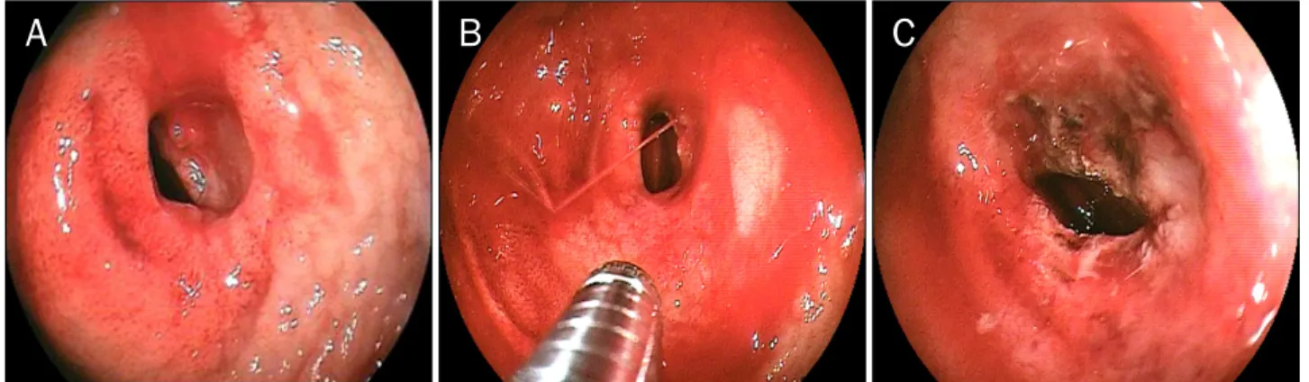
Fig. 2. (A, B) Double balloon endoscopy shows active ulcer with luminal stricture. Arterial spurting was noted at the ulcer base. (C) Argon plasma coagulation with ephinephrine-normal saline mixture injection was used to achieve hemostasis.
examination and abdominal pelvic CT findings performed at previous centers did not detect any abnormalities. He had been taking aspirin for 17 months. On admission, his vital signs were blood pressure 90/60 mmHg, heart rate 96 beats/min, body temperature 36.6oC, and respiratory rate 18 breaths/min. On physical exam, the patient was alert and not pale, and digital rectal examination revealed hemato- chezia. Laboratory findings were as follows; hemoglobin 9.6 g/dL, white blood cells count 7,200/μL (neutrophile 60%, lymphocyte 31.5%, eosinophil 3.3%), platelet 112/μL, PT 14.2 sec (INR 1.09), aPTT 34.9 sec, protein 5.7 g/dL, albumin 3.6 g/dL, AST/ALT/ALP 126/28/103 IU/L, total bilirunbin 1.06 mg/dL. Considering the examinations done in the pre- vious hospital, we performed enteroscopy for hemostasis.
Antegrade DBE (EN450T5; Fujinon, Saitama, Japan) re- vealed arterial spurting from an irregular ulcerative lesion, with severe luminal stricture at the middle ileum. The scope could not pass through this lesion. We used epinephrine in- jection and argon plasma coagulation for bleeding control, and hemostasis was successfully achieved (Fig. 2). Retro- grade DBE was performed, but no other synchronous lesions were found. The whole procedure took about 150 min. DBE guided biopsy revealed an active ulcer. Since he had suffered persistent abdominal pain for 3 years, and the endoscopic appearance was highly suggestive of malignancy, he sub- sequently underwent small bowel resection. The surgical pathology revealed extranodal marginal zone B-cell lympho- ma of MALT type (also known as MALT lymphoma), charac- terized by the appearance of poorly defined follicular areas, which were composed of monocytoid B cells that featured en- larged nuclei and lymphoepithelial lesions (Fig. 3). Post-oper- ative staging work-ups showed no evidence of disseminated disease. Urea breath test for Helicobacter pylori was nega- tive. The patient has been followed up regularly for the last 9 months, and no signs of recurrence have been found.
DISCUSSION
Small bowel tumors are relatively rare disorders, compris- ing less than 10% of all gastrointestinal tumors. The develop- ment of capsule endoscopy and balloon-assisted endoscopy launched a new era for the diagnosis and management of small bowel diseases.6 Several studies have been published on the role of DBE in the diagnosis and treatment of small bowel tumors. Dinesen et al.7 prospectively assessed the di-
Fig. 3. Histopathologic findings of mucosa-associated lymphoid tissue lymphoma. (A) The tumor measured 1.5×1.3 cm across. (B, C) Microscopic examination shows the poorly defined follicular areas that are composed of monocytoid B cells that feature enlarged nuclei (H&E, ×100), and infiltration and distortion of epithelial structures by aggregates of neoplastic lymphoid cells (H&E, ×400).
(D) Immunohistochemistry shows strong positive activity for CD 20 (×400).
agnostic and therapeutic impact of DBE, in patients with sus- pected or documented small bowel neoplasia, seen on VCE or CT scan. Over a 6-year period, a total of 580 DBE proce- dures were carried out. A total of 48 patients were found to have neoplastic disease/masses, and 6% were pathologi- cally proven to be lymphomas. Imaoka et al.8 conducted a ret- rospective chart review in 227 patients who had undergone DBE, and found that small bowel tumor groups contained more symptomatic patients than the non-small bowel tumor group (90% vs. 49%, p<0.05), with a significantly higher rate of gastrointestinal symptoms at presentation (72% vs. 33%, p<0.05). In a Korean multi-center study, among the 112 pa- tients with small bowel tumors who received DBE, benign pol- yp was the most common (n=38, 33.9%), and lymphomas were found in 16.1% (n=18). Small bowel lymphomas were located in the jejunum (50.0%), ileum (38.9%), and duode- num (5.6%), evenly distributed throughout the entire small bowel.6
Because small bowel lymphomas are uncommon, the ma- jority of studies on endoscopic findings have been performed in small bowel tumors, and studies on the endoscopic find- ings of small bowel lymphomas are limited. DBE improved the endoscopic approach to the small bowel, and enabled pre- operative histopatholgic investigations and minimally in- vasive surgery, in small bowel tumors, including lymphomas.
In this case, endoscopic treatment was performed immedi- ately, for active arterial spurting. The endoscopic appear- ances of small bowel lymphoma on DBE included stenosis, ulceration, and a mass or ulcerated mass lesion.8 Endoscopy commonly demonstrates MALT lymphoma of the small intes- tine, presented as either the ulcerative or polypoid type, and was indistinguishable from other high grade B-cell or T-cell lymphomas.9 In a Japanese retrospective study,8 among the 5 patients with small bowel lymphoma who had undergone DBE, the endoscopic findings of 2 cases of MALT lymphoma were multiple whitish nodules.
368 표정의 등. 회장 말트림프종
Clinical manifestations may vary from each patient, who may present with any one, or a combination of any, of the fol- lowing: dyspepsia, epigastric pain, abdominal pain, nausea, vomiting, diarrhea, weight loss, malabsorption, obstruction, anemia, and to a lesser extent ulceration, perforation, and intussusceptions.10-12 The most frequent symptoms for small bowel tumors were obscure gastrointestinal bleeding (57%), and chronic abdominal pain/diarrhea/obstruction (15%).6 Conversely, a few patients were reportedly asymptomatic.13 In our case, the patients had abdominal pain that persisted for 3 years, and hematochezia. Endoscopic finding showed arterial spurting in the ileum, with annular stricture. To date, only a few cases of MALT lymphoma of the small bowel have shown annular stricture. Yanai et al.14 reported a rare case of MALT lymphoma of the small bowel with annular stricture, presumably induced by NSAID.
MALT lymphomas display unusually indolent behavior, re- maining localized to their site of origin for long periods of time without disseminating, a feature which has made them uniquely amenable to cure by local therapy, such as surgical excision, with or without radiation therapy.15 Gastric MALT lymphomas, which represent up to 48% of all primary gastric lymphomas, are associated with H. pylori infection, and the eradication of H. pylori correlates with tumor regression.16 In contrast to gastric MALT lymphoma, the relationship be- tween small bowel MALT lymphoma and H. pylori infection has not been established; and whether H. pylori eradication may, or may not lead to lymphoma regression, has also not been established.16,17 H. pylori test showed negative in our case. Relapses involving the small bowel are rare, unlike gas- tric MALT lymphoma, where, following complete remission af- ter the eradication of H. pylori, the risk of relapse justifies life long follow-up examinations.18
In conclusion, small bowel MALT lymphoma is a relatively rare form of gastrointestinal tumor. To date, only a few cases of small bowel MALT lymphoma have been reported, and to our knowledge, this is the first case to report small bowel MALT lymphoma complicated with annular stricture and ac- tive bleeding. Small bowel tumors are difficult to diagnose, because they are usually asymptomatic in the initial phase;
and traditional methods of investigating the small intestine, such as small bowel follow-through and CT scanning, have low yield for tumors.19 This case represents the successful detection and treatment of arterial bleeding from rare ileal
MALT lymphoma accompanied by annular stricture, using DBE.
REFERENCES
1. Darling RC, Welch CE. Tumors of the small intestine. N Engl J Med 1959;260:397-408.
2. Radaszkiewicz T, Dragosics B, Bauer P. Gastrointestinal malig- nant lymphomas of the mucosa-associated lymphoid tissue:
factors relevant to prognosis. Gastroenterology 1992;102:
1628-1638.
3. Isaacson P, Wright DH. Malignant lymphoma of mucosa-asso- ciated lymphoid tissue. A distinctive type of B-cell lymphoma.
Cancer 1983;52:1410-1416.
4. Harris NL, Jaffe ES, Stein H, et al. A revised European-American classification of lymphoid neoplasms: a proposal from the International Lymphoma Study Group. Blood 1994;84:1361- 1392.
5. Cogliatti SB, Schmid U, Schumacher U, et al. Primary B-cell gas- tric lymphoma: a clinicopathological study of 145 patients.
Gastroenterology 1991;101:1159-1170.
6. Lee BI, Choi H, Choi KY, et al. Clinical characteristics of small bowel tumors diagnosed by double-balloon endoscopy: KASID multi-center study. Dig Dis Sci 2011;56:2920-2927.
7. Dinesen LC, Kaffes AJ, Selby W. Diagnostic and therapeutic ben- efits of double balloon endoscopy in small bowel neoplasia.
Gastroenterology 2011;140(Suppl 1):S118-S119.
8. Imaoka H, Higaki N, Kumagi T, et al. Characteristics of small bow- el tumors detected by double balloon endoscopy. Dig Dis Sci 2011;56:2366-2371.
9. Nakamura S, Matsumoto T, Takeshita M, et al. A clinicopatho- logic study of primary small intestine lymphoma: prognostic sig- nificance of mucosa-associated lymphoid tissue-derived lym- phoma. Cancer 2000;88:286-294.
10. de Leval L, Gaulard P. Pathology and biology of peripheral T-cell lymphomas. Histopathology 2011;58:49-68.
11. Ioannidis O, Cheva A, Kakoutis E, et al. Acute adult intussu- sception caused by primary cecal non Hodgkin lymphoma. Acta Gastroenterol Belg 2011;74:451-453.
12. Ferreri AJ, Montalbán C. Primary diffuse large B-cell lymphoma of the stomach. Crit Rev Oncol Hematol 2007;63:65-71.
13. Mansoor A, Pittaluga S, Beck PL, Wilson WH, Ferry JA, Jaffe ES.
NK-cell enteropathy: a benign NK-cell lymphoproliferative dis- ease mimicking intestinal lymphoma: clinicopathologic fea- tures and follow-up in a unique case series. Blood 2011;
117:1447-1452.
14. Yanai S, Nakamura S, Hirahashi M, Ueki T, Matsumoto T, Kitazono T. Education and imaging. Gastrointestinal: MALT lym- phoma of the small bowel accompanied by NSAID-induced enteropathy. J Gastroenterol Hepatol 2012;27:1126.
15. Schechter NR, Portlock CS, Yahalom J. Treatment of muco- sa-associated lymphoid tissue lymphoma of the stomach with radiation alone. J Clin Oncol 1998;16:1916-1921.
16. Papa A, Cammarota G, Tursi A, Gasbarrini A, Gasbarrini G.
Helicobacter pylori eradication and remission of low-grade gas- tric mucosa-associated lymphoid tissue lymphoma: a long-term follow-up study. J Clin Gastroenterol 2000;31:169-171.
17. Fischbach W, Tacke W, Greiner A, Konrad H, Müller-Hermelink HK. Regression of immunoproliferative small intestinal disease after eradication of Helicobacter pylori. Lancet 1997;349:31- 32.
18. Raderer M, Wöhrer S, Streubel B, et al. Assessment of disease
dissemination in gastric compared with extragastric muco- sa-associated lymphoid tissue lymphoma using extensive stag- ing: a single-center experience. J Clin Oncol 2006;24:3136- 3141.
19. Domizio P, Owen RA, Shepherd NA, Talbot IC, Norton AJ. Primary lymphoma of the small intestine. A clinicopathological study of 119 cases. Am J Surg Pathol 1993;17:429-442.