Received:January 20, 2015, Revised:March 21, 2015, Accepted:March 24, 2015
Corresponding to:Jun Ki Min, Division of Rheumatology, Department of Internal Medicine, The Catholic University of Korea, Bucheon St. Mary’s Hospital, 327 Sosa-ro, Wonmi-gu, Bucheon 14647, Korea. E-mail:[email protected]
pISSN: 2093-940X, eISSN: 2233-4718
Copyright ⓒ 2015 by The Korean College of Rheumatology. All rights reserved.
This is a Free Access article, which permits unrestricted non-commerical use, distribution, and reproduction in any medium, provided the original work is properly cited.
Mikulicz’s Disease with Progressively Transformed Germinal Centers-type Immunoglobulin G4-related Lymphadenopathy Mimicking Sjögren’s Syndrome
Hye Ji Kim1, Jean A Kim2, Jun Ki Min3
1Department of Internal Medicine, The Catholic University of Korea, Catholic Medical Center, Seoul, 2Department of Hospital Pathology,
3Division of Rheumatology, Department of Internal Medicine, The Catholic University of Korea, Bucheon St. Mary’s Hospital, Bucheon, Korea
Immunoglobulin G4-related disease (IgG4-RD) is a systemic disease, and lymphadenopathy is frequently observed in these patients. Among the 5 subtypes of IgG4-related lymphadenopathy, progressively transformed germinal centers (PTGC)-type IgG4-related lymphadenopathy possesses a unique characteristic that differentiates it from the other 4 subtypes. Here, we report on a rare case of PTGC-type IgG4-related lymphadenopathy accompanying Mikulicz’s disease. A 39-year-old female com- plained of a left cervical mass and bilateral upper eyelid hypertrophy. The serum level of IgG4 was elevated, and computed to- mography showed enlargement of the bilateral lacrimal and submandibular glands and left cervical lymph node. Excisional bi- opsy of a submandibular gland and cervical lymph node was performed, and the histopathologic findings revealed Mikulicz’s disease accompanied by PTGC-type IgG4-related lymphadenopathy. After treatment of the patient with oral prednisolone and azathioprine, the patient’s appearance improved. To the best of our knowledge, no case of PTGC-type IgG4-related lymphaden- opathy has been previously reported in Korea. (J Rheum Dis 2015;22:395-400)
Key Words. Immunoglobulin G4-related lymphadenopathy, Progressively transformed germinal centers, Immunoglobulin G4-related disease, Mikulicz’s disease
INTRODUCTION
Immunoglobulin G4-related disease (IgG4-RD) is a novel chronic inflammatory disease, which was first brought into attention in 2000. It is characterized by lym- phoplasmacytic infiltrate, fibrosis with elevation of the serum IgG4 level, and abundant infiltration of IgG4-positive plasma cells to the internal organs [1,2]. IgG4-RD is a systemic disease that can involve almost all organs, in- cluding the pancreas, bile ducts, gallbladder, liver, stom- ach, salivary glands, lacrimal glands or orbit tissues, kid- ney, lung, lymph nodes, meninges, pituitary gland, aorta, breast, prostate, thyroid gland, pericardium, pleura, mes- entery, retro-peritoneum, peripheral nerves, and skin [2,3]. IgG4-related lymphadenopathy can be divided into
5 types: multicentric Castleman disease-like, reactive fol- licular hyperplasia-like, interfollicular hyperplasia-like, progressively transformed germinal centers (PTGC)-type, and inflammatory pseudotumor-like [4]. Among these, no case of PTGC-type IgG4-related lymphadenopathy has yet been reported in Korea. Here, we report a rare case of PTGC-type IgG4-related lymphadenopathy accompany- ing Mikulicz’s disease, which involved both the lacrimal and salivary glands.
CASE REPORT
A 39-year-old female was admitted to our hospital with chief complaints of a left cervical mass and bilateral upper eyelid hypertrophy that had been progressing for 4 years.
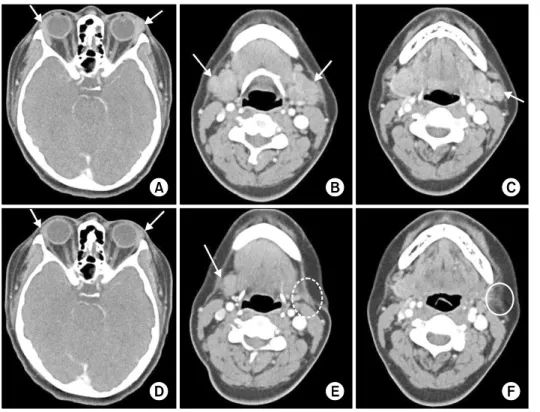
Figure 1. Comparison of radio- logical features before and after excisional biopsy of the left sub- mandibular gland and left cer- vical lymph node and medical treatment. (A∼C) Contrast-enhan- ced computed tomography im- ages revealing enlargement of the bilateral lacrimal and sub- mandibular glands and left cer- vical lymph node. (D, E) After 2 months of medical treatment, the sizes of the bilateral lacrimal glands and right submandibular gland were decreased. (E, F) The dotted circle indicates the surgi- cally removed left submandibular gland and the lined circle in- dicates the surgically removed left cervical lymph node by exci- sional biopsy.
She had a history of treatment with 2 mg methyl- prednisolone, 300 mg hydroxychloroquine, and 7.5 mg methotrexate for Sjögren’s syndrome at another hospital 4 years earlier. Two years ago, she presented to our hospi- tal with a chief complaint of pain at multiple sites. A 1.7-cm left cervical lymphadenopathy was observed upon cervical contrast-enhanced computed tomography (CT).
Compared to the CT findings from the other hospital 4 years ago, no size change in the left cervical lymphaden- opathy was observed. Moreover, a 3.8-cm right axillary lymphadenopathy was found upon CT, and an excisional biopsy was performed at that time. The biopsy results in- dicated reactive follicular hyperplasia with PTGC. There- after, the patient was followed-up conservatively for 1 year. However, as the aforementioned chief complaints progressed, she was consequently readmitted. At the time of admission, the patient showed blood pressure 120/70 mmHg, pulse 68 beats/min, respiration rate 20 breaths/min, and temperature 36.6oC, with clear con- sciousness. Swelling of the bilateral upper eyelids and en- largement of left cervical lymph nodes were observed. All other physical examination findings were unremarkable.
The laboratory findings revealed that the complete blood cell count, blood coagulation test, and erythrocyte sed- imentation rate were normal. The various biochemical and urine test results, and the C-reactive protein (CRP), C3, and C4 levels were also normal. Antinuclear and an-
ti-DNA antibodies were negative, and the levels of Ig were normal for IgE (70.1 IU/mL; normal range: <100 IU/mL), IgA (228.0 mg/dL; normal range: 70 to 400 mg/dL), IgM (90.0 mg/dL; normal range: 40 to 230 mg/dL), and IgG (1,178 mg/dL; normal range: 700 to 1,600 mg/dL. However, the IgG4 subclass was greatly in- creased to 304.6 mg/dL (normal range: 3.9 to 86.4 mg/dL). Upon cervical contrast-enhanced CT, slight en- largements of the bilateral lacrimal and submandibular glands were observed (Figure 1A and 1B), with not much difference in the left cervical lymphadenopathy noted compared to the CT results from 2 years ago (Figure 1C).
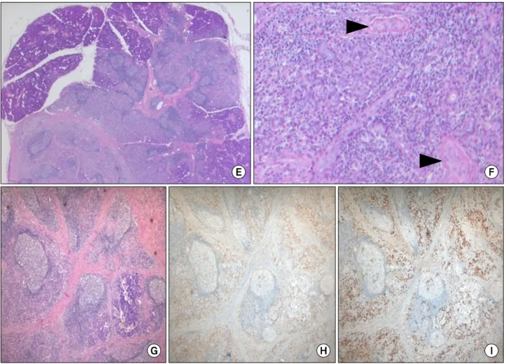
Left submandibular gland and left cervical lymph node excisional biopsy was performed, and IgG/IgG4 im- munohistochemical staining was performed on the above specimens as well as those previously obtained from the right axillary lymph node. In the cervical and axillary lymph nodes, round or oval follicles with a diameter 2 to 3 times larger than other reactive follicles were found (Figure 2A). The IgG4+/IgG+ plasma cell ratio was 90%
and the IgG4+ plasma cells were counted as 180/high power field (HPF) (Figure 2C and 2D). Only a small num- ber of IgG4+ plasma cells were present in the inter- follicular zone, with most residing in the germinal centers (Figure 2C and 2D). Storiform fibrosis and obliterative phlebitis were observed in the submandibular gland specimens (Figure 2F and 2G). Further, the B cell receptor
Figure 2. Histological and immunohistochemical features of the excisonal biopsy specimens of (A∼D) the lymph node and (E∼I) submandibular gland. (A) The excised lymph node shows extensive reactive follicular hyperplasia and progressively transformed germinal centers (PTGCs) (arrowheads) (H&E, ×100). The PTGCs appear as round-to-oval structures, 2 to 3 times the diameter of the other reactive follicles. These large follicles show thickened mantles with inward extensions into the germinal centers but no expansion of the interfollicular zone. (B) The germinal centersare predominantly composed of lymphocytes, centrocytes, centro- blasts, and numerous mature plasma cells and plasmacytoid cells (H&E, ×200). (C, D) The majority of immunoglobulin (Ig)G4+
plasma cells reside in the germinal centers, with a small number presenting in the interfollicular zone. This is a unique feature of PTGC-type IgG4-related lymphadenopathy, which distinguishes it from the other 4 subtypes. The IgG4+/IgG+ plasma cell ratio is 90%, and the IgG4+ plasma cells were counted as 180/high power field (C: IgG-immunostain, ×200; D: IgG4-immunostain,
×200). The features of the cervical lymph node are identical to those of the axillary lymph node specimen. (E) Acinar atrophy and destruction of the salivary gland are observed, along with marked lymphocytic infiltration with lymphoid follicles (H&E, ×40). (F) Veins occluded by inflammatory infiltrate composed of lymphocytes and plasma cells are noted, and indicate obliterative phlebitis (arrowheads) (H&E, ×200). (G) The storiform pattern of fibrosis is present, indicating dense fibrosis within which lymphocytes, plasma cells, and occasional eosinophils are embedded (H&E, ×100). Storiform fibrosis and obliterative phlebitis are usually ab- sent in IgG4-related lymphadenopathy. (H, I) The IgG4+/IgG+ plasma cell ratio is estimated at 90% (G: IgG-immunostain, ×100;
H: IgG4-immunostain, ×100).
and T cell receptor gene rearrangements showed poly- clonal patterns. Based on the clinical findings, distinctive histopathologic results, and laboratory findings of IgG4 increase, the patient was diagnosed with Mikulicz’s dis- ease accompanied by PTGC-type IgG4-related lympha- denopathy and medical treatment was initiated with oral prednisolone 40 mg/d. As a result, the bilateral upper
eyelid hypertrophy was clinically improved and the pre- dnisolone dose was tapered. Follow-up cervical con- trast-enhanced CT after 2 months of treatment also showed decreased sizes of the lacrimal and salivary glands (Figure 1D and 1E), and finally, prednisolone was terminated after 3 months of treatment. However, the bi- lateral upper eyelid hypertrophy recurred, and pre-
Figure 2. Continued.
dnisolone 5 mg/d and azathioprine 100 mg/d were there- fore administered. The patient again showed improve- ment of the symptoms and is currently under medication.
DISCUSSION
Mikulicz’s disease displays similar clinical symptoms to Sjögren’s syndrome; however, unlike Sjögren’s syndrome, Mikulicz’s disease is a multi-organ lymphoproliferative disease characterized by IgG4 increases in the serum and tissues [5]. The patient in the present case showed symp- toms of dry eyes and dry mouth, with a positive Schirmer’s test and focus score of 1 in the histopathologic findings of the minor salivary gland biopsy, whereas the salivary gland scan revealed no abnormal findings and no anti- nuclear antibody and anti-Ro/La antibodies were found in the laboratory data from our hospital and the other hospital 4 years ago, fulfilling four of the six classification criteria for Sjögren’s syndrome. However, a diagnosis of IgG4-RD should be ruled out to confirm Sjögren’s syn-
drome; thereby, the patient can be diagnosed with Miku- licz’s disease from the clinical and either the histopatho- logical or serological criteria.
Hyper-interleukin (IL)-6 syndromes such as multicentric Castleman’s disease and rheumatoid arthritis, as well as other immune-mediated conditions, should be differ- entiated from IgG4-RD, as they also present with elevated serum IgG4 levels secondary to the effects of the high IL-6 levels, sometimes fulfill the histological diagnostic cri- teria for IgG4-RD, and frequently involve the lymph no- des [4]. The laboratory findings are the most important to differentiate between IgG4-RD and hyper-IL-6 syndromes.
In hyper-IL6 syndromes, the levels of IgG, IgA, IgM, and CRP are mostly elevated, and thrombocytosis, anemia, hypoalbuminemia and hypocholestrolemia are observed due to the effects of the high IL-6 levels.
It has been reported that lymphoma is relatively fre- quently found in IgG4-RD patients [6], indicating that the activation and proliferation of lymphocytes and chronic antibody stimulation in IgG4-RD may increase
the risk of lymphoma development. In particular, the PTGC has been reported to be associated with the onset of nodular lymphocyte predominant Hodgkin lymphoma [7]. Differentiating PTGC from this disease based only on the histological findings can be difficult; however, im- munohistochemical staining using B cell and T cell mono- clonal antibody panels is helpful for the differentiation [8]. In the present case, B cell receptor and T cell receptor gene rearrangements were performed, resulting in poly- clonal patterns, thus excluding lymphoma.
PTGC-type IgG4-related lymphadenopathy possesses a unique characteristic that differentiates it from the other 4 subtypes [9]; according to a study with patients who satisfied the diagnosis of PTGC-type IgG4-related lym- phadenopathy, it occurred most frequently in the sub- mandibular lymph nodes, and some patients showed ac- companying cervical lymph node involvement. Although the reason for the submandibular lymph nodes being fre- quently involved is not clear, they receive lymph fluids cir- culating the orbit tissues, nose, oral cavity, paranasal si- nuses, and salivary glands. Therefore, it can be hypothe- sized that the submandibular lymph nodes relevant to lymphatic circulation are also influenced by this disease.
In addition, during the follow-up, involvement of other organs besides the lymph nodes, or transition to systemic diseases, are observed in approximately half of the pa- tients, with the most commonly involved organs being the lacrimal and salivary glands. In the present case, the cervical and axillary lymph nodes were involved, and IgG4-RD was also observed in the lacrimal and salivary glands.
Sato et al. [9] reported no cases with involvement of the axillary lymph nodes in PTGC-type IgG4-related lympha- denopathy patients, which suggests that the site of lym- phadenopathy in this case is highly rare and that this case is therefore significant. In addition, IgG4+ plasma cells are found in the interfollicular zone in the other 4 sub- types of IgG4-related lymphadenopathy, while they are found in the intra-germinal centers in PTGC-type IgG4-re- lated lymphadenopathy.
In terms of the mechanism of IgG4-RD occurrence, the immune responses of type 2 helper T (Th2) and regu- latory T (Treg) cells play very important roles [1,10]. To- gether with the increases in the IL-4, IL-5, IL-10, and IL-13 levels, increased levels of the Th2-type cytokine can lead to elevation of the serum IgE and eosinophil levels.
Moreover, IL-10 and transforming growth factor-β are produced by activated Treg cells, which in turn can result
in increases in the IgG4 levels and in fibrosis [10]. Among the 40 cases with PTGC-type IgG4-related lymphadenop- athy reported by Sato et al. [9], the serum IgE levels were elevated in 12 of 13 cases (92%), and the peripheral blood eosinophil count was increased in 18 of 34 (53%).
However, in the present case, the IgE and eosinophil lev- els were in the normal ranges.
The diagnosis of IgG4-RD is based on the clinical pat- terns, histopathologic features, and serologic tests [11].
CT scan and ultrasonography can also be helpful [2]. The proposed diagnostic criteria are as follows [12]: clinical signs of organ involvement or damage, histopathologic findings of IgG4+/IgG+ plasma cell ratio >40% and IgG4+ plasma cells >10/HPF, and blood test results with an IgG4 value >135 mg/dL. Criterion 1 is essential in the diagnosis of IgG4-RD; when satisfying both criteria 2 and 3, the diagnosis can be confirmed as IgG4-RD. The pres- ent case satisfied all 3 criteria, and was thereby diagnosed as IgG4-RD.
As for the treatment of IgG4-RD, oral prednisolone is considered the primary treatment in the presence of symptomatic disease with organ involvement [13]. After maintaining the initial dose of 0.6 mg/kg for 2 to 4 weeks, the dose is generally reduced to 5 mg/d for 3 to 6 months, followed by 2.5 to 5 mg/d for 3 years. Alternatively, after maintaining the initial dose of 40 mg/d for 4 weeks, the dose can be reduced to 5 mg/d for 11 weeks and sub- sequently terminated. Recurrence is common during the reduction or termination of prednisolone, even when fa- vorable treatment effects are noted. Such patients can be treated by administering 2.0 to 2.5 mg/kg of azathio- prine, 750 mg of mycophenolate mofetil 2 times/d, or methotrexate as means of achieving additional immu- nosuppression and sparing the patients the effects of long-term glucocorticoid administration [1,13]. Rituximab, an anti-CD20 antibody that possesses B lymphocyte in- hibition functions, may also represent an effective drug for patients with recurrence or no response to other drugs [3,13]. Because there are some cases of lymphadenop- athy without symptoms and without changes for several years, or even decades, follow-up can be carried out through careful observation prior to commencing medication [1].
The present case showed appearance changes with in- volvement of the lacrimal and salivary glands; therefore, the patient received glucocorticoid treatment. However, she developed recurrence of the disease during the gluco- corticoid tapering and was consequently treated with glu- cocorticoid plus azathioprine. Hart et al. [14] recently
showed in their observational study that immunomo- dulators and rituximab are reasonable alternatives for treatment of type 1 autoimmune pancreatitis. However, with no prospective, controlled studies currently avail- able, the decision regarding what kind of immunomo- dulator to use for the treatment of recurrent and re- fractory disease must be made on an individual, case-to-case basis.
SUMMARY
In the presence of lymphadenopathy in IgG4-RD, organ involvement other than to the lymph nodes can occur be- fore, after, or concurrently as the lymphadenopathy; how- ever, the order and exact mechanism of this occurrence are unclear. We here experienced a highly rare case of PTGC-type IgG4-related lymphadenopathy that pre- sented as cervical and axillary lymphadenopathy with in- volvement of the lacrimal and salivary glands in IgG4-RD, known as Mikulicz’s disease, and here reported the find- ings of this case along with a literature review.
CONFLICT OF INTEREST
No potential conflict of interest relevant to this article was reported.
REFERENCES
1. Stone JH, Zen Y, Deshpande V. IgG4-related disease. N Engl J Med 2012;366:539-51.
2. Yamamoto M, Takahashi H, Shinomura Y. Mechanisms and assessment of IgG4-related disease: lessons for the rheu-
matologist. Nat Rev Rheumatol 2014;10:148-59.
3. Mahajan VS, Mattoo H, Deshpande V, Pillai SS, Stone JH.
IgG4-related disease. Annu Rev Pathol 2014;9:315-47.
4. Sato Y, Yoshino T. IgG4-related lymphadenopathy. Int J Rheumatol 2012;2012:572539.
5. Yao Q, Wu G, Hoschar A. IgG4-related Mikulicz's disease is a multiorgan lymphoproliferative disease distinct from Sjögren's syndrome: a Caucasian patient and literature review. Clin Exp Rheumatol 2013;31:289-94.
6. Ferry JA. IgG4-related lymphadenopathy and IgG4-related lymphoma: moving targets. Diagn Histopathol 2013;19:
128-39.
7. Jones D. Dismantling the germinal center: comparing the processes of transformation, regression, and fragmentation of the lymphoid follicle. Adv Anat Pathol 2002;9:129-38.
8. Nguyen PL, Ferry JA, Harris NL. Progressive transformation of germinal centers and nodular lymphocyte predominance Hodgkin's disease: a comparative immunohistochemical study. Am J Surg Pathol 1999;23:27-33.
9. Sato Y, Inoue D, Asano N, Takata K, Asaoku H, Maeda Y, et al. Association between IgG4-related disease and pro- gressively transformed germinal centers of lymph nodes.
Mod Pathol 2012;25:956-67.
10. Takeuchi M, Sato Y, Ohno K, Tanaka S, Takata K, Gion Y, et al. T helper 2 and regulatory T-cell cytokine production by mast cells: a key factor in the pathogenesis of IgG4-related disease. Mod Pathol 2014;27:1126-36.
11. Cheuk W, Chan JK. Lymphadenopathy of IgG4-related dis- ease: an underdiagnosed and overdiagnosed entity. Semin Diagn Pathol 2012;29:226-34.
12. Umehara H, Okazaki K, Masaki Y, Kawano M, Yamamoto M, Saeki T, et al. Comprehensive diagnostic criteria for IgG4-related disease (IgG4-RD), 2011. Mod Rheumatol 2012;22:21-30.
13. Khosroshahi A, Stone JH. Treatment approaches to IgG4- related systemic disease. Curr Opin Rheumatol 2011;23:
67-71.
14. Hart PA, Topazian MD, Witzig TE, Clain JE, Gleeson FC, Klebig RR, et al. Treatment of relapsing autoimmune pan- creatitis with immunomodulators and rituximab: the Mayo Clinic experience. Gut 2013;62:1607-15.