Fusiform Superior Cerebellar Artery Aneurysm Treated with Endovascular Treatment
Joon Bok Jeon, Se-yang Oh, Dong-Keun Hyun, Yu Shik Shim
Department of Neurosurgery, Inha University School of Medicine and Hospital, Incheon, Korea
An aneurysm of the distal superior cerebellar artery (SCA) is a highly rare disease. Fusiform aneurysms of the distal SCA are particularly challenging to treat. Clipping, trapping with or without bypass using microsurgery or endovascular treatment (EVT) were used to treat this condition. We de- scribe the case of fusiform distal SCA aneurysms treated successfully with endovascular coiling with a 3-month follow-up. A 39 year-old male was presented with subarachnoid hemorrhage (SAH) and a 15 mm fusiform aneurysm of the ambient segment of the left distal SCA. EVT for parent artery occlusion and packing of the aneurysm was done. Left sixth nerve palsy appeared after 1 day of EVT. The symptom completely recovered within 1 week of the post-procedural period. No neurological deficit was seen during the clinical 3-month follow-up. EVT of fusiform distal SCA aneurysms with coils is a safe and feasible option to manage this rare condition. However, the treatment options must be carefully selected de- pending on the neurologic condition, development of collateral circu- lation, and configuration of the dissection.
J Cerebrovasc Endovasc Neurosurg.
2016 September;18(3):276-280 Received : 29 April 2016
Revised : 5 September 2016 Accepted : 6 September 2016 Correspondence to Se-yang Oh
Department of Neurosurgery, Inha University School of Medicine and Hospital, 27 Inhang-ro, Jung-gu, Incheon 22332, Korea
Tel : 82-32-890-2946 Fax : 82-32-890-3467 E-mail : [email protected]
ORCID : http://orcid.org/0000-0001-7261-4632 Poster, the 55rd Annual Meeting of the Korean Neurosurgical Society, republic of Korea, October 15-18, 2015.
This is an Open Access article distributed under the terms of the Creative Commons Attribution Non- Commercial License (http://creativecommons.org/li- censes/by-nc/3.0) which permits unrestricted non- commercial use, distribution, and reproduction in any medium, provided the original work is properly cited.
Keywords Endovascular techniques, Intracranial aneurysm, Subarachnoid hemorrhage Journal of Cerebrovascular and Endovascular Neurosurgery
pISSN 2234-8565, eISSN 2287-3139, http://dx.doi.org/10.7461/jcen.2016.18.3.276
Case Report
INTRODUCTION
Aneurysms of the distal superior cerebellar artery (SCA) are very rare and account for less than 0.2% of all treated intracranial aneurysms.1) Fusiform aneur- ysms of the distal SCA are particularly challenging to treat. Clipping, trapping with or without bypass using microsurgery or endovascular surgery were used to treat this condition. We describe the case of fusiform SCA aneurysms treated successfully with endovas- cular coiling with 3-month follow-up.
CASE REPORT
A 39 year-old male without past notable medical history was admitted with a sudden headache and
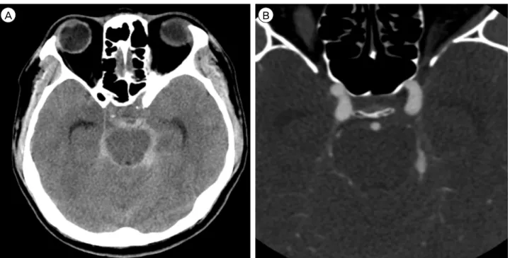
neck stiffness. Brain computed tomography (CT) dem- onstrated evidence of Fisher Grade III subarachnoid hemorrhage (SAH) predominance of the pre-pontine, left ambient and quadrigeminal cistern (Fig. 1A), and a fusiform aneurysm of a left distal SCA was revealed on the CT angiography (Fig. 1B). Subsequent digital subtraction angiography (DSA) revealed the fusiform dilatation (12 mm length; 4.5 mm width) involving the ambient segment of the left distal SCA. The left SCA was duplicated ahead of the aneurysm and there was sufficient collateral circulation from the ipsilateral anterior inferior cerebellar artery (AICA) and posterior inferior cerebellar artery (PICA) (Fig. 2). The aneurysm was treated by endovascular occlusion of the aneurysm sac and the involved branch. Under general anesthesia an Excelsior SL-10 microcatheter was used to access
A B
Fig. 1. (A) Brain computed tomography (CT) image. The brain CT shows a Fisher Grade III SAH in the pre-pontine, left ambient and quadrigeminal cistern. (B) Brain computed tomography angiography (CT angiography) image. The brain CT angiography shows fusi- form dilatation of the left distal SCA. SAH = subarachnoid hemorrhage; SCA = superior cerebellar artery.
Fig. 2. Digital subtraction angiography (DSA) images. The DSA images show the fusiform aneurysm of the left superior cerebellar artery. There was sufficient collateral circulation from the ipsilateral AICA and PICA. AICA = anterior inferior cerebellar artery; PICA = posterior inferior cerebellar artery.
the aneurysm. Seven detachable platinum coils were used to obliterate the lesion. Post-procedural angiog-
raphy showed complete occlusion of the aneurysm sac and involved the SCA branch (Fig. 3). There was
ENDOVASCULAR TREATMENT FOR DISTAL SUPERIOR CEREBELLAR ARTERY ANEURYSM
Fig. 3. Pre-operative 3-dimensional (D) CT angiography (left) and post-operative 3D DSA images (right). The 3D CT angiography re- veals fusiform dilatation of the left distal SCA (left). The 3D DSA shows complete occlusion of the aneurysm sac and involved the distal SCA branch. The ipsilateral duplicated SCA is intact. CT = computed tomography; DSA = digital subtraction angiography; SCA = superior cerebellar artery.
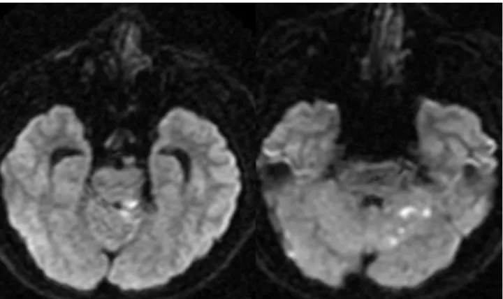
no immediate complication after embolization. Diffusion MRI was done 8 hours after embolization. MRI re- vealed scattered high signal change on the left poste- rior tegmentum, superior vermis, and superior part of the left cerebellum (Fig. 4). Left sixth nerve palsy oc- curred in the patient 1 day after EVT. The symptom completely recovered within 1 week of the post-pro- cedural period. No neurological deficit was seen dur- ing the clinical 3-month follow-up.
DISCUSSION
Non-saccular aneurysms are often referred to as fusiform, dissecting, or trunkal aneurysms.2) According to some authors, the majority of these spontaneous, non-saccular aneurysms originate from vessel dissection.4-6) Fusiform aneurysms of the distal SCA are extremely rare and
only 17 cases are reported in the literature. SAH was the most common clinical presentation.11) However, some reported cases were also presented with ische- mic events.7) Most of the aneurysms in the distal SCA usually present with SAH. A hematoma predom- inantly in the pre-pontine cistern should raise suspi- cion of the probability of an SCA aneurysm. The exact rate of re-bleeding is not known but re-bleeding has been reported in the literature. Due to re-bleeding risk, DSA for accurate evaluation of a distal SCA aneurysm and early treatment in the acute phase is recommended in some studies.3)5)
The fusiform aneurysms of the distal SCA are par- ticularly challenging to treat. First, surgical treatments of these aneurysms were suggested such as parent ar- tery occlusion, clipping of the aneurysm body, trap- ping with or without bypass, and wrapping.9) These
Fig. 4. Diffusion weighted images (DWIs) 1 day post-treatment. The DWIs show scattered high signal change, suggesting acute in- farction on the left posterior tegmentum, superior vermis, and superior part of the left cerebellum.
surgical treatments are associated with good results, however, an invasive approach should be considered for clipping or trapping of aneurysms and injury of cranial nerves III and IV, originating from the brain stem adjacent to the SCA. Second, EVT for emboliza- tion of the sac or parent arteries with platinum coils or other embolic materials is suggested as a good therapeutic option to treat these aneurysms. These techniques were used in recently reported cases with favorable results.1)4)5)9) Maintenance of the parent ves- sel patency is an important issue associated with these procedures as flow preservation demands more risky and complex techniques.
In our case, the SCA was duplicated ahead of the aneurysm and collateral circulation of the cerebellum from the AICA and PICA was sufficient. We selected EVT treatment of the distal SCA aneurysm, regardless of bypass surgery. The distal SCA is frequently duplicated. Furthermore, good collaterals can exist be- tween the SCA and both the AICA and PICA through
the vermian arcade and also with the paramedian branches and perforators of the basilar artery.8)10) If a patent artery with total SCA occlusion is necessary, these collateral circulations must be considered.
However, in the current case, acute infarction in the SCA territory after EVT of total occlusion and sixth nerve palsy complication occurred 1 day after the procedure. Evaluation of collateral circulation and possibility of bypass surgery should be considered for total occlusion of the distal SCA that includes the aneurysm sac by EVT.
CONCLUSION
A fusiform distal SCA aneurysm is rare and present with infarction or SAH by rupture of the aneurysm.
Surgical treatment must consider the anatomical structure, brain swelling, and technical challenge of a bypass. In this instance, parent artery total occlusion by EVT can be a feasible treatment option. However,
ENDOVASCULAR TREATMENT FOR DISTAL SUPERIOR CEREBELLAR ARTERY ANEURYSM
a pre-operative evaluation, including the location of the aneurysm, presence of duplicated SCA, and suffi- cient collateral circulation should be carefully consid- ered to minimize regional infarction.
Disclosure
The authors report no conflict of interest concerning the materials or methods used in this study or the findings specified in this paper.
REFERENCES
1. Alurkar A, Karanam LSP, Nayak S, Oak S. Endovascular management of fusiform superior cerebellar artery aneur- ysms: a series of three cases with review of literature. J Clin Imaging Sci. 2012 July;2:47.
2. Biondi A. Trunkal intracranial aneurysms: dissecting and fusiform aneurysms. Neuroimaging Clin N Am. 2006 Aug;16(3):453-65, viii.
3. Bozboğa M, Canbolat A, Savaş A, Türker K. Aneurysm arising from the medial branch of the superior cerebellar artery. Acta Neurochir (Wien). 1996 Aug;138(8):1013-4.
4. Briganti F, Marseglia M, Leone G, Briganti G, Piccolo D, Napoli M, et al. Endovascular treatment of a small aneur- ysm of the superior cerebellar artery with a flow-diverter
device. A case report. Neuroradiol J. 2013 Jun;26(3):327-31.
5. Chaloupka JC, Putman CM, Awad IA. Endovascular therapeutic approach to peripheral aneurysms of the su- perior cerebellar artery. AJNR Am J Neuroradiol. 1996 Aug;17(7):1338-42.
6. Chang SW, Abla AA, Kakarla UK, Sauvageau E, Dashti SR, Nakaji P, et al. Treatment of distal posterior cerebral artery aneurysms: a critical appraisal of the occipital ar- tery-to-posterior cerebral artery bypass. Neurosurgery.
2010 Jul;67(1):16-25; discussion 25-6.
7. Danet M, Raymond J, Roy D. Distal superior cerebellar artery aneurysm presenting with cerebellar infarction: re- port of two cases. AJNR Am J Neuroradiol. 2001 Apr;22(4):717-20.
8. Haruma J, Sugiu K, Shimazu Y, Michiue H, Tokunaga K, Date I. Surgical and endovascular treatment for supe- rior cerebellar artery aneurysms: report of two cases. No Shinkei Geka. 2013 Jan;41(1):45-51.
9. Ikeda K, Shoin K, Taguchi H, Yamano J, Kawahara R.
Postpartum dissecting aneurysm of the superior cer- ebellar artery--case report. Neurol Med Chir (Tokyo).
1999 Nov;39(12):852-7.
10. Jin SC, Park ES, Kwon DH, Ahn JS, Kwun BD, Kim CJ, et al. Endovascular and microsurgical treatment of supe- rior cerebellar artery aneurysms. J Cerebrovasc Endovasc Neurosurg. 2012 Mar;14(1):29-36.
11. Lamis FC, De Paiva Neto MA, Cavalheiro S. Fusiform superior cerebellar artery aneurysm treated with STA-SCA bypass and trapping. Surg Neurol Int. 2014 Jun;5(Suppl 4):S139-42.