INTERNATIONAL JOURNAL OF MOLECULAR MEDICINE 13: 691-696, 2004
Carboxyl-terminus of the amyloid protein precursor and
ERB are required for estrogenic effect in activating
mitogen-activated protein kinase
Abstract. Estrogen influences the processing of the amyloid
6 precursor protein (APP) in the pathogenesis of Alzheimer's disease, and this effect is mediated by estrogen receptors (ERs) in activating mitogen-activated protein kinase (MAPK)-signaling pathway. To test whether the estrogenic effect on both carboxyl-terminal amino acid fragment (C-terminal) of APP (APP-C105)- and ERB-mediated MAPK activation in
in vitro, two hybrid genes containing each human ERB and
APP-C105 gene fused to the neuron-specific enolase (NSE) promoter were constructed and were transfected to the neuronal SK-N-MC cells. Western blot shows that the activation of JNK-signaling pathway, but not p38 and ERK, is dependent on ERB through estrogen treatment and APP-C105 is also mediated through estrogen in activating MAPK-signaling pathway. The results suggest that ERB and APP-C105 derived from APP are necessary for estrogenic effect in activating MAPK-signaling pathway.
Introduction
Alzheimer's Disease (AD), the most common cause of dementia in elderly humans, occurs when neurons in the memory and cognition of the brain are accompanied by massive accumulation of abnormal fibrous amyloid B-protein (AB). AB are deposited as the extracellular senile plaque composed of the 39 to 43 amino acid long peptide derived from the amyloid precursor protein (APP) by cleavage with B- and y-secretase enzyme. An a-secretase cleaves the middle of the AB region containing the first 16 amino acids of the AB (sAPPa), releasing a C-terminal 83 amino acid-long membrane
Correspondence to: Dr Yong Kyu Kim, Division of Laboratory
Animal Resources, Korea FDA, National Institute of Toxicological Research, 5 Nokbun-dong Eunpyng-ku, Seoul 122-704, Republic of Korea
E-mail: [email protected]
Key words: estrogen, estrogen receptor, mitogen-activated protein
kinase, APP C-terminal
anchored fragment termed C83. A B-secretase cleaves between Met 671 and Asp 672, producing the amino-terminal end of the AB and releasing a C-terminal 99 amino acid-long membrane-anchored fragment termed C99 composed a truncated form ending with Met 671 (sAPPB), the substrate of y-secretase.
Estrogen is known to decrease the production of AB (1) and increase the generation of a-secretase derived APP (2). Clinical trials of estrogen substitution in AD patients show an improvement in enhancing cognitive functions, which may be related to modulation of neurotransmitters and neuro protective effect (3-5). Estrogen also initiates rapid signaling events including activation of the mitogen-activated protein kinases (MAPK) (6,7). This activation by estrogen is mediated by estrogen receptors (ERs) (8,9). Of the ERs, the ERB was observed in neuronal cells in the hippocampus subfields (CA1-4), in astrocytes and in extracellular deposits both in controls and AD patients, but no ERa immuno-reactivity was observed (10,11). In particular, the brains of ER6-knockout mice show a severe neuronal deficit in the somatosensory cortex, especially layers II, III, and V at 2 months of age, suggesting that ERB play an important role in the development of degenerative diseases-like Alzheimer Disease (11). These results provide information to characterize the role of ERB in mediating the activation of signaling pathways and neuroprotection.
The MAPK pathway was activated in the brain of these transgenic mice expressing mutant presenilin-1 gene (M146L), and co-expressing Swedish mutant APP695/(Tg2576) and mutant presenilin-1 (P264L) (12,13). Since, these activations were coincident with the increase in amyloid deposition, it is possible that C-terminal of APP is also correlated with activation of the MAPK pathway. The APP-C99, APP-C100, APP-C104, and APP-C105 include the AB as well as the transmembrane and intracellular domains and are sub sequently cleaved by a y-secretase at two sites to produce either the AB residue 1-40 (AB40) or a longer, more amyloido-genic form containing residues 1-42 (AB-42) (14-17). During apoptosis, the C-terminal domain can be cleaved at amino acid 664 by caspases-3, -6, and -8 and can thus generate two peptides N- and C-terminal to amino acid 664 (C31) (18). It was noted that treatment of two human neuroblastoma cell lines HWAJ. LIM1, CHULJ. LIM1, DAE Y.HWANG1, SU H.LEE1, SAEH. MIN1, YOUNS. SONG1, SUJ. SEO1,
HYE K.PARK1, YHUN Y. SHEEN2, JUNGS. CHO1 and YONG K.KIM1
'Division of Laboratory Animal Resources, Korea Food and Drug Administration, National Institute of Toxicological Research, Seoul 122-704; 2College of Pharmacy, Ewha Womans University, Seoul 120-750, Republic of Korea
692 LM et ai. ESTROGENIC EFFECT ON ERB AND APP-C105-MEDIATED MAPK ACTIVATION (SH-SY-5Y and IMR32) with A6-17-42 activated caspase-3
and caspase-8 leading to neuronal apoptosis, and moderately phosphorylated JNK pathway (19). Moreover, estrogen influences the processing of the APP, and this effect is mediated through estrogen via the phosphorylation of ERK1/2, a prominent members of the MAPK pathway (20). Thus, intracellular accumulation of the APP-C-terminal fragment including AB-42 and C31 might play a potential role in activation of MAPK pathway through estrogen.
We have previously found that AB-42 and ERs were highly expressed, and MAPK was also activated in the brains of 8-month old double transgenic mice co-expressing neuron-specific enolase (NSE)-controlled human mutant presenilin-2 (hPS2m) and APPsw (unpublished data). In order to examine the effect of estrogen on carboxyl-terminal (C-terminal) of APP and ER6-mediated MAPK activation, two hybrid genes containing each human ER6 and APP-C105 gene linked to the NSE were constructed. We demonstrate that the activation of the MAPK-signaling pathway is dependent on ERB and APP-C105 mediated through estrogen.
Materials and methods
Gene construction. We constructed three plasmids,
pNSE-Splice, pNSE/hERB and pNSE/APP-C105. First, pNSE-Splice was constructed by inserting the rat neuron-specific enolase (NSE) sequence into the pTet-Splice (Gibco, BRL), which tetracycline operator sequence (Tet) had been eliminated by digestion with Xhol/Spel enzymes. Rat NSE promoter was amplified by PCR, using sense primer (5'-CGTCGACTATGG TGGTATGGCTGA-3') with a nucleotide 37-55, and antisense primer (5'-TCGAGGACTGCAGACTCAG-3') with a nucleotide 1786-1804 using pNSECAT (22) as a template. The primers were added with the recognition sequence for
Sail and Spel, to the 5' and 3' of the PCR products,
respectively. The pNSE/CAT was a gift from Dr J. Gregor-Sutcliffe at the Research Institute of Seriffs Clinic. Second, pNSE/ERB containing human estrogen receptor B (hERB) under the control of NSE promoters was constructed. The hERB sequence was amplified by the RT-PCR using 5 u.g of total RNA from human testis (Clontech. Cat# 6535-1) as a template. Primers used for the amplification were: hERB sense primer, 5'-CAGCTGTTATCTCAAGACATGG-3' (corresponding to nucleotide 81-102 of hERB), and anti-sense primer, 5'-GTCACTGAGACTGTGGGTTCTG-3' (corresponding to nucleotide 1671-1691 of hERB). The amplified hERB was 1627-bp in length, and the product was cloned into the pGEM-T (Promega A36600) (phER6-T). The hERB fragment isolated from the phERB-T digested with
Notl/Spel enzymes was cloned into the NoiUSpel sites of the
pCMVB, which lacZ fragment had been eliminated by digestion with NoiUSpel enzymes (pCMV-hERB). The NSE fragment obtained from pNSE-T by digestion with EcoRl was then cloned into the EcoRl site of pCMV-hERB, lacking the CMV promoter sequence (pNSE/ hERB). The pNSE-T used here was inserted into the amplified NSE product (1777-bp). Third, pNSE/hAPP-C105 were constructed by inserting of APP-C105 linked to the NSE promoter. The CMVAPP695SW was a gift from Dr Tae-Wan Kim at the Columbia University. The APP-C105 sequence was amplified
by PCR, with a full-length of APP695sw as a template. Primers used for the amplification were: sense primer, 5'-TCTAGATCGCGATGCTG-3' (corresponding to nucleotide
143-154 of APPsw), antisense primer, 5'-GTCTAGAGTC TAGTTCTGCATC-3' (corresponding to nucleotide 2223-2238 of APPsw). The recognition sequence for the Xbal and Spel enzymes were added, to the 5' and 3', of the PCR products, respectively. The amplified APP-C105 product was cloned into the pGEM-T (pAPP-C105-T). Finally, the APP-C105 fragment obtained from digestion of pAPP-C105-T with Spel was then cloned into the Spel site of pNSE-Splice (pNSE/APP-C105).
Cell cultures and DNA transfection. Cells (4xl05) were plated in 100-mm plastic culture dishes in 10 ml of medium supplemented with 10% of fetal bovine serum. The cultures were maintained from 24 to 36 h in Dulbecco's modified eagle's medium (Gibco-BRL, USA) containing 1% non-essential amino acids, 2 ml L-glutamine, 100 IU/ml penicillin, and 100 ng/ml streptomycin. At 66 h, each dish of cells was washed with Opti-MAM and exposed to a mixture containing 50 |iil-Lipofectamine (Life Technologies Inc), with 2 \ig of DNA, as specified by the manufacturer. After incubation of the cells for a further 24 h, the Lipofectamine DNA mixture was removed, and the cultures incubated for 2 h with 10 ml of complete growth medium containing the inducers only. Inducers were added as a 1,000-fold stock in ethanol. Cells were co-transfected with a total of 10 (ig of DNA (per dish), consisting of a control group (pNSE-Splice) and two experimental groups (pNSE/APP-C105 and pNSE/hERB). The pNSE-Splice and pNSE/APP-C105 plasmids were digested with Notl enzyme, and pNSE/hERB plasmid was digested with Hindlll, generating the linear fragments. After an pre incubation period for 30 min at 10 nM of 17B-estradiol, the media was aspirated, and the cells were transfected with these constructs into SK-N-MC cells. Transfected cells were then exposed to various concentration of 178-estradiol for 15 min. Since, MAPK was activated within 15 min of exposure to
10 nM 17B-estradiol (21), we used this experimental condition with 10% FBS.
RT-PCR analysis. The cells of each group were homogenated
in RNAzol B solution (Biogenesis), and the RNA isolated was quantified by UV spectroscopy. RT-PCR analysis was performed using 5 fi.g of the total RNA. The oligo-dT primer (Gibco BRL) (500 ng) was annealed at 62°C for 10 min. Complementary DNA, serving as template for further amplification, was synthesized by the addition of dATP, dCTP, dGTP, and dTTP, and 200 unit of reverse transcriptase at 42°C for 50 min. Thereafter, 10 pmoles of the sense and antisense primers were added, and the reaction mixture subjected to 30 cycles of amplifications. The amplification was carried out in a Perkin-Elmer Cycler, using the following cycles: 30 sec at 94°C; 30 sec at 62°C; 60 sec at 72°C. For each case, minus-RT controls were included to distinguish the DNA from the RNA products. RT-PCR was performed using primers specific for 6-actin, the sense primer 5'-GTGGGGCGCCCCAGGCACCAGGGC-3', and the antisense primer 5'-CTCCTTAATGTCACGCA CGATTTC-3'.
INTERNATIONAL JOURNAL OF MOLECULAR MEDICINE 13: 691-696, 2004 693
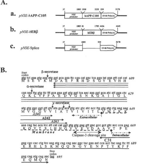
Figure 1. Gene construction. A, Three constructs. The APP-C105 and hERB were placed under the control of the NSE gene promoter (pNSE/hAPP-C105 and pNSE/hERB) (a and b). Control plasmid (pNSE-Splice) with eliminated hAPP-C105 or hERB sequence is shown (c). B, Feature of APP-C105 nucleotide and their amino acid sequences. Sequences for AB-40, A6-42, AB-43 and (<->) are indicated. Secretase (a, B, y) cleavage sites (J.), caspase-3 cleavage site («), start-and stop-codon (-), beginning of extracellular- start-and intracellular membrane (-. - .->), start-and numbers of sequences are shown, respectively.
Western blot analysis. Cells were lysed in buffer containing
50 mM Tris (pH 8.0), 50 mM NaCl, 0.5 mM EDTA, and 1 mM PMSF and lysed cells were centrifuged at 5,000 x g for 15 min. The final supernatants were aliquoted and stored at -80°C. Protein content was determined by the bicinchoninic (BCA) method with a BSA standard. Protein (50 |ig) was separated in a 10 to 20% gradient gel of polyacrylamide electrophoresis for 3 h and was transferred to a nitrocellulose membrane using an electroblot. Membranes were then incubated with primary antibodies [anti-JNKl (1:500); anti-phospho-JNKl (1:330); anti-ERK (ERK1 and ERK2) (1:2000); anti-phospho-ERK (p-ERK-1 and pERK2) (1:2,000); 38 (1:2,000) and anti-phospho-38 (1:1,000); and ERB (1:500)]. Here, anti-phospho JNK1 specifically recognizes JNK that are phosphorylated at Thr-183 and Tyr-185. The monoclonal antibody anti-p38 MAP kinase reacts specifically with the active double-phosphorylated form of MAP kinase. In addition, activation of ERK1 and ERK2 are directly mediated by MAP kinase kinase (MAPKK or MEK) that phosphorylates serine/ threonine/tyrosine residues in the regulatory sites of MAP kinase. Each complex of antigen-antibody were visualized with biotinylated secondary antibody (goat
anti-rabbit)-conjugated HRP streptavidin (Zymed, Histostain-Plus Kit) diluted 1:1,500 in PBS blocking buffer.
Statistical analysis. Tests for significance were performed
using one-way analysis of variance (SPSS for Window, Release 10.01, Standard Version, Chicago, IL). All values are reported as the mean ± standard deviation. Statistical significance was set at p<0.05.
Results
Expressions ofhERfi and APP-C105 in transfected cells.
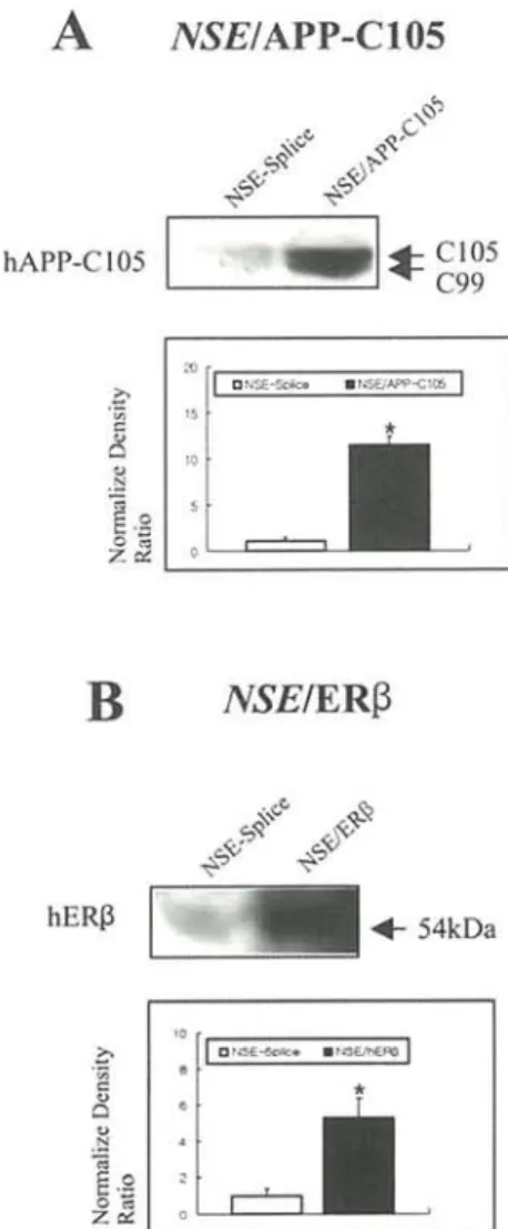
SK-N-MC cells were transfected with three constructs (Fig. 1A) to detect expressions of hERB and APP-C105. The region of the APP-C105 contained potential structure features, as shown in Fig. IB. The NSE promoter and the polyadenylation signal provide the start and termination of APP-C105 transcript, respectively. Immuno-reactive proteins of the hERB and APP-C105 were detected in transfected SK-N-MC cells by immunoblotting (Fig. 2). In the expression of APP-C105, two bands were visible with APP-C105 and C99, one (C99) of which was believed to be the cleaved by B-secretase (Fig. 2A).
694 LIM et al: ESTROGENIC EFFECT ON ERB- AND APP-C105-MEDIATED MAPK ACTIVATION
A yVS£/APP-C105
Figure 2. Expressions of hERB and APP-C105 in transfected cells. Protein (50 (xg) were analyzed by Western blot using anti-APP antibodies (Zymed) (A) and polyclonal rabbit anti-ERB (Santa Cruz) (B). Quantifications of bands through A and B are shown. Three experiments were assayed in triplicate on Western blot. Values are mean ± SD. p<0.05 versus control (pNSE-Splice).
These results confirmed that both hERG and APP-C105 constructs were highly over-expressed in transfected cells.
Effect of 17fi-estradiol on hER-mediated activation of MAPK.
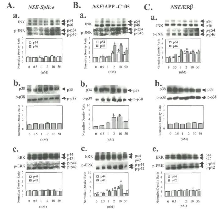
To examine the role of estrogen on hERB-mediated MAPK activation, cells were pretreated with 176-estradiol and cells were then transfected with NSE/hERfi. Transfected cells were treated with an increasing concentration of 176-estradiol for 15 min. Proteins were then prepared from transfected cells for Western blot analysis and were assayed for MAPK activation with anti-active MAPK activities (Fig. 3C). Although, the levels of phospho-JNK-1 and total JNK-1 migrating at two molecular weight, p54 and p46, were slightly increased, the levels in active form from a density ratio of phospho-p38 were not increased in the 176-estradiol-treated cells when normalized to the corresponding total p38. In addition, there was no increase in activity of ERK, p42 and p44, when the brains were examined for phospho-ERK on Western blot and normalized to ERK. These results suggest that ERB transfection led to clear change through estrogen only in activating JNK
compared to that of control cells transfected with pNSE-Splice (Fig. 3A).
Effect of 17fi-estradiol on APP-C105-mediated MAPK activation. To test whether estrogen lead to
APP-C105-mediated MAPK activation, an NSE/APP-C105 combining APP-C105 and NSE promoter (pNSE/APP-C105) was constructed (Fig. IB). Cells were pretreated with 10 nM 176-estradiol for 30 min, and were transfected with this plasmid. Transfected cells were then treated with various doses of 176-estradiol for 15 min. Proteins were prepared from the transfected cells and were assayed for the measurement of MAPK activation by Western blot analysis with anti-activated JNK-1, anti-ERK, and anti-p38 polyclonal antibodies against the phosphorylation site of JNK-1, ERK, and p38. The levels of activation for ERK and p38 gradually increased, with clear changes apparent between 1 and 2 nM or 10 nM and there after it exhibited decreased levels of their phosphorylation (Fig. 3B). Moreover, JNK activation is also dependent on doses of 176-estradiol, reaching a maximum near 2 to 10 nM relative to those of non-treated control cells. This result suggests that APP-C105 succeeds in activating the MAPK family in an estrogen-dose-dependent manner, but no activation was observed in NSE-transfected control cells (Fig. 3A).
Discussion
In our previous study, up-regulation of both ERs were observed in the brains of doubly transgenic progeny at 8 months of age by Western blot analysis (unpublished data). It was much higher in the double transgenic progenies than those in single transgenic and non-transgenic progeny. The levels of ER expression and MAPK activation were not changed by ovariectomy of double transgenic mice, indicating that estrogen is not responsible for up-regulation of ERs and thus, maintaining MAPK activation.
The aim of this study was to charaterize the role of estrogen in ER6-mediated effect on activation of MAPK pathway using in vitro cell system expressed hER6. This study provides information to test more clearly the dose of estrogen suitable for ER6-mediated activation of MAPK family. It clarifies the role of ER6 in estrogen-induced activation of the MAPK pathway in the in vitro system. We found that treatment of the hER6-transfected cells with different doses of estrogen led to an induction of the MAPK signaling pathway. This result suggests that ER6 is required for estrogen-mediated JNK activation, but not for p38 and ERK activation. It was demonstrated that both ERa and ER6-transfected cells activated MAPK within 15 min of exposure to 10 nM 176-estradiol, and estrogen receptor is necessary to detect MAPK activation (22). However, activation of MAPK was independent of estrogen receptor expression (20), and ER6 is presumed to mediate an inhibitory effect of estrogens (22). It is not clear yet why transfectant cells can respond to the estrogen on MAPK activation in a different manner. The biological actions of estrogens are manifest only in cells expressing a specific high-affinity ER (24). In fact, ER is a ligand-dependent transcription factor, accounting for the latency of most estrogenic responses in target tissues (25).
INTERNATIONAL JOURNAL OF MOLECULAR MEDICINE 13: 691-696, 2004 695
A.
NSE-Splice
B.
Atf£/APP-C105
C.
NSE/ERfi
Figure 3. Effect of estrogen on hERB- and APP-C105-mediated activation of the MAPK-signaling pathway. A, Activations of three members (JNK, p38, and ERK) of the MAPK family in pNSE-transfected cells, treated with 17B-estradiol. a, JNK and JNK. b, p38 and p38. c, ERK and ERK. B, Activations of three members of MAPK in pNSE/hERB-transfected cells treated with 1713-estradiol. a, JNK and JNK. b, p38 and phospho-p38. c. ERK and phospho-ERK. C, Activations of three members of MAPK in pNSE/hAPP-C105 transfected cells treated with 17B-estradiol. a, JNK and phospho-JNK. b, p38 and phospho-p38. c, ERK and phospho-ERK. In the A, B, and C, 50 ug of proteins per sample were immunoblotted with antibodies for JNK and phospho-JNK, p38 and phospho-38, and ERK and phospho-ERK. Quantification of bands through A, B, and C are shown. Three experiments were assayed in triplicate on Western blot. Values are mean + SD. *p<0.05 versus control (A).
Recent genetic, biochemical, and pharmacological dissection of the estrogen signal transduction pathway like the three members of MAPK has led to the identification of numerous proteins and processes that impinge on ER function, revealing an unexpected level of complexity in the action of this hormone (26). Based on our in vitro studies, the 176-estradiol and ER complex contributes to the characterization of ER-mediated JNK activation, but not p38 and ERK activation. Thus, proteins that impinge on ER6 function exist differently in each three members of the MAPK signal pathway.
The effect of 176-estradiol on APP-C105-mediated MAPK activation has not been documented. It was only demonstrated that there was an increase in the activation of MAPK family
in the cultured rat hippocampal and cortical neurons treated with A6 (27). Addition of 17B-estradiol to the SK-N-MC cells transfected with APP-C105 resulted in an induction of the JNK, p38 and ERK activities at concentration between 0.5 and 10 nM of 176-estradiol, and thereafter it was gradually declined at concentration of 10 or 50 nM 176-estradiol. Clearly, 176-estradiol at 2- or 10 nM concentration induces a much higher level of phospho-JNK, phospho-p38 and phospho-ERK, and treatment with high doses of 176-estradiol (50 nM) resulted in inhibition of JNK, p38 and ERK phosphorylation. It is likely linked to the apoptotic cells exposed by high doses of 176-estradiol. In fact, 176-estradiol excess is not more neuroprotective in physiological levels of this estradiol (28). However, the effect of doses of
176-696 LIM et al: ESTROGENIC EFFECT ON ER6- AND APP-C105-MEDIATED MAPK ACTIVATION
estradiol (2-10 nM) on APP-C105-mediated MAPK activation is correlated with a report that 176-estradiol induce a rapid secretion of sAPPct-non-amyloidogenic APP via the MAPK pathway (20), releasing A8-40, which is not induced apoptosis. Our data provide the first evidence that estrogen activates the MAPK pathway through APP-C105, raising a possibility that estrogen increases the production of a-secretase-derived APP fragment in the status of MAPK activation that is released A6 when a-secretase cuts the APP-C105 substrate. Although, a c t i v a t i o n of M A P K f a m i l y h a s b e e n s t u d i e d for t h e protective effect of estrogen on A6 toxicity, it is clear that APP-C105 as well as A6 are linked to activation of MAPK p a t h w a y at 2 or 10 n M d o s e s of e s t r o g e n . T h u s , it is necessary to test whether these activations are likely to be m e d i a t e d by activation of u p s t r e a m k i n a s e s . This may correlate with the result that AB is mediated by the activation of MAPK (13).
Acknowledgments
Financial support for this research was provided by grants to Dr Yong K. Kim from the Korea Ministry of Health and Welfare (01-PJ1-20500-0129) and the Korea Food and Drug Administration.
References
1. Xu H, Gouras GK, Greenfield JP, Vincent B, Näslund J,
Mazzarell L, Fried G, Jovanovic JN, Seeger M, Relkin NR, Liao F, Checler F, Buxbaum JD, Chait BT, Thinakaren G, Sisodia SS, Wang R, Greengard P and Gandy S: Estrogen reduces neuronal generation of Alzheimer beta-amyloid peptides. Nat Med 4: 447-451, 1998.
2. Jaffe AB, Toran-Allerand CD, Greengard P and Gandy SE: Estrogen regulates metabolism of Alzheimer amyloid beta precursor protein. J Biol Chem 269: 13065-13068, 1994. 3. Henderson VW: Estrogen replacement therapy for the
prevention and treatment of Alzheimer's disease. CNS Drugs 8: 343-351, 1997.
4. Gibbs RB and Aggarwal P: Estrogen and basal forebrain cholinergic neurons: implications for brain aging and Alzheimer's disease-related cognitive decline. Horm Behav 34: 98-111,1998.
5. Green PS and Simpkins JW: Neuroprotective effects of estrogens: potential mechanisms of actions. Int J Dev Neurosci 18: 347-358, 2000.
6. Watters JJ, Capbell JS, Cunningham MJ, Krebs EG and Dorsa DM: Rapid membrane effects of steroid in neuroblastoma cells: effect of estrogen on mitogen activated protein kinase signaling cascade and c-fos immediate early gene transcription. Endocrinology 138: 4030-4033, 1997.
7. Singh M, Setalo G, Guan X, Warren M and Toran AC: Estrogen-induced activation of mitogen-activated protein kinase in cerebral cortical explant: convergence of estrogen and neuro-tropin signaling pathway. J Neurosci 19: 1179-1188, 1999. 8. Migliaccio A, Di Domenico M, Castoria G, de Falco A,
Bontempo P, Nola E and Auricchio F: Tyrosine kinase/p21 ras/MAP-kinase pathway activation by estradiol-recerptor complex in MCF-7 cells. EMBO J 15: 1292-1300, 1996.
9. Razand M, Pedram A, Greene GL and Levin ER: Cell membrane and nuclear estrogen receptors (ERs) originate from a single transcript: studies of ER alpha and ER beta expressed in Chinese hamster ovary cells. Mol Endocrinol 13: 307-319, 1999. 10. Savaskan E, Olivieri G, Meier F, Ravid R and Muller-Spahn F:
Hippocampal estrogen B-receptor immunoreactivity is increased in Alzheimer's disease. Brain Res 908: 113-119, 2001.
11. Wang L, Andeerson S, Warner M and Gustafsson JA: Morpho logical abnormalities in the brains of estrogen beta knockout mice. Proc Natl Acad Sci USA 98: 2792-2796, 2001.
12. Shoji M, Iwakami N, Takeuchi S, Waragai M, Suzuki M, Kanazawa I, Lippa CF and Ono Satoshi Pkazawa H: JNK activation is associated with intracellular beta-amyloid accumulation. Mol Brain Res 85: 221-233, 2001.
13. Savage MJ, Lin YG, Ciallella JR, Flood DG and Scott RW: Activation of c-Jun N-terminal kinase and p38 in an Alzheimer's disease model is associated with amyloid deposition. J Neurosci 22: 3376-3385, 2002.
14. Haass C, Schlossmacher MG, Hung AY, Vigo-Pelfrey C, Mellon A, Ostaszewski BL, Lieberburg I, Koo EH, Schenk D, Teplow DB and Selkoe DE: Amyloid-peptide is produced by cultured cells during normal metabolism. Nature 359: 32-35, 1992.
15. Citron M, Diehl TS, Gordon G, Biere AL, Seubert P and Selkoe DJ: Evidence that the 42- and 40-amino acid forms of amyloid B protein are generated from the B-amyloid precursor protein by different protease activities. Proc Natl Acad Sci USA 93: 13170-13175, 1996.
16. Klafki H, Abramowski D, Swoboda R, Paganetti PA and Staufenbiel M: The carboxyl-termini of B amyloid peptides 1-40 and 1-42 are generated by distict y-secretase activities. J Biol Chem 271: 28655-28659, 1996.
17. Murphy MP, Hickman LJ, Eckman CB, Uljon SN, Wang R and Golde TE: y-secretase, evidence for multiple proteolytic activities and influence of membrane positioning of substrate on generation of amyloid B-peptide of varying length. J Biol Chem 274: 11914-11923, 1999.
18. Gervais FG, Robertson GS, Vaillancourt JP, Zhu Y, Huang J, LeBlanc A, Smith D, Rigby M, Shearman MS, Clarke EE, Zheng H, van der Ploeg LH, Ruffolo SC, Thrnberry NA, Xanthoudakis S, Zamboni RJ, Roy S and Nicolson DW: Involvement of caspases in proteolytic cleavage of Alzheimer's amyloid-B precursor protein and amyloidogenic AB peptide formation. Cell 97: 395-406, 1999.
19. Wei W, Norton DD, Wang X and Kusiak JW: AB 17-42 in Alzheimer's disease activates JNK and caspase-8 leading to neyronal apoptosis. Brain 125: 2036-2043, 2002.
20. Manthey D, Heck S and Behi C: Estrogen induces a rapid secretion of amyloid B precursor protein via mitogen-activated protein kinase pathway. Eur J Biochem 268: 4285-4291, 2001. 21. Forss S, Denielson PE, Catsicas S, Battenberg E, Price J,
Nerenberg M and Sutcliffe M: Transgenic mice expressing B-galactosidase in mature neuron under neuron-specific enolase promoter control. Cell 5: 187-197, 1990.
22. Paech K, Webb P, Kuiper GGJM, Nilson S, Gustafson JA, Kushner PJ and Scanlan TS: Differential ligand activation of estrogen receptors ERa and ERB at API site. Science 277: 1508-1510, 1997.
23. Fitzpatrick JL, Mize AL, Wade CB, Harris JA, Shapiro JL and Dorsa DM: Estrogen-mediated neuroprotection against 6-amyloid toxicity requires expression of estrogen receptor a or B and activation of the MAPK pathway. J Neurochem 82: 674-682, 2002.
24. McKenna NJ and O'Malley: Nuclear receptor, coregulator, ligands, and selective receptor modulators: making sense of the patch work guilt. Ann N Y Acad Sci 949: 3-5, 2001.
25. Means AR, Comstock JP, Rosenfield GC and O'Malley BW: Ovalbumin messenger RNA of chick oviduct partial charaterization, estrogen dependence, and translation in vitro. Proc Natl Acad Sci USA 69: 1146-1150, 1972.
26. McDonnell DP, Chang CY and Norris JD: Capitalizing on the complexities of estrogen receptor pharmacology in the quest for the perfect SERM. Ann N Y Acad Sci 949: 16-35, 2001. 27. Abe K and Saito H: Amyloid B neurotoxicity not mediated by
the mitogen-activated protein kinase cascade in cultured and hippocampal and cortical neurons. Neurosci Lett 292: 1-4, 2000.
28. Liu Z, Gastard M, Verina T, Bora S, Mouton PR and Koliatsos VE: Estrogens modulate experimentally induced apoptosis of granule cells in the adult hippocampus. J Comp Neurol 44: 441-448, 2001.