322 책임저자:이원섭, 660-702, 진주시 칠암동 90
경상대학교 의과대학 내과학교실 Tel: 055-750-8733, Fax: 055-758-9122
E-mail: [email protected], [email protected] 최영현, 614-052, 부산시 진구 양정동 산45 동의대학교 한의과대학 생화학교실
Tel: 051-850-7413, Fax: 051-853-4036 E-mail: [email protected]
접수일:2009년 12월 4일, 게재승인일:2009년 12월 14일
Correspondence to:Won Sup Lee
Department of Internal Medicine, Gyeongsang National University School of Medicine, 90, Chilam-dong, Jinju 660-702, Korea
Tel: +82-55-750-8733, Fax: +82-55-758-9122 E-mail: [email protected], [email protected] Co-Corresponding author:Yung Hyun Choi
Department of Biochemistry, Dongeui University College of Oriental Medicine, San 42, Yangjung-dong, Busan 614-052, Korea
Tel: +82-51-850-7413, Fax: +82-51-853-4036 E-mail: [email protected]
Anti-cancer Activities of Anthocyanins of Vitis Coignetiae Pulliat in Human Hepatocelluar Carcinoma Cells
Dong Yeok Shin1, Won Sup Lee2, Jing Nan Lu2, Chi Young Jung3, Dong Hoon Kim4, Gi Young Kim5, Ho Sung Kang6, Chung Ho Ryu7, Jin Myung Jung8, Soon Chan Hong3, Sung Chul Shin9,
Yung Hyun Choi1 and Woo Song Ha3
1Department of Biochemistry, Dongeui University College of Oriental Medicine and Department of Biomaterial Control (BK21 Program), Dongeui University Graduate School, Busan 614-052, Departments of 2Internal Medicine, 3Surgery,
4Emergency Medicine and 8Department of Neurosurgery Institute of Health Sciences and Gyeongnam Regional Cancer Center, Gyeongsang National University School of Medicine, Jinju 660-702, 5Department of Applied Marine Science, Cheju National University, Jeju 690-756, 6Department of Molecular Biology, Pusan National University, Busan 609-735, 7Division of Applied Life Science (BK 21 Program), Institute of Agriculture and Life Science, 9Department of Chemistry, Research
Institute of Life Science, Gyeongsang National University, Jinju 660-701, Korea
Anthocyanins belong to a class of flavonoids, exhibiting some of the anti-tumor activities: anti- angiogenesis and anti-invasive activity. Recently, the anthocyanins from the Fruit of Vitis coignetiae Pulliat (Meoru in Korea, AIMs) have been reported to have anti-cancer activities. Here, we tested the anti-cancer effects of AIMs in human hepatocelluar carcinoma cells (Hep3B and HepG2 cells). The AIMs inhibited the proliferation of the cells in a dose dependent manner. Hep3B cells were more sensitive to AIMs.
The AIMs induced apoptosis through the loss of MMP (ΔΨm). The AIMs inhibited the motility and invasion of the cells in wound healing test and Matrigel-invasion assay, respectively. The anti-migratory and anti-invasive activities of AIMs were superior to the anthocyanins isolated from black bean. In conclusion, this study indicates that AIMs have apoptotic and anti-invasive effects on human hepatocellular carcinoma cells. AIMs had stronger anti-invasive activity than the anthocyanins from coat of black bean on both Hep3B and HepG2 cells. This study provides evidence that the anthocyanins isolated from Meoru might be useful in the treatment of human hepatocelluar carcinoma. (Cancer Prev Res 14, 322-328, 2009) Key Words: Anthocyanins, Apoptosis, Invasion, Hep3B cell, HepG2, Cancer
INTRODUCTION
Hepatocellular carcinoma is one of the most common malig- nant tumors worldwide.1) For the management of this disease, few effective systemic chemotherapeutic drugs are available.
Therefore, the development of chemotherapeutic or chemopre- ventive agents is crucial for the control of this disease.
Dietary agents are known to safely modulate physiological function and enhance anti-cancer activity.2,3) In addition, with an ecological frame of mind, natural products have become more popular for the prevention or treatment of cancer. This is demonstrated by the increase in the frequencies of the researches focusing on natural products. Vitis coignetiae Pulliat (Known as Meoru in Korea) belongs to the grape family. The intense dark red color of the fruit is due to an abundance of
anthocyanin pigments which belong to a class of flavonoids.
Recently, in vitro and in vivo anti-cancer activities of an- thocyanins have been reported regarding anti-angiognesis and cancer invasion.4)
We investigated anticancer effects of AIMs in hepatocelluar carcinoma cells and compare the efficacy of AIMs with that of the anthocyanins isolated from black bean in terms of invasive activities.
MATERIALS AND METHODS 1. Cell culture and chemicals
Human hepatoma Hep3B and HepG2 cells from the American type culture collection (Rockville, MD, USA) were cultured in RPMI 1,640 medium (Invitrogen Corp, Carlsbad, CA, USA) supplemented with 10% (v/v) fetal bovine serum (FBS) (GIBCO BRL, Grand Island, NY, USA), 1 mM L- glutamine, 100 U/ml penicillin, and 100μg/ml streptomycin at 37°C in a humidified atmosphere of 95% air and 5% CO2. An enhanced chemiluminescence (ECL) kit was purchased from Amersham (Arlington Heights, IL, USA). 5,5',6,6'-tetrachloro- 1,1',3,3'-tetraethylbenzimidazolylcarbocyanine iodide (JC-1) was obtained from Calbiochem (San Diego, CA, USA). All other chemicals which are not specifically cited here were purchased from Sigma Chemical Co. (St. Louis, MO, USA). We prepared two kinds of anthocyanins; one is isolated from Meoru and the other is from coat of black bean. The anthocyanin of isolated from Meoru were composed of delphinidin-3,5-diglucoside, cyanidin-3,5-diglucoside, petunidin-3,5-diglucoside, delphinidin- 3-glucoside, malvdin-3,5-diglucoside, peonidin-3,5-diglucoside, cyanidin-3-glucoside, petunidin-3-glucoside, peonidin-3-gluco- side, and malvidin-3-glucoside with the ration of 3.5:3.4:
7.1:23.9:8.0:9.6:9.1:16.1:5.7:13.4. The anthocya- nins from coat of black bean were composed of cyanidin-3- glucoside (72%), delphinidin-3-glucoside (20%), and petunidin- 3-glucoside (6%).
2. Cell proliferation assays
For the cell viability assay, the cells were seeded onto 24-well plates at a concentration of 5×10⁴ cells/ml and treated with AIMs for 48 h and the number of surviving cells was counted using trypan blue exclusion methods.
3. Flow cytometry analysis for measurement of sub-G1 phase
The DNA content of the cells was measured using a DNA staining kit (CycleTestTM Plus Kit, Becton-Dickinson, San Jose, CA). Propidium iodide (PI)-stained nuclear fractions were obtained using the instructions provided in the kit. Flow cytometric analyses were carried out using a FACS flow cytometer (FACSCalibur, Becton-Dikinson). The sub-G1 population was calculated to estimate the apoptotic cell population.
4. Western blotting
The concentrations of cell lysate proteins were determined by means of the Bradford protein assay (Biorad lab, Ricmond, CA, USA) using bovine serum albumin as the standard.
Molecular mass markers for proteins were obtained from Pharmacia Biotech (Saclay, France). Antibodies against Bid, poly (ADP-ribose) polymerase (PARP), pro-caspase-3 were purchased form Santa Cruz Biotechnology (Santa Cruz, CA, USA). Antibody against β-actin was from Sigma. Peroxidase- labeled donkey anti-rabbit and sheep anti-mouse immunoglo- bulin were purchased from Amersham. For the Western blot- ting, 30 micrograms of proteins were resolved by electro- phoresis, elctrotransferred to a polyvinylidene difluoride mem- branes (Millipore, Bedford, MA, USA), and then blotted with primary antibodies followed by secondary antibody conjugated to peroxidase. Blots were developed with an ECL detection system.
5. Mitochondrial membrane potential (MMP, Δ Ψm) assay
The MMP (ΔΨm) in living cells was measured by flow cytometry with the lipophilic cationic probe JC-1, which is a ratiometric, dual-emission fluorescent dye. There are two excitation wavelengths, 527 nm (green) for the monomer form and 590 nm (red) for the J-aggregate form. Quantitation of green fluorescent signals reflects the amount of damaged mitochondria. Hep3B cells were trypsinized and the cell pellets were resuspended in 500μl of PBS, incubated with 10μM JC-1 for 20 min at 37°C. The cells were subsequently washed once with cold PBS, suspended in a total volume of 500μl and analyzed using flow cytometry.
Fig. 1. Growth inhibition AIMs in human hepatocellular carcinoma cell lines, Hep3B and HepG2. The cells were seeded at the density of 5×10⁴ cells per ml. The cells were treated with indicated concentrations of AIMs for 48 h. (A) Cell number was determined by hemocytometer counts of trypan blue-excluding cells. The data are shown as means±SD of three independent experiments.
*p<0.05 between the treated and the untreated control group. (B) To quantify the degree of AIMs-induced apoptosis, sub-G1 DNA content, which represents the fractions undergoing apoptotic DNA degradation, was analyzed by flow cytometry. The results are from one representative experiment of two independent experiments that showed similar patterns.
6. Wound healing assay
The wound healing assays were conducted according to the methods described previously.5) Hep3B cells were grown on 35 mm dish plate to 100% confluent monolayer and then scratched to form a 100-μm "wound" using sterile pipette tips.
The cells were then cultured in the presence or absence of AIMs (500μg/ml) in serum-free media for 24 h. The images were recorded at 12 h and 24 h after scratch using an Olympus photomicroscope.
7. Cell invasion assay
For the cell invasion assays, Hep3B cells were cultured in serum-free media overnight. The cells (5×10⁴ cells) were loaded on pre-coated Matrigel 24-well invasion chambers (BD Biosciences, San Jose, CA, USA) in the presence or absence of AIMs (500μg/ml). Then 0.5 ml of 5% fetal calf serum medium was added to the wells of the plate to serve as the chemoattractant for the cells. The Matrigel invasion chambers were incubated for 24 h. The invading cells were fixed with 10% formalin, stained with Harris modified hematoxylin (Sigma), and analyzed according to manufacturer's instruc- tions.14)
8. Statistics
Each experiment was performed in triplicate. The results
were expressed as means±SD. Significant differences were determined using the Student's t test. Statistical significance was defined as p<0.05.
RESULTS
1. AIMs inhibited proliferation of human hepa- tocelluar carcinoma cells, Hep3B and HepG2
To investigate the anti-tumor activity of AIMs, the human hepatocellular cell lines Hep3B and hepG2 were treated with various concentrations of AIMs for 48 h and evaluated their viability using a trypan blue assay. As shown in Fig. 1A, AIMs inhibited the viability of Hep3B cells in a dose-dependent manner. Treatment with 500μg/ml of AIMs caused appro- ximately 65% and 20% inhibition of cell growth in Hep3B and HepG2, respectively, when compared with each control.
This finding suggests that Hep3B is more sensitive to AIMs treatment (Fig. 1A).
To investigate how AIMs affects the growth of Hep3B cells, we performed flow cytometry analysis. As shown in Fig. 1B, treatment with AIMs resulted in a significant accumulation of cells with sub-G1 DNA content in a dose-dependent manner.
This finding suggested that the cytotoxic effects observed in response to AIMs are associated with the induction of apoptosis.
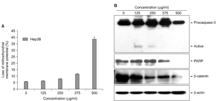
Fig. 2. The loss of mitochondrial membrane potentials (MMP, ΔΨm) and caspase-3 activation induced by AIMs in Hep3B cells.
Hep3B cells were incubated at indicated concentrations of AIMs for 48 h. (A) The cells were stained with JC-1 and incubated at 37°C for 20 min. The mean JC-1 fluorescence intensity was detected using a flow cytometer. Data are expressed as the means±SD of representative experiments performed at least three times. (B) Total cell lysates were resolved by SDS-polyacrylamide gels and transferred onto nitrocellulose membranes. The membranes were probed with the anti-procaspase-3, β-catenin and anti-PARP antibodies. The proteins were visualized using an ECL detection system. β-Actin was used as an internal control.
2. AIMs induced loss of mitochondrial mem- brane potentials (MMP, ΔΨm) and caspase- 3 activation
Mitochondria play a central role in apoptosis. As the early event of apoptosis, mitochondrial depolarization occurs. There- fore, we measured MMP (ΔΨm) using a fluorescent cationic dye JC-1. As shown in Fig. 2A, AIMs increased the degree of MMP (ΔΨm) loss in Hep3B cells in a dose dependent man- ner after the AIMs treatment and the levels of MMP (ΔΨm) loss reached up to 40% at 500μg/ml for 48 h, evidencing that AIMs increase depolarization of the MMP (ΔΨm). This mitochondrial dysfunction elicited by AIMs lead to activation of caspase-3, which has been shown to play a pivotal role in the terminal execution phase of apoptosis. Therefore, we determined that AIMs-induced apoptosis was associated with the activation of caspases. As shown in Fig. 2B, the treatment of AIMs decreased the expression levels of pro-caspase-3 in a concentration-dependent manner. We also found that AIMs caused the proteolytic cleavage of PARP protein, which is a downstream target of the activated caspase-3, in a concentration-dependent manner (Fig. 2B). These findings
suggest that AIMs may induce apoptotic death through a caspase-dependent pathway.
3. AIMs inhibit the migration and invasion of Hep3B cells
Cancer cell migration and invasion are the key events in metastasis; therefore, we further tested the effects of AIMs on cell migration and invasion in vitro. Our results showed that AIMs at 500μg/ml significantly inhibited Hep3B cell migra- tion in wound healing assays (Fig. 3A). In Matrigel invasion assays, AIMs inhibited cell invasion at the concentration of 500 μg/ml by about 80%, as compare to control (Fig. 3B). To determine that AIMs have better anti-invasive effects that anthocyanins from another sources, we compared the efficacy of AIMs with the anthocyanins isolated from the husks of black beans. We found that AIMs showed stronger anti-migratory and anti-invasive effects than the anthocyanins isolated from the husks of black beans on Hep3B cells.
4. AIMs inhibited the migration and invasion of HepG2 cells
To confirm the above finding in Hep3B cells, we also
Fig. 3. Effects of the anthocyaninns from Meoru and black bean on the migration and invasion of Hep3B human hepatocellular carcinoma cells. (A) Cells were grown to 100% confluency on 30-mm cell culture dishes coated with rat tail collagen and then treated with and without anthocyanins (500μg/ml) for 24 h. A scratch was made through the cell layer using a pipette tip. After washing with PBS, serum-free media with and without the anthocyanins was added. Photographs of the wounded area were taken at the interval of 0 h, 12 h, and 24 h after the scratch to evaluate cell movement into the wounded area. (B) The cells were exposed to AIMs for 6 h. The cells (5×10⁴ cells) were loaded on pre-coated Matrigel 24-well invasion chambers (BD Biosciences) with and without the anthocyanins (500μg/ml). Medium containing 20% FBS was placed in the basolateral chamber to act as a chemoattractant. After 48 h, the cells on the apical side were wiped off using a Q-tip. Next, the cells on the bottom of the filter were stained using hematoxylin and then counted. The effects of the anthocyanins were represented as percentages of the values of untreated control cells (% of control). *p<0.05 versus control, **p<0.05 versus the anthocyanins from black bean.
Fig. 4. Effects of the anthocyaninns from Meoru and black bean on the migration and invasion of HepG2 human hepatocellular carcinoma cells. (A) Cells were grown to 100% confluency on 30-mm cell culture dishes coated with rat tail collagen and then treated with and without anthocyanins (500μg/ml) for 24 h. A scratch was made through the cell layer using a pipette tip. After washing with PBS, serum-free media with and without the anthocyanins was added. Photographs of the wounded area were taken at the interval of 0 h, 12 h, and 24 h after the scratch to evaluate cell movement into the wounded area. (B) The cells were exposed to AIMs for 6 h. The cells (5×10⁴ cells) were loaded on pre-coated Matrigel 24-well invasion chambers (BD Biosciences) with and without the anthocyanins (500μg/ml). Medium containing 20% FBS was placed in the basolateral chamber to act as a chemoattractant. After 48 h, the cells on the apical side were wiped off using a Q-tip. Next, the cells on the bottom of the filter were stained using hematoxylin and then counted. The effects of the anthocyanins were represented as percentages of the values of untreated control cells (% of control). *p<0.05 versus control, **p<0.05 versus the anthocyanins from black bean.
compared the efficacy of AIMs with the anthocyanins isolated from the husks of black beans in HepG2. Similar to the above findings, the results showed that AIMs at 500μg/ml signi- ficantly inhibited HepG2 cell migration in wound healing tests than did the anthocyanins isolated from the husks of black beans (Fig. 4A). In Matrigel invasion assays, AIMs inhibited cell invasion at the concentration of 500μg/ml by about 70%, as opposedto 40% inhibition of invasion by the anthocyanins isolated from the husks of black beans (Fig. 4B). The difference in anti-invasive activity between the two anthocyanins was statistically significant.
DISCUSSION AND CONCLUSION
This study was designed to investigate the anticancer effects on human hepatocelluar carcinoma cell lines, Hep3B and HepG2, and compare the efficacy with anthocyanins from another source. We found that AIMs significantly attenuated the proliferation and induced apoptosis in Hep3B cells, which was more sensitive to AIMs treatment. Moreover, AIMs had anti-invasive activities on both Hep3B and HepG2 cells. The anti-invasive activity of AIMs was significantly stronger than the anthocyanins from black bean. These findings suggest that the composition of anthocyanins might be an important in showing anti-cancer activities. This finding is consistent with the previous studies.6,7)
Apoptosis, a type of programmed cell death, should be an underlying mechanism by which various natural compounds exert anti-cancer effects.2,3) Some of the data from the present study are consistent with previous studies demonstrating that anthocyanins had the remarkable cytotoxic effects on malignant cells.6,7) Evidences indicated that apoptosis could be triggered by the activation of a set of caspases and their activation played important roles during apoptosis. In the most of apoptotic processes, caspase-3 has been shown to play a pivotal role in the terminal and execution phase of apoptosis induced by diverse stimuli.8,9) We examined whether the caspase-3 protease is involved in AIMs-induced cell death response. Furthermore, this study suggested that the activation of caspase-3 through mitochondrial dysfunction elicited by AIMs might cause the cleavage of PARP. This finding is consistent with previous studies on the anthocyanins from Vitis coignetiae Pulliat (Meoru in Korea).10,11) In this study, we did not investigate the detailed pathway of AIMs-induced apoptosis. However, the previous
studies suggested that AIMs induce apoptosis though intrinsic pathway.10,11) Here, we compared the sensitivity of Hep3B to AIMs treatment with that of HepG2.
However, this finding differs from that of previous study,12) where the anthocyanidines exhibited stronger growth inhibitory effects against human hepatoma HepG2 cells than against Hep3B cells. In that study, anthocyanidins, mainly cyanidin, delphinidin, and malvidin caused strong growth inhibition in hepatoma cell line HepG2, whereas the aglycoside anthocya- nins, including cyanidin 3-glucoside, peonidin 3-glucoside, pelargonidin 3-glucoside, and malvidin 3-glucoside, had lower inhibitory activities in HepG2. The discrepancies between the results of the two studies may be explained by two ways; one reason is that the anthocyanins they used are not the same as the major components of AIMs. The substitution pattern of aglycon of anthocyanins may affect anti-cancer activity.13) The other is that the previous study did not compared the efficacy of the five anthocyanidins with that of aglycoside anthocyanin with the same aglycone because the activities of anthocyanidins might be altered by glycosylation.
We demonstrated that AIMs had inhibitory activities of the migration and invasion on cancer cells. The process includes proteolytic digestion of the ECM, and cell migration through the basement membranes to develop metastasis.15) Therefore, tumor migration and invasion is the first essential step for the metastasis.
Data presented here indicated that AIMs in the concentra- tion of 500μg/ml had the apoptotic activity in human hepa- tocellular carcinoma cell lines, Hep3B and HepG2. The con- centrations used in the present study is consistent with those in many other studies on the anti-tumor effect of anthocyanins in culture cells.6,7,14) Our data revealed that AIMs possessed growth-inhibitory ability against human hepatocellular carcino- ma cell lines, Hep3B and HepG2 whereas a normal human liver cell line (chang liver cells) was completely resistant to the cytotoxic activity at the high concentration (500μg/ml) of AIMs (data not shown). This is consistent with results in a previous study showing selective toxicity of anthocyanins on cancer cells.12) Hep3B cells express hepatitis virus B antigen and produce mostly plasma major proteins. A majority of the hepatomas developed in Korea are related to hepatitis virus B.
Therefore, these results are much valuable in the clinical aspects.
In conclusion, AIMs have anti-proliferative, anti-invasive, cy-
totoxic effects on human hepatocellular carcinoma cell lines, Hep3B and HepG2. AIMs had stronger anti-invasive activity than the anthocyanins from coat of black bean on both Hep3B and HepG2 cells. This study provides evidence that AIMs might be useful in the treatment of human hepatocellular carcinoma.
ACKNOWLEDGEMENT
This study was supported by a grant of the National R&D Program for Cancer Control, Ministry for Health, Welfare &
Family Affairs, Republic of Korea (0820050).
REFERENCES
1) El-Serag HB, Mason AC. Rising incidence of hepatocellular carcinoma in the United States. N Engl J Med 340, 745-750, 1999.
2) Sandur SK, Ahn KS, Ichikawa H, Sethi G, Shishodia S, Newman RA, Aggarwal BB. Zyflamend, a polyherbal pre- paration, inhibits invasion, suppresses osteoclastogenesis, and potentiates apoptosis through down-regulation of NF-kappa B activation and NF-kappa B-regulated gene products. Nutr Cancer 57, 78-87, 2007.
3) Park C, Jin CY, Kim GY, Choi IW, Kwon TK, Choi BT, Lee SJ, Lee WH, Choi YH. Induction of apoptosis by esculetin in human leukemia U937 cells through activation of JNK and ERK. Toxicol Appl Pharmacol 227, 219-228, 2008.
4) Middleton E Jr, Kandaswami C, Theoharides TC. The effects of plant flavonoids on mammalian cells: implications for inflammation, heart disease, and cancer. Pharmacol Rev 52, 673-751, 2000.
5) Favot L, Martin S, Keravis T, Andriantsitohaina R, Lugnier C. Involvement of cyclin-dependent pathway in the inhibitory effect of delphinidin on angiogenesis. Cardiovasc Res 59, 479-
487, 2003.
6) Kamei H, Kojima T, Hasegawa M, Koide T, Umeda T, Yukawa T, Terabe K. Suppression of tumor cell growth by anthocyanins in vitro. Cancer Invest 13, 590-594, 1995.
7) Koide T, Kamei H, Hashimoto Y, Kojima T, Hasegawa M.
Antitumor effect of hydrolyzed anthocyanin from grape rinds and red rice. Cancer Biother Radiopharm 11, 273-277, 1996.
8) Ashkenazi A. Targeting death and decoy receptors of the tumour-necrosis factor superfamily. Nat Rev Cancer 2, 420- 430, 2002.
9) Thornberry NA, Lazebnik Y. Caspases: enemies within. Scien- ce 281, 1312-1316, 1998.
10) Lee SH, Park SM, Park JH, Shin DY, Kim GY, Ryu CH, Shin SC, Jung JM, Kang HS, Lee WS, Choi YH. Induction of apoptosis in human leukemia U937 cells by anthocyanins through down-regulation of Bcl-2 and activation of caspases.
Int J Oncol 34, 1077-1083, 2009.
11) Shin DY, Ryu CH, Lee WS, Kim DC, Kim SH, Hah YS, Lee SJ, Shin SC, Kang HS, Choi YH. Induction of apoptosis and inhibition of invasion in human hepatoma cells by anthocyanins from meoru. Ann NY Acad Sci 1171, 137-148, 2009.
12) Yeh CT, Yen GC. Induction of apoptosis by the Anthocy- anidins through regulation of Bcl-2 gene and activation of c-Jun N-terminal kinase cascade in hepatoma cells. J Agric Food Chem 53, 1740-1749, 2005.
13) Marko D, Puppel N, Tjaden Z, Jakobs S, Pahlke G. The substitution pattern of anthocyanidins affects different cellular signaling cascades regulating cell proliferation. Mol Nutr Food Res 48, 318-325, 2004.
14) Meiers S, Kemeny M, Weyand U, Gastpar R, von Angerer E, Marko D. The anthocyanidins cyanidin and delphinidin are potent inhibitors of the epidermal growth-factor receptor. J Agric Food Chem 49, 958-962, 2001.
15) Liabakk NB, Talbot I, Smith RA, Wilkinson K, Balkwill F.
Matrix metalloprotease 2 (MMP-2) and matrix metalloprotease 9 (MMP-9) type IV collagenases in colorectal cancer. Cancer Res 56, 190-196, 1996.