Identification of a Bioactive Compound, Violacein, from Microbulbifer sp. Isolated from a Marine Sponge
Hymeniacidon sinapium on the West Coast of Korea
Nam-Il Won
1, Ga-Eun Lee
2, Keebeom Ko
3, Dong-Chan Oh
3, Yang Ho Na
4*, and Jin-Sook Park
2*
1
Water Resources Research Center, K-water Institute, Daejeon 34045, Republic of Korea
2
Department of Bioscience and Biotechnology, Hannam University, Daejeon 34430, Republic of Korea
3
Natural Products Research Institute, College of Pharmacy, Seoul National University, Seoul 08826, Republic of Korea
4
Department of Advanced Materials, Hannam University, Daejeon 34430, Republic of Korea
Received: February 10, 2017 / Revised: March 28, 2017 / Accepted: March 28, 2017
Introduction
Food and bioactive materials are part of most import- ant ecosystem services. These services can be produced by photosynthetic organisms and originate from carbon dioxide, water, and sunlight, which are being affected by climate change. There have been increasing reports on
critical environmental stressors on marine ecosystems from both anthropogenic and climate forcing [1, 2].
Remarks on the environmental impact of the marine ecosystem have been made often in terms of ecosystem structure (e.g., species range shift) [3 −5]. Services pro- vided by marine ecosystems, such as fisheries and bio- materials, are important areas that will be affected by climate change, which could lead to possible threats against access to and the sustainable use of bioactive compounds from marine organisms.
Natural products have become important resources for human well-being amidst recent global concerns, such as increasing outbreaks of epidemics including avian influ- enza and the emergence of antibiotic resistance, as well Microbial secondary metabolites of marine organisms are regarded as major sources of structurally and biologically novel compounds with numerous potential uses. Sponge-microbe associations are among the most interesting sources for exploring bioactive compounds. In this study, the bacterial strain Microbulbifer sp. (127CP7-12) was isolated from the Asian marine sponge Hymeniacidon sinapium collected at an inter- tidal zone on the west coast of Korea. Cultured bacteria produced a violet pigment, and optimal culture conditions for violet pigment production were investigated. Maximum production of the violet pigment from the strain culture was observed under the conditions of 25 ℃, pH 6.0, and 3% NaCl. Acetone provided better extraction of the pigment from fermented broth compared with ethanol and methanol. The proposed structure of the major component in the extracted crude pigment was determined via high-performance liquid chromatography, nuclear magnetic resonance, mass spectrometry, and UV spectra analyses, which showed that the metabolite was the promising bioactive compound violacein. This study describes the examination of marine bioactive materials from microbe-engaged metabolites and the ecological implica- tions of the sponge-microbe association in a changing ocean.
Keywords: Marine sponge, bacterial production, violacein, violet pigment, Korean waters
*Corresponding authors Y. H. N.
Tel: +82-42-629-8905, Fax: +82-42-629-8853 E-mail: [email protected]
J.-S. P.
Tel: +82-42-629-8771, Fax: +82-42-629-8751 E-mail: [email protected]
© 2017, The Korean Society for Microbiology and Biotechnology
as for clinical trials for anticancer and antimicrobial agents [6, 7]. Many candidate bioactive products have been reported from various taxa, including from bacteria and plants. Marine organisms are one of the groups attracting the most focus for the examination of natural products [8, 9]. Especially marine benthic organisms have been a major target group because they harbor use- ful bacterial communities for the production of bioactive materials owing to complex prey-predator interactions among large marine organisms as well as the microor- ganisms present in biofilms. Recent attention has focused on the bioactive materials produced by microor- ganisms living on marine benthic organisms and in bio- film. Among them, violacein is one promising natural product that has been reported from diverse microbe- organism interactions as well as from a microbe-sponge interaction, as recently reviewed by Choi et al. [10].
Despite the well-documented studies on violacein, there is still limited understanding of the ecological function of this material for both producer microbes and host organ- isms, and many studies are needed to investigate its clinical applications. This study examined microbial pro- duction originating from a marine sponge, and violacein was one of the natural products expected to be found during the research.
Marine sponges (phylum Porifera) are among the old- est and simplest animals, and they grow in every ocean and have a great capacity for withstanding extreme tem- peratures and pressures. They have no true tissues or organs and are just constructed with layers of cells with- out even a nervous system. As filter feeders, their bodies are full of pores and channels that allow water to circu- late through them, allowing interactions with various organisms. These structural features for sequestering food by filtering make sponge a suitable habitat for sym- biotic microorganisms. From their interactions with these various species as well as their long biological his- tory, sponges are well-known for their production of sec- ondary metabolites that constitute an effective defense mechanism against foreign predators [11]. Since the beginning of the exploration of marine natural products in the 1970s, investigations of secondary metabolites from marine sponge have been reported throughout the world. The barrel sponge, belonging to Xestospongia sponges (family Petrosiidae), is one of the most studied
species [12] with many studies having been carried out successively in several regions around the world. This sponge has been recognized as a rich source of different chemical classes and its crude extracts and isolated com- pounds display remarkable bioactivities [13].
Among sponge-related biological interactions, sponge- microbe associations have been important sources for exploring natural products. Various microorganisms have been found in sponges. These microorganisms may occupy more than half of the sponge body volume, exceeding microorganisms in seawater by 2 −4 orders of magnitude, and include a diverse range of green algae, heterotrophic bacteria, cyanobacteria, archaea, crypto- phytes, red algae, dinoflagellates, and diatoms. The sym- biotic microbial community is a highly diverse society.
One host sponge can possess diverse symbionts. Some of the symbionts inhabit specific sponges while others do not. Moreover, sponge-associated microbes are import- ant in terms of marine biodiversity because some microbes are found only in a sponge and not in seawater, as also reported for biofilm-associated microbes [14, 15].
For example, a species of α-proteobacteria dominates in sponge Rhopaloeides odorabile over various habitats, but it is not detected in seawater, which is an indication that the symbiont is sponge-specific. Analogously, the violacein-producers living on biofilm are prevalent in the ocean, but the violacein gene cluster has not been detected in the currently largest metagenomics sequence database of pelagic ocean waters [16, 17].
As part of continued interest in identifying bioactive compounds from microorganisms isolated from marine benthic organisms, we thoroughly investigated the pig- ments produced from a sponge-microbe association. The marine sponge Hymeniacidon sinapium used in this study is native to Korea and Japan [18, 19]. It is reported to inhabit other countries as an invasive species [20]. In this paper, we report the production and characteriza- tion of pigment extract produced by microbial activity isolated from the marine sponge H. sinapium collected in Korean coastal water. Also, the growth characteriza- tion of the isolated strain and the production of pigment are evaluated against various culturing parameters.
Finally, we discuss briefly the marine sponge-microbe
association in terms of the changing ocean around
Korean coastal waters.
Materials and Methods
Marine sponge collection and microorganism
The sponge, H. sinapium, was collected by hand from an intertidal rocky shore on the western coast of Korea.
The organism was transported to the laboratory after washing with sterilized artificial seawater and used for isolation of bacteria within 12 h [21]. The isolated marine bacterium was examined and maintained by subculturing at a regular interval of 3 days on Marine Broth 2216 (MB; Difco, USA) at 25 ℃ and stored at 4℃.
Fermentation of pigment
Influence of process parameters on pigment produc- tion by the bacterium was studied in a medium [22]. The MB was used for the production of violet pigment by the bacterium. The pre-culture was carried out by inoculat- ing the cells into 0.1 L of medium in a 0.5 L conical flask.
After incubation on a shaker (100 rpm) at 25 ℃ for 3 days, the culture was poured into a 2 L conical flask containing 1 L of the fresh medium. For the study on the optimum concentration of NaCl, the microbial solution was cultured on modified agar (MgSO
4· 7H
2O 4.8 g, MgCl
2· 6H
2O 3.5 g, KCl 1.0 g, CaCl
2· 2H
2O 0.18 g, NaH- CO
30.03 g, NaBr 0.013 g, Bacto Peptone (Difco, USA) 2.5 g, yeast extract (Difco, USA) 5.0 g, glucose (Sigma, USA) 1.0 g, D.W. 1 L, pH 7.2), which was based on a pre- vious report [23].
Extraction of pigment from the fermented broth
For extraction of pigment, the bacterium was incu- bated on a shaker in MB for 3 days. Supernatant was removed from culture broth by centrifugation for 20 min at 11,200 ×g and extracted using ethanol unless stated.
This process was repeated three times. Extracted crude pigment was filtered through a filter paper (BioFACT Membrane Filter, Biofact, Korea). Filtered pigment extracts were dried at 35 ℃ under reduced pressure in a rotary evaporator (EYELA, Japan) and stored at −20℃
until analysis.
Determination of pigment concentration
Pigment concentration in the supernatant was deter- mined by measuring the absorption at k
max(573 nm) using a UV-visible spectrophotometer (UV-3600, Shimadzu, Japan).
Characterization of the pigment by HPLC, NMR, MS, and UV
UV and MS spectra were obtained on an Agilent Tech- nologies 6130 quadrupole mass spectrometer coupled with an Agilent Technologies 1200-series HPLC using a reversed-phase C
18(2) column (Phenomenex Luna, 100 × 4.6 mm). LC/MS analysis was performed under the gra- dient solvent conditions from 10% acetonitrile in water to 100% acetonitrile with 0.1% formic acid over 20 min.
The major pigment was purified using reversed-phase HPLC through a Kromasil column (5 μm, C
18, 250 × 10 mm) under isocratic conditions (39:61 acetonitrile/
water, UV 360-nm detection, flow rate: 2 ml/min). The entire purification procedure was repeated three times and pigment was isolated as pure compound at the retention time of 29 min under the HPLC conditions.
1H NMR spectra were recorded on a Bruker Avance 600 MHz spectrometer at the National Center for Inter-Uni- versity Research Facilities (NCIRF) at Seoul National University.
Results
Characteristics of bacterial strain 127CP7-12
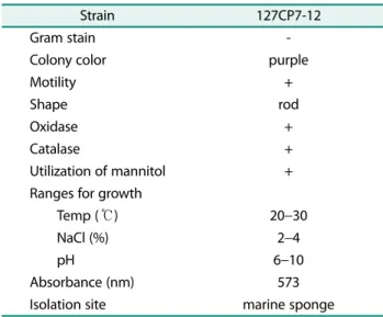
The characteristics of the bacterium isolated from the marine sponge H. sinapium are shown in Table 1. Sev- eral taxonomic characteristics of the pigment-producing strain were consistent with the bacterium Microbulbifer sp. reported in previous studies [21, 24]. Colonies form-
Table 1. Taxonomic characteristics of the pigment-producing Microbulbifer sp.
Strain 127CP7-12
Gram stain -
Colony color purple
Motility +
Shape rod
Oxidase +
Catalase +
Utilization of mannitol +
Ranges for growth
Temp ( ℃) 20 −30
NaCl (%) 2 −4
pH 6 −10
Absorbance (nm) 573
Isolation site marine sponge
ing on Marine Broth 2216 (MB; Difco, USA) are rodlike, smooth, raised, and dark purple in color. The isolated strain (127CP7-12) is kept in the bacterial culture collec- tion of the Laboratory of Microbial Taxonomy and Ecol- ogy, Hannam University.
Effect of pH on pigment production
Pigment production was found to be strongly depen- dent on the cell growth of the bacterium Microbulbifer sp., implying that the pigment production originates from the bacterial production. The bacterium showed the best growth performance around pH 6 and 7 with almost no growth at pH 4 and 5, indicating a relatively narrow optimal range of pH (Fig. 1). Above the optimum pH, growth gradually decreased until pH 10, the tested upper limit. Over all the tested pH range, pigment pro- duction was closely synchronized with bacterial growth.
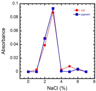
Effect of NaCl concentration on pigment production The peak harvest was shown at 3% NaCl and the intermediate harvest at 2% NaCl among the tested con- centrations from 0 to 7% NaCl (Fig. 2).
Effect of temperature on pigment production
For bacterial pigment production, a good harvest was shown in high temperature regimes (28 ℃ and 30℃) com- pared with a low temperature regime (20 ℃) with an
intermediate feature at 25 ℃ (Fig. 3). After 24 h of bacte- rial inoculation, pigment production was detected from all the temperature treatments and showed the first peak production at 30 h. An abrupt increase was only noted at the three high temperature treatments, while the lowest 20 ℃ treatment showed a gradual increase.
Effect of solvents on pigment extraction
Different solvents, namely distilled water (D.W.), eth- anol, methanol, acetone, and methanol:acetone (7:2, v/v), Fig. 1. Effect of pH on cell growth and pigment production
(Temperature: 25 ℃, NaCl concentration: 3%, Culturing time: 104 h).
Fig. 2. Effect of NaCl concentration on cell growth and pig- ment production (Temperature: 25 ℃, pH: 6, Culturing time: 60 h).
Fig. 3. Effect of temperature on pigment production in the
dark at 20, 25, 28, and 30 ℃ (pH 6).
were used to extract the pigment from the broth. The pigment was extracted with every type of organic sol- vent used in this study, indicating that the produced pig- ment is lipid-soluble. Although the extraction efficiencies were similar among the tested solvents, acetone showed the highest efficiency.
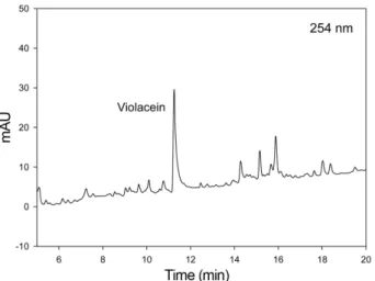
Analysis of the pigment by HPLC, NMR, MS, and UV The chemical profile of the extracted pigment mixture by LC/MS indicated the existence of a major compound (Fig. 4). The major pigment was further purified and analyzed by LC/MS to obtain its UV and MS spectra.
The UV spectrum of the purified compound showed λ
maxvalues at 210, 250, 360, and 570 nm (Fig. 5), which is consistent with those of violacein based on our in-house UV spectral library and the literature [25]. The molecu- lar mass of the major pigment was deduced to 343 on the
Fig. 4. HPLC chromatogram of extracted pigment acquired in LC/MS analysis (UV detection: 254 nm). Violacein was detected as a major compound at the retention time at 11.5 min
Fig. 5. Observed UV spectrum of the major pigment (127CP7- 12). UV spectrum was consistent with that of violacein
Fig. 6. ESI mass spectra (MSD1 in the positive ionization mode and MSD2 in the negative ionization mode) of the major pigment (127CP7-12). The mass spectral data also showed good agreement with the mass of violacein
Fig. 7.
1H NMR spectrum of the major pigment (127CP7-12)
in DMSO-d
6. The
1H NMR spectrum was consistent with that of
violacein
basis of electrospray ionization (ESI) low-resolution mass spectrometric data ([M+H]
+at m/z 344 and [M-H]
−at m/z 342) (Fig. 6). The
1H NMR spectrum showed that the pigment structure was coincident with that of viola- cein. The molecular mass also supported that the major pigment is violacein [25, 26]. For further unequivocal identification, the
1H NMR spectrum of the purified pig- ment was analyzed (Fig. 7). Based on the
1H NMR spec- trum, the pigment was clearly identified as violacein [25, 26]. The molecular structure of violacein is shown in Fig. 8.
Discussion
Violet pigment as bacterial production
The overall production level indicates that bacterial pigment production is optimal at high temperature;
however, the highest production peak was found at 25 ℃ with a gradual increase shown after the abrupt increase and first peak. Furthermore, the two highest and the lowest temperatures showed a similar level of pigment production at the end of the experiment, while the inter- mediate temperature showed a slightly higher final pro- duction. The relatively similar production levels imply that the pigment production is limited to the broth quan- tity available for bacterial growth. The overall patterns of pigment production such as peak appearance were closely synchronized and the final production level was also similar among all the temperature treatments. This result indicates that the pigment production depends on bacterial activities limited by the availability of organic resources in the natural environment. The optimal pro- duction in relatively high temperatures implies that the presented bacterial pigment production could work effec- tively in the warming ocean.
The major component in the extracted crude pigment was violacein, as supported by the experimental results of NMR, MS, and UV spectra analyses. To our best knowledge, this is the first report of violacein from a marine bacterium, Microbulbifer sp., associated with a marine sponge H. sinapium, which is native to Korea and other East Asian countries.
This optimum range of bacterial growth falls into the range from previous studies [24, 27] on the same bacte- rium genus Microbulbifer. It is also consistent with the narrow optimum range reported for the bacterium M.
elongates [27]. With a similar result for the pH effect, the close relationship between bacterial growth and pigment production indicates that this bacteria-pigment associa- tion is present in relatively stable environmental condi- tions such as those of biofilm and cellular fluid rather than in the changing conditions of the open ocean.
The specific production of violacein as a primary com- ponent of violet pigment, mg product per gram of cells, was not presented in this study and is beyond the scope to be discussed from the presented results; however, the production level can be inferred from the production of violet pigment as a proxy of violacein. In the study, pig- ment production was closely related with bacterial growth. The optimal incubation conditions were consis- tent with previous results using Microbulbifer species [24]. In a study on violacein production by a psychro- trophic bacterium, the optimal conditions for violet pig- ment production were reported as 20 ℃ and pH 6 and the maximum concentration and the productivity of violet pigment were 3.7 g/l and 0.12 g· l
-1· h
-1, respectively [26].
As reviewed by Choi et al. [10], there are many factors affecting the violacein production level, and these should be considered carefully in comparing various published values. During the present study, the preliminary yield of violet pigment was 0.25 g/l (unpublished) and further studies need to be performed to suggest both the maxi- mum concentration and the productivity of violacein from the suggested Microbulbifer strain.
Ecological implications of sponge-microbe association in a changing ocean
Many bioactive products are derived from microorgan- ism-engaged activities. In the marine environment, chemical defense mechanisms of attached organisms are prevalent and various microorganism-mediated pro- Fig. 8. Molecular structure of violacein (molecular mass =
343.34).
cesses occur in biofilm. This study reports the successful production of a bioactive pigment produced by a marine bacterium Microbulbifer sp., which was isolated from a marine sponge H. sinapium native to Asian coastal waters. The yield of the violet pigment was found to be dependent on bacterial growth after testing under vari- ous culturing conditions, thereby indicating that viola- cein is produced by the bacterium Microbulbifer sp. The main component of the pigment was identified as viola- cein, which has been reported as one of the potent antibi- otic materials from marine bacteria [10].
The sponge community has been considered as one of the important benthic fauna for indicating environmen- tal impacts such as climate changes. Sponge-microbe associations have been one of the most popular marine resources for obtaining natural products, and changing environmental conditions also could have a considerable effect on optimal bacterial activities and production as well as the ecological status of the sponge community [28]. Therefore, climate effect studies on well-known sponge-microbe interactions and latitudinal sponge dis- tribution can provide valuable insights for understand- ing how climate changes will affect ecosystem services that provide bioactive resources as well as primary marine production.
In the context of the issue of general ocean acidifica- tion (OA), the sponge community as one of the marine benthic communities has been reported as being affected by OA [29], and the impact of OA on marine microorgan- isms was also discussed in consideration of benthic organisms as host organisms [30]; however, the results of this study for the acidic optimum pH range of the bac- terium and its pigment harvest indicate that it is unlikely to be affected directly by recent OA concerns about global ocean conditions. Additionally, considering the usually acidic condition of the sponge cellular fluid, the experimental results on pH dependency indicate that this bacteria-pigment relationship could be rela- tively stable in the host sponge in the coming acidic ocean.
There are still increasing numbers of reports and prog- ress on natural products obtained from marine ecosys- tems. The identification of a bioactive material is not a novel approach in scientific communities; however, marine symbiotic microbial diversity is still not fully explored. Pharmaceutical metabolites from the marine
ecosystem are some of the most promising and challeng- ing subjects. As reviewed by Li [13], there are many use- ful cases of marine sponge-microbe related pharmaceutical metabolites from the South China Sea. Korean marine waters are one of the most affected and changing oceans in the world, providing increasing evidence of the north- ward range shift of tropical marine organisms. In this context, the present evidence of violacein production from a sponge-microbe association is not only one case report of many typical relations to be found in the Korean coastal ecosystem, but it is also a result that may be valuable for exploring marine-origin bioactive and pharmaceutical compounds in the changing ocean.
Acknowledgments
This work was partially supported by the 2016 Hannam University Research Fund, Korea and by the Ministry of Education, Korea through the fostering project of “Hannam University Industry-Academy Coop- eration Campus”. The author (N.-I.W) was also supported by the research project “Study on coastal marine ecosystem structures and suspended sediment in the EEZ sand mining zone” funded by K-water.
References
1. Halpern BS, Walbridge S, Selkoe KA, Kappel CV, Micheli F, D'Agrosa C, et al. 2008. A global map of human impact on marine ecosystems. Science 319: 948-952.
2. Belkin IM. 2009. Rapid warming of large marine ecosystems.
Progress in Oceanography 81: 207-213.
3. Poloczanska ES, Brown CJ, Sydeman WJ, Kiessling W, Schoeman DS, Moore PJ, et al. 2013. Global imprint of climate change on marine life. Nature Climate Change 3: 919-925.
4. Morán XAG, Alonso-Sáez L, Nogueira E, Ducklow HW, González N, López-Urrutia Á, et al. 2015. Presented at the Proc. R. Soc. B.
5. Barton AD, Irwin AJ, Finkel ZV, Stock CA. 2016. Anthropogenic cli- mate change drives shift and shuffle in North Atlantic phyto- plankton communities. Proceedings of the National Academy of Sciences 113: 2964-2969.
6. Dias DA, Urban S, Roessner U. 2012. A historical overview of natu- ral products in drug discovery. Metabolites 2: 303-336.
7. Harvey AL, Edrada-Ebel R, Quinn RJ. 2015. The re-emergence of natural products for drug discovery in the genomics era. Nature Reviews. Drug Discovery 14: 111-129.
8. D'Orazio N, Gammone MA, Gemello E, De Girolamo M, Cusenza S, Riccioni G. 2012. Marine bioactives: pharmacological proper- ties and potential applications against inflammatory diseases.
Marine Drugs 10: 812-833.
9. Skropeta D, Wei L. 2014. Recent advances in deep-sea natural
products. Natural Product Reports 31: 999-1025.
10. Choi SY, Kim S, Lyuck S, Kim SB, Mitchell RJ. 2015. High-level pro- duction of violacein by the newly isolated Duganella violacein- igra str. NI28 and its impact on Staphylococcus aureus. Scientific Reports 5: 15598.
11. Laport MS, Santos OC, Muricy G. 2009. Marine sponges: potential sources of new antimicrobial drugs. Curr. Pharm. Biotechnol. 10:
86-105.
12. El-Shitany NA, Shaala LA, Abbas AT, Abdel-Dayem UA, Azhar EI, Ali SS, et al. 2015. Evaluation of the anti-inflammatory, antioxi- dant and immunomodulatory effects of the organic extract of the red sea marine sponge xestospongia testudinaria against carrageenan induced rat paw inflammation. PLoS One 10:
e0138917.
13. Li Z. 2009. Advances in marine microbial symbionts in the china sea and related pharmaceutical metabolites. Marine Drugs 7:
113-129.
14. Matz C, Kjelleberg S. 2005. Off the hook--how bacteria survive protozoan grazing. Trends Microbiol. 13: 302-307.
15. Matz C, Webb JS, Schupp PJ, Phang SY, Penesyan A, Egan S, et al.
2008. Marine biofilm bacteria evade eukaryotic predation by tar- geted chemical defense. PLoS One 3: e2744.
16. Rusch DB, Halpern AL, Sutton G, Heidelberg KB, Williamson S, Yooseph S, et al. 2007. The Sorcerer II Global Ocean Sampling expedition: northwest Atlantic through eastern tropical Pacific.
PLoS Biol. 5: e77.
17. Seshadri R, Kravitz SA, Smarr L, Gilna P, Frazier M. 2007. CAMERA:
a community resource for metagenomics. PLoS Biology 5: e75.
18. Park MH, Sim CJ, Baek J, Min GS. 2007. Identification of genes suitable for DNA barcoding of morphologically indistinguishable Korean Halichondriidae sponges. Mol. Cells 23: 220-227.
19. Hoshino S, Saito DS, Fujita T. 2008. Contrasting genetic structure of two Pacific Hymeniacidon species. Hydrobiologia 603: 313- 326.
20. Fuller T, Hughey J. 2013. Molecular investigation of the invasive
sponge Hymeniacidon sinapium (de Laubenfels, 1930) in Elkhorn Slough, California. Aquatic Invasions 8: 59-66.
21. Jeong J-B, Park J-S. 2012. Seasonal differences of bacterial com- munities associated with the marine sponge, hymeniacidon sina- pium. The Korean J. Microbiol. 48: 262-269.
22. Yang LH, Xiong H, Lee OO, Qi SH, Qian PY. 2007. Effect of agita- tion on violacein production in Pseudoalteromonas luteoviolacea isolated from a marine sponge. Lett. Appl. Microbiol. 44: 625-630.
23. Ventosa A, Márquez MC, Ruiz-Berraquero F, Kocur M. 1990. Salini- coccus roseus gen. nov., sp. nov., a New Moderately Halophilic Gram-Positive Coccus. Syst. Appl. Microbiol. 13: 29-33.
24. Wakabayashi M, Sakatoku A, Noda F, Noda M, Tanaka D, Nakamura S. 2012. Isolation and characterization of Microbulbi- fer species 6532A degrading seaweed thalli to single cell detritus particles. Biodegradation 23: 93-105.
25. Rettori D, Durán N. 1998. Production, extraction and purifica- tionof violacein: an antibiotic pigment producedby Chromobac- terium violaceum. World J. Microbiol. Biotechnol. 14: 685-688.
26. Nakamura Y, Sawada T, Morita Y, Tamiya E. 2002. Isolation of a psychrotrophic bacterium from the organic residue of a water tank keeping rainbow trout and antibacterial effect of violet pig- ment produced from the strain. Biochem. Eng. J. 12: 79-86.
27. Yoon JH, Kim H, Kang KH, Oh TK, Park YH. 2003. Transfer of Pseu- domonas elongata Humm 1946 to the genus Microbulbifer as Microbulbifer elongatus comb. nov. Int. J. Syst. Evol. Microbiol. 53:
1357-1361.
28. Webster NS, Cobb RE, Negri AP. 2008. Temperature thresholds for bacterial symbiosis with a sponge. The ISME J. 2: 830-842.
29. Goodwin C, Rodolfo-Metalpa R, Picton B, Hall-Spencer JM. 2014.
Effects of ocean acidification on sponge communities. Mar. Ecol.
35: 41-49.
30. Das S, Mangwani N. 2015. Ocean acidification and marine micro-
organisms: responses and consequences. Oceanologia 57: 349-
361.
국문초록
한국 서해안에 서식하는 주황해변해면에서 분리된 해양세균 Microbulbifer sp.으로부터 생리활성물질 비올라세인의 규명 원남일
1, 이가은
2, 고기범
3, 오동찬
3, 나양호
4*, 박진숙
2*
1
한국수자원공사 K-water 연구원
2
한남대학교 생명시스템과학과
3
서울대학교 약학대학 천연물과학연구소
4