Parenchyma-preserving hepatectomy including segments I + IV resection and bile duct resection in a patient with type IV perihilar cholangiocarcinoma:
A case report with video clip
Shin Hwang
Department of Surgery, Asan Medical Center, University of Ulsan College of Medicine, Seoul, Korea
Case Report
It has been reported that parenchyma-preserving hepatectomy (PPH) might lower surgical curability with an increased likelihood of bile duct resection margins (BDRMs). Apparently, PPH is indicated for patients expected to achieve curative resection. The author herein presents a case of a 77-year-old male patient with type IV perihilar cholangiocarcinoma and decreased cardiac function treated with hepatic segments I + IV resection and bile duct resection. During the operation, he underwent two hepatic parenchymal transec- tions matched with right trisectionectomy and left hepatectomy. After removing segments VI and I and extrahepatic bile duct, six he- patic duct openings were exposed at the left and right hila. As some of them were conjoined, two hepaticojejunostomies at the right liv- er and one hepaticojejunostomy at the left lateral section were performed consecutively. This operation took 7 hours. Eight sessions of intraoperative frozen-section biopsy were performed. All BDRMs were tumor-negative. According to the 8th edition of the American Joint Committee on Cancer staging system, the extent of the tumor was pT2bN2M0. It was regarded as stage IVA tumor. The patient recovered uneventfully. He was discharged on the 18th postoperative day. The patient underwent concurrent chemoradiation therapy and adjuvant chemotherapy. The patient has been doing well without tumor recurrence for the past 24 months to date. In conclusion, PPH can lead to curative resection and improved outcomes through reasonable adjustment of the extent of hepatectomy.
Key Words: Parenchyma-preserving hepatectomy; Perihilar cholangiocarcinoma; Klatskin tumor; Bile duct resection margin; Hepatic failure
INTRODUCTION
It is generally accepted that aggressive surgical approaches with extended hepatectomy may result in an improved prog- nosis for patients with perihilar cholangiocarcinoma (PHCC) [1-5]. Extended hepatectomy can enhance surgical curability
by demonstrating tumor-negative bile duct resection margins (BDRMs) compared to local resection [5-7]. However, extended hepatectomy can increase postoperative morbidity and mor- tality because it often requires massive liver resection which is closely associated with post-hepatectomy liver failure (PHLF), hepatic decompensation, and secondary septic complications [8-10]. PHLF in patients with obstructive jaundice is dependent on the volume of the resected hepatic mass [8,9]. Thus, paren- chyma-preserving hepatectomy (PPH) can be considered in patients with high risk of PHLF [10]. It has been reported that PPH may lower surgical curability with an increased likelihood of tumor-positive BDRMs. Apparently, PPH is indicated only for patients expected to achieve curative resection. However, in real-world practice, the decision to determine surgical curabil- ity for PHCC is often difficult before surgery or even during surgery. Preoperative right portal vein embolization (PVE) for right hepatectomy or more extended hepatectomy can decrease
Received: December 29, 2020, Revised: January 6, 2021, Accepted: January 7, 2021
Corresponding author: Shin Hwang
Department of Surgery, Asan Medical Center, University of Ulsan College of Medicine, 88 Olympic-ro 43-gil, Songpa-gu, Seoul 05505, Korea
Tel: +82-2-3010-3930, Fax: +82-2-3010-6701, E-mail: [email protected] ORCID: https://orcid.org/0000-0002-9045-2531
Copyright Ⓒ The Korean Association of Hepato-Biliary-Pancreatic Surgery
This is an Open Access article distributed under the terms of the Creative Commons Attri- bution Non-Commercial License (http://creativecommons.org/licenses/by-nc/4.0) which permits unrestricted non-commercial use, distribution, and reproduction in any medium, provided the original work is properly cited.
Shin Hwang 420
the hepatic parenchymal resection rate [11,12]. However, PVE can also have a negative effect on surgical curability if it is not relevantly applied. When surgical curability is not determined in Bismuth-Corlette type IV PHCC, central hepatectomy in the form of PPH instead of PVE can lead to tailored expansion of hepatic resection to obtain tumor-free BDRMs as it can permit extended resection towards the left, right, or both sides of the liver. The author herein present a case of a patient with type IV PHCC treated with hepatic segments I+IV resection and bile duct resection (BDR). Detailed surgical procedures are present- ed in a Supplementary Video of 7 minutes.
CASE
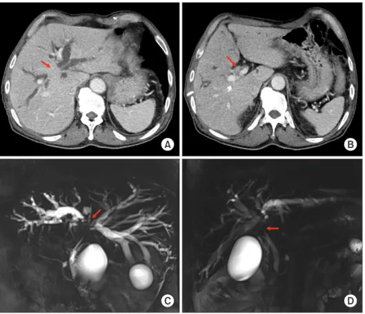
A 77-year-old male patient with obstructive jaundice was admitted under the diagnosis of Bismuth-Corlette type IV PHCC. The patient underwent percutaneous transhepatic bili- ary drainage at another hospital. Preoperative imaging studies revealed that the tumor was compatible with type IV PHCC without vascular invasion (Fig. 1). Preoperative workup re- vealed dysfunction of the left ventricle with deceased ejection fraction. Obstructive jaundice resolved slowly with repeated episodes of cholangitis despite multiple external biliary drain- ages. The operation was performed after biliary decompression with serum total bilirubin concentration of 3.0 mg/dL.
Considering the old age of this patient, two surgical plans were prepared regarding the extent of hepatectomy: If the left hepatic BDRM was tumor-positive, extended left hepatectomy with caudate lobectomy would be performed. If the left hepatic BDRM was tumor-negative, segments I + VI resection with or without additional hepatic resection would be performed
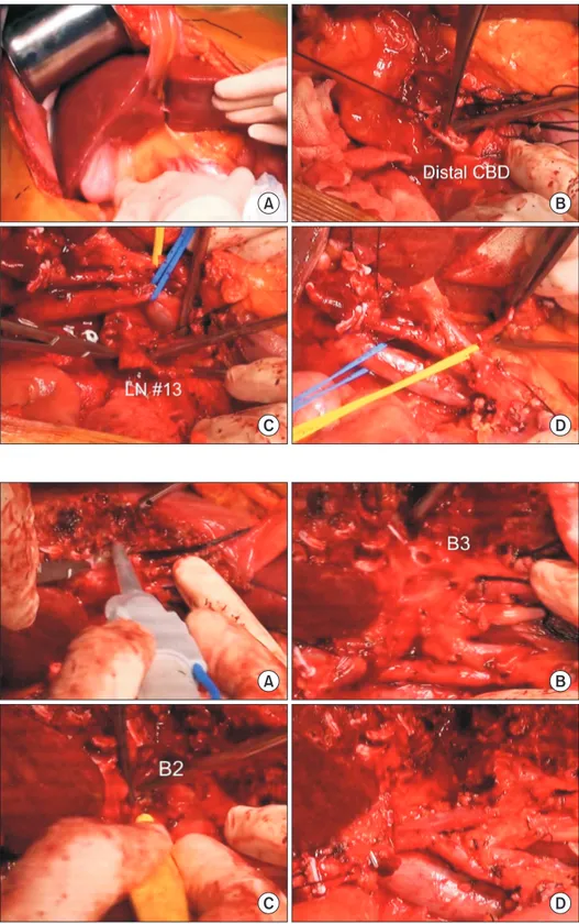
After laparotomy, surgical resectability was assessed by man- ual palpation (Fig. 2A). The distal bile duct was dissected first and transected to assess the status of tumor invasion. The dis- tal BDRM was found to be tumor-free (Fig. 2B). Enlarged ret- ropancreatic lymph nodes (LNs) were dissected (Fig. 2C), and intraoperative frozen-section biopsy showed nodal metastasis.
Dissection was continued towards the hepatic hilum. The he- patic artery and portal vein branches were successfully isolated (Fig. 2D), indicating the absence of hilar vascular invasion.
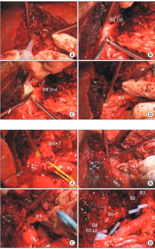
The liver parenchyma was transected along the falciform ligament (Fig. 3A). Segment III duct (B3) and segment II duct (B2) were consecutively transected (Fig. 3B, 3C), in which their BDRMs were tumor-negative. The left portal vein and the hepatic artery were further dissected to expose the umbilical portion of the left portal vein. Hepatic transection continued towards the dorsal part of the left caudate lobe and the rem- nant left lateral segment was completely separated from the left caudate lobe (Fig. 3D).
The right-sided hepatic parenchymal transection was per-
Fig. 1. Preoperative imaging study findings.
Computed tomography images (A, B) and magnetic resonance cholangiography images (C, D) show extensive hilar bile duct obstruction, suggesting perihilar cholangiocarcinoma of Bismuth-Corlette type IV. Arrows indicate tumor.
A B
C D
formed along the hemi-liver discoloration line (Fig. 4A). At the right hepatic hilum, two segment V ducts (B5s) were transected (Fig. 4B, 4C), which were found to be tumor-negative. The pus drained from B5s was collected for bacterial culture. Consecu-
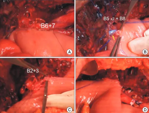
tively, segment VIII duct (B8) was transected (Fig. 4D), which was also found to be tumor-negative. The right posterior duct, which was a conjoined portion of segment VI and VII ducts (B6 + 7), was transected (Fig. 5A), and found to be tumor-negative.
Fig. 2. Intraoperative photographs showing dissection of the hepatoduodenal ligament.
Surgical resectability is assessed through manual palpation (A). The distal common bile duct (CBD) is transected (B). Enlarged regional lymph nodes (LNs) are dissected (C). Intraoperative frozen-section biopsy showed nodal metastasis. The hepatic artery and portal vein branches are isolated without macroscopic vascular invasion (D).
A B
C D
A B
C D
Fig. 3. Intraoperative photographs showing left liver transection. The liver parenchyma is transected along the falciform ligament (A). The segment III duct (B3) and segment II duct (B2) are consecutively transected (B, C). The left portal vein and hepatic artery are further dissected to expose the umbilical portion of the left portal vein, and the remnant left lateral segment is separated from the left caudate lobe (D).
Shin Hwang 422
The paracaval portion and the left caudate lobe were removed along with segment IV resection (Fig. 5B, 5C). Considering that all left and right hepatic BDRMs were tumor negative and retropancreatic LNs were metastatic, the extent of resection
was thought to be sufficient for this patient.
There were six hepatic duct openings at the left and right hila (Fig. 5D). A redundant Roux-Y jejunal limb was prepared.
First, the B6 + 7 was reconstructed through a single hepaticoje- Fig. 4. Intraoperative photographs showing right liver transection. Right-sided hepatic parenchymal transection is performed along the hemi-liver discoloration line (A). Two segment V ducts (B5) are transected (B, C).
Segment VIII duct (B8) is transected (D).
A B
C D
Fig. 5. Intraoperative photographs showing right liver transection. Conjoined segments VI and VII duct (B6 + 7) is transected (A). The paracaval portion and the left caudate lobe (S1) are removed along with segment IV (S4) resection (B, C). There are six hepatic duct openings at the left and right hila (D).
A B
C D
junostomy (HJ) (Fig. 6A) because it was located at the deepest portion. Two B5 and one B8 were conjoined with 5-0 monofila- ment sutures to make a single opening, and then single HJ was performed (Fig. 6B). Thereafter, B2 and B3 were conjoined and a single HJ was performed (Fig. 6C, 6D). Consequently, biliary reconstruction included two HJs at the right liver and one HJ at the left lateral section. This operation took 7 hours. Eight sessions of intraoperative frozen-section biopsy (6 for DBRMs and two for LNs) were performed. Detailed surgical procedures are presented in a Supplementary Video of 7 minutes.
The pathology report revealed that the tumor was cholan- giocarcinoma measuring 3.8 cm in size; moderately differen- tiated with extension beyond the bile duct (depth of invasion was 7 mm from the surface epithelia) and involvement of the hepatic parenchyma; the presence of lymphovascular invasion and perineural invasion, and involvement of the distal and B3 BDRMs by high-grade biliary intraepithelial neoplasia. Me- tastasis was present in 5 of 22 LNs (LN #12, 2/6; LN #8 and 13, 2/10; and pericholangiotic LN, 1/6) (Fig. 7). According to the 8th edition of the American Joint Committee on Cancer stag- ing system, the extent of the tumor was pT2bN2M0. Thus, it was regarded as stage IVA tumor.
The patient recovered uneventfully. He was discharged from the hospital on postoperative day 18 (Fig. 8A, 8B). Because of high risk of tumor recurrence, the patient underwent concur- rent chemoradiation therapy and adjuvant chemotherapy with oral uracil-tegafur plus leucovorin. No surgical complications occurred during follow-up to date (Fig. 8C, 8D). The patient
has been doing well without any evidence of tumor recurrence for the past 24 months after the operation.
DISCUSSION
It is well known that curative resection is of great prognos- tic significance after surgical resection for PHCC [5,13]. To
Fig. 6. Intraoperative photographs showing biliary reconstruction. Segments VI and VII duct (B6 + 7) is reconstructed through a single hepaticojejunostomy (HJ) (A). Two segment V ducts (B5) and one segment VIII duct (B8) are conjoined and a single HJ is performed with stent insertion into the small B5 duct (arrow) (B). Segment II and III ducts (B2 + 3) are conjoined and a single HJ is performed (C, D).
A B
C D
Fig. 7. Gross photograph of the resected specimen with segments VI and I and the extrahepatic bile duct.
Shin Hwang 424
achieve curative resection for PHCC, extended hepatic resec- tion is usually required. However, extended hepatectomy in patients with obstructive jaundice can lead to high surgical morbidity and mortality, even after sufficient biliary decom- pression. It has been suggested that surgical morbidity and mortality after hepatic resection are dependent on the volume of the resected hepatic mass in patients with obstructive jaun- dice [8,9,14]. To avoid PHLF following extended hepatectomy, preoperative PVE has been performed to decrease the paren- chymal resection rate [11,12].
It has been hypothesized that if hepatic resection is limited as much as possible to what is necessary for curative resection, fewer postoperative complications in patients with PHCC might occur. It has been advocated to perform customized limited hepatic resection according to individual tumor extent of each patient [4]. Because segment I resection is known to be a requisite for curative resection of PHCC [15], resection of the entire segment I could be the most limited hepatectomy in surgical resection for PHCC [16,17]. Limited hepatic resection of segments I and IV as PPH for PHCC in which the extent of cancer is localized around the confluence of bilateral hepatic ducts has been previously demonstrated [18].
The clinical implications of PPH for PHCC has not been ad- dressed or clarified yet. It has been suggested that PPH may de- crease the rate of curative surgical resection. However, several studies have revealed that surgical curability is not impaired by
PPH. On the other hand, PPH could elicit favorable prognosis after surgical resection similar to that of extended hepatecto- my [10,19]. Thus, PPH is known to have beneficial effects on surgical morbidity and mortality as an operative procedure for PHCC [10].
However, it has also been reported that PPH is unsuitable as an operative procedure to obtain curative resection for the majority of patients with PHCC. A Japanese study has revealed that only 15% of patients could be selected to undergo PPH as a limited hepatectomy procedure to obtain curative resection [10]. PPH might have beneficial effects for patients with PHCC extending only into bile duct branches of segments I and IV without lymph nodal involvement. It might also have beneficial effects for those who do not require vascular resection, espe- cially for patients with high operative risk.
In contrast with the conventional concept of PPH, we have also performed PPH in patients with more advanced PHCCs, as shown in the present case with type IV tumor, especially when right or left hemihepatectomy might not ensure surgical curability. For type IV PHCC, left or right trisectionectomy may provide the highest surgical curability. However, its sur- gical indication is usually very limited even after preoperative PVE. In real-world practice, irrelevant right PVE can result in R1 resection if the tumor is involved deeply at the left he- patic duct and if right trisectionectomy is not permitted. On the contrary, customized central hepatectomy in the form of Fig. 8. Postoperative computed tomography findings. Images taken 10 days (A, B) and 18 months (C, D) after surgery show no abnormal findings.
A B
C D
PPH can provide a chance to expand the extent of resection to obtain tumor-free BDRMs because it can permit extended re- section towards the left, right, or both sides of the liver. Based on our experience, the extent of PPH for PHCC ranges from segment I+IV resection to central bisectionectomy + segment I resection, depending on the extent of tumor involvement.
However, PPH requires more complex surgical procedures than extended hepatectomy. It is mandatory to perform mul- tiple bilio-enteric anastomoses after PPH. Thorough preoper- ative anatomic recognition, accurate evaluation of the cancer extent, and skilled surgical techniques for meticulous hepatec- tomy are required to perform PPH procedures [10].
In conclusion, PPH can lead to curative resection and im- proved outcomes of patients with PHCC localized at the hepat- ic duct confluence. PPH may also provide beneficial effects for selected patients according to the extent of the tumor and for patients with high operative risk.
SUPPLEMENTARY DATA
Supplementary data related to this article can be found at https://doi.org/10.14701/ahbps.2021.25.3.419.
CONFLICT OF INTEREST
No potential conflict of interest relevant to this article was reported.
REFERENCES
1. Baer HU, Stain SC, Dennison AR, Eggers B, Blumgart LH. Improve- ments in survival by aggressive resections of hilar cholangiocarcino- ma. Ann Surg 1993;217:20-27.
2. Childs T, Hart M. Aggressive surgical therapy for Klatskin tumors.
Am J Surg 1993;165:554-557.
3. Tsuzuki T, Ueda M, Kuramochi S, Iida S, Takahashi S, Iri H. Carci- noma of the main hepatic duct junction: indications, operative mor- bidity and mortality, and long-term survival. Surgery 1990;108:495- 501.
4. Nimura Y, Hayakawa N, Kamiya J, Kondo S, Shionoya S. Hepatic seg- mentectomy with caudate lobe resection for bile duct carcinoma of the hepatic hilus. World J Surg 1990;14:535-543; discussion 544.
5. Miyazaki M, Ito H, Nakagawa K, Ambiru S, Shimizu H, Shimizu Y, et al. Aggressive surgical approaches to hilar cholangiocarcinoma:
hepatic or local resection? Surgery 1998;123:131-136.
6. Iida S, Tsuzuki T, Ogata Y, Yoneyama K, Iri H, Watanabe K. The long-term survival of patients with carcinoma of the main hepatic duct junction. Cancer 1987;60:1612-1619.
7. Hayashi S, Miyazaki M, Kondo Y, Nakajima N. Invasive growth pat- terns of hepatic hilar ductal carcinoma. A histologic analysis of 18 surgical cases. Cancer 1994;73:2922-2929.
8. Blumgart LH, Hadjis NS, Benjamin IS, Beazley R. Surgical approach- es to cholangiocarcinoma at confluence of hepatic ducts. Lancet 1984;1:66-70.
9. Miyazaki M, Itoh H, Ambiru S, Shimizu H, Togawa A, Gohchi E, et al. Radical surgery for advanced gallbladder carcinoma. Br J Surg 1996;83:478-481.
10. Miyazaki M, Ito H, Nakagawa K, Ambiru S, Shimizu H, Okaya T, et al. Parenchyma-preserving hepatectomy in the surgical treatment of hilar cholangiocarcinoma. J Am Coll Surg 1999;189:575-583.
11. Lee SG, Hwang S. How I do it: assessment of hepatic functional re- serve for indication of hepatic resection. J Hepatobiliary Pancreat Surg 2005;12:38-43.
12. Hwang S, Ha TY, Ko GY, Kwon DI, Song GW, Jung DH, et al. Preop- erative sequential portal and hepatic vein embolization in patients with hepatobiliary malignancy. World J Surg 2015;39:2990-2998.
13. Klempnauer J, Ridder GJ, Werner M, Weimann A, Pichlmayr R.
What constitutes long-term survival after surgery for hilar cholan- giocarcinoma? Cancer 1997;79:26-34.
14. Klempnauer J, Ridder GJ, von Wasielewski R, Werner M, Weimann A, Pichlmayr R. Resectional surgery of hilar cholangiocarcinoma: a multivariate analysis of prognostic factors. J Clin Oncol 1997;15:947- 954.
15. Mizumoto R, Kawarada Y, Suzuki H. Surgical treatment of hilar car- cinoma of the bile duct. Surg Gynecol Obstet 1986;162:153-158.
16. Hwang S, Ha TY, Kim JS, Kim KH, Lee SG, Lee YJ, et al. Isolated cau- date lobectomy with bile duct resection performed in a patient with type IV hilar bile duct cancer. J Korean Surg Soc 2003;64:441-446.
17. Hwang S, Moon DB, Park EH, Kim MH, Lee YJ, Lee SG. S4a+S5 with caudate lobe (S1) resection as a parenchyma-preserving liver resec- tion for a patient with type IIIb hilar bile duct cance. J Korean Surg Soc 2003;64:515-520.
18. Miyazaki M, Ito H, Nakagawa K, Ambiru S, Shimizu H, Shimizu Y, et al. Segments I and IV resection as a new approach for hepatic hilar cholangiocarcinoma. Am J Surg 1998;175:229-231.
19. Lee SG, Song GW, Hwang S, Ha TY, Moon DB, Jung DH, et al. Sur- gical treatment of hilar cholangiocarcinoma in the new era: the Asan experience. J Hepatobiliary Pancreat Sci 2010;17:476-489.