Copyrights © 2015 The Korean Society of Radiology
423
Case Report
pISSN 1738-2637 / eISSN 2288-2928 J Korean Soc Radiol 2015;72(6):423-426 http://dx.doi.org/10.3348/jksr.2015.72.6.423
INTRODUCTION
Encephaloceles consist of herniated cerebral tissue through the skull or dural defect (1). Encephaloceles may develop spon- taneously as congenital maldevelopments or may occur subse- quent to acquired processes, such as infection, trauma, neopla- sms, and surgical procedures. The incidence of this malformat- ion has been estimated at one in every 3000 to 10000 live births (2). These lesions are a rare complication of skull fractures and rarely occur in adults. They can be difficult to distinguish from congenital encephaloceles in patients with no history of head trauma.
We report a rare case of parietal intradiploic encephalocele incidentally found on computed tomography (CT) and magnet- ic resonance imaging (MRI). Cerebral angiographic imaging was useful to define the abnormal vascular lesion, such as arteriove- nous malformation, arteriovenous fistula, and hyper- or hypo- vascular tumor.
CASE REPORT
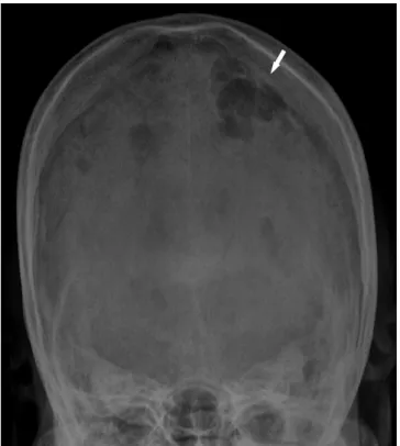
A previously healthy 50-year-old man presented with inter- mittent headache for the past 6 months. He had no history of head trauma and surgery. The patient was alert and had normal vital signs. The neurological and general physical examinations were normal. Results of routine laboratory blood tests includ- ing C-reactive protein and erythrocyte sedimentation rate were within their normal ranges. Tumor markers were negative. Skull radiography showed osteolytic changes involving the left pari- etal bone (Fig. 1). CT demonstrated a defect of the inner table, widened diploic space with outer table bony erosion, and thin- ning of the left parietal parasagittal bone (Fig. 2). MRI revealed a lesion in the parietal parasagittal intradiploic space continuous with the left parietal lobe appearing isointense to the normal br- ain on the axial T2-weighted image (Fig. 3A). Sagittal T2-wei- ghted MRI showed a defect in the left parietal bone, which con- tained cerebrospinal fluid and herniated cerebral tissue (Fig. 3B).
Intradiploic Encephalocele of the Left Parietal Bone: A Case Report
1 좌측 두정골의 판간내 뇌류: 증례 보고1Hyung-Seok Kim, MD
1, Choon-Woong Huh, MD
1, Dal-Soo Kim, MD
1, Jin-Ho Mok, MD
1, In-Soo Kim, MD
1, Geun-Seok Yang, MD
2Departments of 1Neurosurgery, 2Radiology, Myongji St. Mary’s Hospital, Seoul, Korea
Encephaloceles are generally regarded as midline abnormalities. A 50-year-old man presented with a parietal intradiploic encephalocele manifesting as intermittent headache for the past 6 months. Computed tomography (CT) showed bone destruc- tion associated with a left parietal lesion. Magnetic resonance imaging (MRI) dem- onstrated brain herniation within the intradiploic space. Cerebral angiographic im- aging showed a normal cerebral vessel pattern within the herniated brain lesion. In this case, surgical treatment may not be necessary in the absence of concurrent symptoms and neurologic deficit. We report the CT, MRI, and angiographic findings of an extremely rare case of parietal intradiploic encephalocele in adulthood.
Index terms
Parietal Intradiploic Encephalocele Computed Tomography
Magnetic Resonance Imaging Cerebral Angiographic Imaging
Received December 15, 2014 Accepted April 11, 2015
Corresponding author: Hyung-Seok Kim, MD
Department of Neurosurgery, Myongji St. Mary’s Hospital, 156 Dorim-ro, Yeongdeungpo-gu, Seoul 150-814, Korea.
Tel. 82-2-829-7831 Fax. 82-2-829-7888 E-mail: [email protected]
This is an Open Access article distributed under the terms of the Creative Commons Attribution Non-Commercial License (http://creativecommons.org/licenses/by-nc/3.0) which permits unrestricted non-commercial use, distri- bution, and reproduction in any medium, provided the original work is properly cited.
424
Intradiploic Encephalocele of the Left Parietal Bone
jksronline.org
J Korean Soc Radiol 2015;72(6):423-426 We performed transfemoral cerebral angiography to identify
vascular abnormalities. A baseline control angiogram of the left internal carotid artery showed a normal cerebral vessel pattern for the herniated brain in the left parietal bony defect area and
no vascular abnormalities, such as arteriovenous malformation, arterioveous fistula, hemangioma, and hyper- or hypo-vascular tumor (Fig. 4). After the headache was resolved symptomatically with medical therapy, the patient was discharged.
Fig. 1. Skull film: town view shows the left parietal defect (white ar- row).
Fig. 2. Coronal image of bone window CT shows a defect of the inner table, widened diploic space (black arrow). There is marked thinning and erosion of the outer table of the left parietal parasagittal bone (white arrow).
Fig. 3. MRI shows a lesion in the parietal parasagittal intradiploic space continuous with the left parietal lobe appearing isointense with the nor- mal brain on axial T2-weighted (A) image. Sagittal T2-weighted MRI shows a defect in the left parietal bone, which contains cerebrospinal fluid and herniated cerebral tissue (white arrow) (B).
A B
425
Hyung-Seok Kim, et al
jksronline.org J Korean Soc Radiol 2015;72(6):423-426
DISCUSSION
A cephalocele is defined as a protrusion of intracranial con- tents through a defect in the skull or dura. The herniating neural tissue may include meninges, brain parenchyma, ventricles, and vascular structures. Encephaloceles consist of herniated cere- bral tissue through the skull or dural defect, and are generally re- garded as midline cerebral abnormalities (3).
During the 8th week of development, the two parietal bones undergo membranous ossification from two primary centers for each bone. At 4 months, the fusion between these centers is com- plete. At birth, the parietal bones are unilaminar and are sepa- rated by the sagittal suture. At the age of 4 years, differentiation between the inner and outer tables is evident, and the ossification of the sagittal suture begins at the same time for the two layers.
A complete fusion takes place by the age of 20 years. Defects oc- curring as the sagittal suture closes allow the herniation of the brain, but they involve both tables (4). Therefore, these sponta- neous lesions usually occur at the site of a cranial suture, and most of these lesions represent primary or secondary midline closure defects of the neural tube. Parietal cephaloceles are very rare (10% of cephaloceles), and if they are congenital, they are usually associated with anomalies, such as corpus callosum agen- esis and Dandy-Walker malformation (5).
Intradiploic encephaloceles are also very rare, with only a few
cases having been reported with diagnosis based on surgical bi- opsy (1, 3). Paters et al. (5) and Dobrin et al. (6) reported a glob- al defect of both inner and outer tables in all cases of congenital parietal encephaloceles. This unusual lesion resembles the mech- anism of a growing skull fracture (6). Although the majority of reported intradiploic encephaloceles are of traumatic origin, several reports found no certain cause for these lesions. In this lesion, increased intracranial pressure results in further hernia- tion of the involved site, which can cause symptoms that in- clude headache, seizure, motor weakness, and aphasia (7). The differential diagnosis includes head trauma, multiple myelo- mas, dermoids, epidermoids, metastatic bone tumor, arachnoid cyst, eosinophilic granuloma, and brain tumor (3, 8). CT, MRI, and cerebral angiography have been used in the evaluation of encephaloceles. Surgery is indicated for both diagnosis and treatment of symptomatic encephaloceles (3). However, the sur- gical treatment for asymptomatic incidental lesions remains controversial. In the present case, we considered that maybe the lesion was an acquired lesion following an insignificant head trauma in early childhood because congenital encephaloceles usually occur in the midline and extend through both the inner and outer tables, and there were no other malformations (5).
In conclusion, we report a rare case of intradiploic encepha- locele in the left parietal lobe incidentally discovered on CT and MRI. Cerebral angiography was useful in the differential diag- Fig. 4. Left internal carotid artery angiogram with arterial (A) and venous (B) phase films show normal cerebral vessel pattern for herniated brain in left parietal bony defect area (black arrowheads).
A B
426
Intradiploic Encephalocele of the Left Parietal Bone
jksronline.org
J Korean Soc Radiol 2015;72(6):423-426 nosis of an abnormal vascular lesion.
REFERENCES
1. Kosnik EJ, Meagher JN, Quenemoen LR. Parietal intradiploic encephalocele. Case report. J Neurosurg 1976;44:617-619 2. Mealey J Jr, Dzenitis AJ, Hockey AA. The prognosis of en-
cephaloceles. J Neurosurg 1970;32:209-218
3. Tsuboi Y, Hayashi N, Noguchi K, Kurimoto M, Nagai S, Endo S. Parietal intradiploic encephalocele--case report. Neurol Med Chir (Tokyo) 2007;47:240-241
4. Sadler TW. Langman’s medical embryology, 10th ed. Balti-
more: Lippincott Williams & Wilkins, 2006:126-143, 286-317 5. Peters J, Raab P, Marquardt G, Zanella FE. Intradiploic me-
ningoencephalocele. Eur Radiol 2002;12 Suppl 3:S25-S27 6. Dobrin N, Ba˘linis¸teanu M, Costa˘chescu B, Tudorache C, Chi-
riac A, Poeata˘ I. Acquired parietal intradiploic encephalocele.
Case report and review of the literature. Rom S Neurosurg 2011;18
7. Loumiotis I, Jones L, Diehn F, Lanzino G. Symptomatic left intradiploic encephalocele. Neurology 2010;75:1027 8. Kumar R, Chandra SP, Sharma BS. Giant intradiploic pseu-
domeningocele of occipital bone. J Neurosurg Pediatr 2012;
9:82-85
좌측 두정골의 판간내 뇌류: 증례 보고1
김형석
1· 허춘웅
1· 김달수
1· 목진호
1· 김인수
1· 양근석
2뇌류는 일반적으로 정중선을 따라 생기는 기형이다. 6개월 동안 지속되는 간헐적인 두통증상을 호소하던 50세 남자 환자 는 좌측 두정골의 판간내 뇌류가 발견되었다. 전산화단층촬영 소견상 좌측 두정골의 골 파괴 소견이 있고, 자기공명영상 소견상 두정골의 판간 공간에 뇌실질 탈출소견이 있었다. 뇌혈관조영술 소견상 탈출된 뇌의 혈관은 정상 뇌혈관 소견을 보 였다. 이 환자는 상기 병변과 일치된 증상과 신경학적 손상이 없었기 때문에 수술적인 치료는 필요하지 않았다. 저자들은 성인 두정부위에 발생된 판간내 뇌류의 매우 드문 증례를 전산화단층촬영, 자기공명영상, 혈관조영술 소견을 중심으로 소 개하고자 한다.
명지성모병원 1신경외과, 2영상의학과