Gender pride in male may greatly be attributable to the penis or testicles. The preservation of these organs may be the utmost priority for a male patient, even while faced with other life threatening conditions. To date, despite such importance, there has not been a single suitable substitute for the penile or urethral component.1,2 The best alternative for reconstructive surgeons to consider have included abdominal tubed flap, gracilis myocutaneous flap, scrotal flap, tubed groin flap, skin flap with split or full thickness skin graft, bladder flap, anterolateral thigh flap, radial and ulnar forearm free flap, and etc.1-8 Recent advancements in surgical techniques and the emergence of microsurgery have introduced such a variety of choices;3 however, complicated and multi-staged operations are still unacceptable to some patients, who are traumatized by such condition. The authors used a conventional groin flap
method, and have extended its capability to reconstruct a long penile urethra along with ventral penis.
CASE REPORT
Case (Fig. 1-6)
A 45-year-old male was admitted to the urology department of a local hospital to remove the paraffin self-injected on penile body and scrotum 20 years ago. During the foreign body excision, an iatrogenic penile urethral injury had put caused multiple complications and numerous reconstructive surgeries for this patient. Collaborating with the plastic surgery department, the patient has undergone several attempts of conventional surgeries and unsuccessful outcomes. Such conventional surgical methods included simple urethral repair
Reconstruction of Penile and Long Urethral Defect Using a Groin Flap
So-Min Hwang*, On Lim, Hyung-Do Kim, Dong-Gil Shin1
Department of Plastic and Reconstructive Surgery, Good Moonhwa Hospital, 1Department of Urology, Pusan National University Hospital, Busan, Korea
CC This is an open-access article distributed under the terms of the Creative Commons Attribution Non-Commercial License (http://creativecommons.org/licenses/by-nc/4.0) which permits unrestricted noncommercial use, distribution, and reproduction in any medium, provided the original work is properly cited.
Copyright © 2016 by the Korean Society for Microsurgery. All Rights Reserved.
Received November 29, 2015 Revised March 8, 2016 Accepted April 24, 2016
*Correspondence to: So-Min Hwang Department of Plastic and Reconstructive Surgery, Good Moonhwa Hospital, 119 Beomil-ro, Dong-gu, Busan 48735, Korea Tel: +82-51-630-0198
Fax: +82-51-630-0145 E-mail: [email protected]
Financial support: None.
Conflict of interest: None.
Urethral reconstruction is a problematic issue, thus its management can be challenging.
Different methods using various materials were introduced for urethral reconstruction.
The authors have made some changes in the groin flap surgery, affording more successful urethral reconstruction for defects of long urethra and penile soft tissue. A 45-year-old male requested both functional and cosmetic reconstruction of his defected penis, caused by an iatrogenic urethral injury and chronic infection following removal of paraffin self- injected on the penile shaft. The defect affected the full length of the penile urethra, corpus spongiosum, and prepuce. A groin flap was designed, measuring 28×10 cm. The most distal flap was utilized for the construction of the luminal surface of the neourethra;
relaxed length measuring 8 cm, and the lumen wide enough. Competent external meatus and neourethra was confirmed by retrograde cystogram and the patient voided with sufficient urine caliber up to 2 years follow-up. This operative technique has advantages.
Donor sites have non-hair bearing skin for the neourethra and minimal or almost not- recognizable donor site morbidity. After surgery, the patient was relieved from voiding difficulties combined with psychological stress. The author would like to introduce a unique approach for the urethral and ventral phalloplasty using the groin flap.
Key Words: Urethra, Genitalia, Reconstruction, Groin, Flap
ARMS
Archieves of Reconstructive Microsurgery http://dx.doi.org/10.15596/ARMS.2016.25.1.19under microscopic exploration, a urethral reconstruction using split thickness skin graft, and again by full thickness skin graft and even the scrotal flap surgery.
By the time of the referral to Department of Aethetic Plastic and Reconstructive Surgery of Center at Good Moonhwa Hospital, several months have already passed, and the degree of serious infectious condition alongside the extensive penile necrosis required an exceptional choice of partial phalloplasty and long urethral reconstruction.
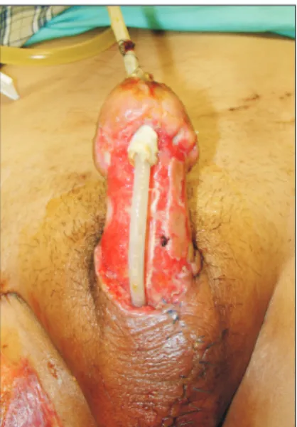
The extent of the involved lesion presented nearly total necrosis of prepus, ventral penis, cavernous spongiosum, and penile urethra (Fig. 1). The patient had sustained an indwelling Foley catheter during the treatment period period—
several months—at the previous hospital, which caused an ascending infection to the bladder, functioning as a depository for continuous infectious source, in this case, the nosocomial infection of Pseudomonas aerosinosa.
The state of the patient’s general condition was pathetic.
The blood test revealed mild leukocytopenia and elevated liver profiles possibly from chronic use of cephalosporin antibiotics.
Several months of wasteful improvident medical attention has created the patient with a trend of mutual distrust regarding medicine in general. Psychologic trauma of a missing penile function and persisting long term stress condition lead to a serious depressive mood, with occasional suicidal tendencies.
The authors have attempted to focus not only on the technical aspect but also on the functional result and the reliability of its outcome in choosing the method of urethral and penile reconstruction.
Surgical technique
The authors have discovered a new technique of long penile urethral reconstruction with concurrent ventral phalloplasty using a groin flap.
Before surgery, branching point of the superficial circumflex iliac artery was traced with a Doppler (Bi-directional Doppler ES-100V3; Hadeco, Kawasaki, Japan) probe and marked on the skin. Spinal block and concurrent intravenous sedation allowed for a safe and comforting operative environment for the patient and surgery team. Under an exaggerated lithotomy position, suprapubic cystostomy was performed to diversify the infectious source, which also resolved the aggravated voiding difficulties.
Fig. 1. Initial wound, nearly total necrosis of prepus, ventral penis, cavernous spongiosum and penile urethra.
A B
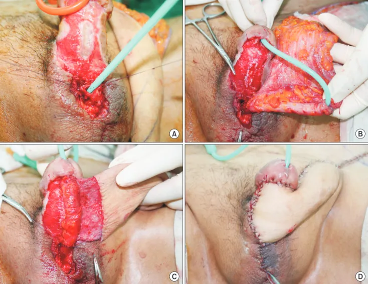
Fig. 2. (A) Dimension of the groin skin flap was 28×10 cm. (B) Neourethra reconstruction. By encircling the distal flap over 12 Fr. Foley catheter, neourethra was created and measured 8 cm at relaxed state.
A limited mid-line incision was made on the ventral surface of the penile root and scrotum. Skin flap was carefully elevated at the sub-Dartos fascia plane. The root of the penile urethra, surrounded by the infectious granulation tissue, was exposed and the urethral mucosal layer was confirmed by removing the previously indwelling Foley catheter (Fig. 3A).
The penile urethra surrounded by the corpus spongiosum beneath the Buck’s fascia from the penile root to the glans tip was completely vanished, leaving the ventral penis exposed directly to the intercavernous septum of Buck’s fascia. The total length of the urethral defect was the entire penile division, 10 cm at the relaxed state, including the glans.
To utilize the groin flap, an oblique elliptical flap design was made, 11 cm beyond the anterior superior iliac spine by the adequate width of 10 cm. The flap dimension measured
28×10 cm (Fig. 2A). The most distal flap was elevated as a thin skin flap, enough for a neourethral reconstruction. In order to preserve the superior circumflex iliac artery as the supplying pedicle to the flap, the groin flap was elevated thicker as the dissection proceeded medially, down to the level of the deep fascia. The donor site was primarily closed.
The utmost lateral skin flap was encircled over the 12 Fr Foley catheter to construct a long neourethra (Fig. 2B). The luminal surface of the neourethra, lined with epithelial layer was closed with 4-0 Chromic, followed by 5-0 PDS in multi-layer water tight fashion. Distal groin flap now consists of the neourethra and adjacent deepithelized portion. Followed by anastomosis of the neourethra to the membranous urethra proximally, and to the glans base distally, a continuity was maintained by an insertion of the Release-NF® Foley catheter (Rochester Medical
A B
C D
Fig. 3. Anastomosis of neourethra to the normal membranous urethra (A, B) and glans base (C). (D) Immediate postoperative clinical photo.
Co., Stewartville, MN, USA) (Fig. 3).
De-epithelized portion, just proximal to the neourethra, can be used for ventral phalloplasty, and at the same time, supply reliable blood flow to the neourethra, offering more secure prognosis. Proximal epithelial lining was advanced and
sutured to the lateral penile skin. Remaining proximal flap was initially tubed as a delay procedure. As the first stage is concluded, the patient was able to wear normal size underwear, permit orthostatic micturition through the neourethra after removal of the Foley catheter at postoperative 3 weeks (Fig. 4).
Most importantly, location of the donor site allowed minimal disturbance to daily life. Nearly full recovery with normal voiding function allowed rapid release from the psychologic depression as well. This surgery is completed by an office-based flap division operation at 3 months after the 1st procedure.
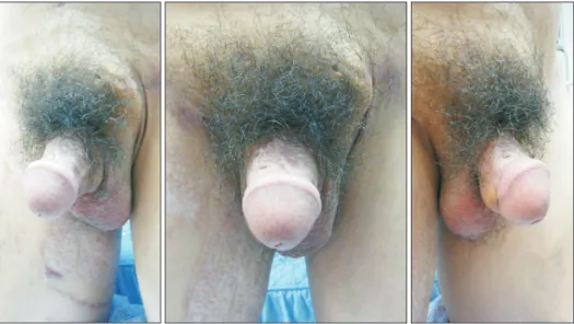
The long term follow-up after 3 years has shown excellent result without any complication (Fig. 5, 6).
DISCUSSION
In this case, a lustful attempt in complete removal of the foreign material has injured the functional portion, which resulted in a disaster. Despite awareness of the potentially damaging effects of paraffin injection, they are still being used by layman in attempts to increase the circumference of the penile shaft. Scholten et al.3 had stated state the following aims
A B
Fig. 5. (A) Photo obtained at follow-up 15 months, showing healthy urine caliber. (B) Uroflowmetry. Maximal flow rate of 10.5 mL/s, average flow rate 7.1 mL/s and voided volume 261 mL.
Fig. 4. After first stage operation, the patient was able to permit orthostatic micturition through the neourethra after removal of the Foley catheter at postoperative 3 weeks.
in a reconstructive surgery of a self-injection injury involving the penis, scrotum, and suprapubic region: the preservation of penile and testes functions should be considered while completely removing the skin and subcutaneous tissues infiltrated with the foreign material.9
Various authors suggested many different techniques in urethral reconstruction and phalloplasty, however they only differ in their methods. The anatomic concept of one structured homogenous organ of ‘penile urethra within the penis’ has not been mentioned in any article. In the history of phalloplasty, Gillies and Farina rolled a tube on the abdominal wall to produce a urethra and surrounded this with another tube pedicle, thereby producing a composite homogenous penis, which tends to be too large and disfavored by later reconstructive surgeons.10 Afterwards, separate concepts between urethral reconstruction and phalloplasty was discovered, and had drawn a bold line in surgical trend of penile reconstruction upto date.
Phalloplasty has been performed generally using a radial forearm free flap or by osteocutaneous free flap, which supported erectile function.6 Urethral reconstruction is most commonly performed using split or full thickness skin grafting, but recently, cutaneous free flap surgery had been introduced.
The outcome of free flap surgery relies greatly on the survival, which depends greatly upon the surgeon’s operation technique.
Use of vascularized free flap even accompanies a possibility of ischemia of the donor area. It has been reported that 16.6%
of cases in radial forearm free flaps and 3% in ulnar forearm free flaps had experienced ischemia of the hand.8 Thus, in the obvious case that requires a free flap, such as in this case, reconstructive surgeons are confronted with the risk of flap failure or donor site ischemia, and should consider the impact of the failure in patients combined with unstable psychological mood and emotional status. It is unnecessary to emphasize the importance in guarantee of the surgical outcome.
Groin flap provides a relatively non-hair-bearing skin for use in the reconstruction of the neourethra, and also provides the external skin and subcutaneous fat needed for phallus reconstruction. Different from the conventional tubed groin flap for the construction of male genitalia in the transsexual introduced by Puckett and Montie7 in 1978, a distal portion was not simply inset at the mons pubis, but rather encircled around the large diameter Foley catheter to construct a more reliable neourethra at its initial operation.
Not different from any other groin flaps, the donor site can primarily be closed. Another advantage of this surgical technique comes from the minimal or maybe almost not- recognizable donor site morbidity. According to Felici and Felici,8 the free radial forearm flap and suprapubic flap are more frequently used, but with low acceptance by patients of a vivid donor site scar, and recommended free anterolateral thigh flap as an alternative surgical modality. Thus, from both perspectives of site morbidity and scarring, the groin flap surpasses any surgical techniques published thus far.
Fig. 6. Final postoperative clinical photo at 3 years.
Beside from having to keep a Foley catheter for few weeks, patients can immediately return to daily life. Ideally speaking, the epithelial layer of neourethra should completely be approximated at postoperative 3 to 5 days, which suggests safe removal of the Foley catheter after postoperative 7 days.
However, avoidance of urethro-cutaneous fistula or stricture formation was assisted by the maturation of neourethral track under presence of a large-diameter Foley catheter for 3 weeks.
This surgical method should only fall into patients with preserved erectile portions as in our case. If in a case of defective corpus spongiosum, it is recommended to place an inflatable penile prosthesis into the newly constructed penis or perform secondary free iliac or fibular osseous flap insertion6,7 during the second stage flap division operation.
REFERENCES
1. Dubin BJ, Sato RM, Laub DR. Results of phalloplasty. Plast Reconstr Surg 1979;64:163-70.
2. Barabás J, Kelemen Z, Bánfi G, Németh Z, Romics I, Nyirády P.
Penis covering and simultaneous urethral replacement by scrotal
skin for severe penile and urethral necrosis. Int Urol Nephrol 2009;41:537-40.
3. Scholten E, Nanhekhan LV, Oudit D, Hage JJ. Scrotal and penile reconstruction after massive self-injection of liquid paraffin and petroleum jelly. Plast Reconstr Surg 2005;115:2168-9.
4. David D. Phalloplasty. South Afr Med J 1957;31:990-1.
5. Lee HB, Hur JY, Song JM, Tark KC. Long anterior urethral reconstruction using a sensate ulnar forearm free flap. Plast Reconstr Surg 2001;108:2053-6.
6. Edgerton MT, Gillenwater JY, Kenney JG, Horowitz J. The bladder flap for urethral reconstruction in total phalloplasty.
Plast Reconstr Surg 1984;74:259-66.
7. Puckett CL, Montie JE. Construction of male genitalia in the transsexual, using a tubed groin flap for the penis and a hydraulic inflation device. Plast Reconstr Surg 1978;61:523-30.
8. Felici N, Felici A. A new phalloplasty technique: the free anterolateral thigh flap phalloplasty. J Plast Reconstr Aesthet Surg 2006;59:153-7.
9. Cavadas PC. Secondary free fibular flap for providing rigidity in a radial forearm phalloplasty. Plast Reconstr Surg 2008;122:101e- 2e.
10. Casoli V, Verolino P, Castede JC, Pelissier P, Martin D, Baudet J. One-stage complete phalloplasty with forearm free flap after severe electrical burns. Plast Reconstr Surg 2004;113:313-6.