• 검색 결과가 없습니다.

Minimally Invasive Approach to Esophageal Perforation after Endoscopic Ultrasound-Guided Fine-Needle Aspiration: A Report of 2 Cases

N/A
N/A
Protected

Academic year: 2021

Share "Minimally Invasive Approach to Esophageal Perforation after Endoscopic Ultrasound-Guided Fine-Needle Aspiration: A Report of 2 Cases"

Copied!
4
0
0

로드 중.... (전체 텍스트 보기)

전체 글

(1)

313 www.kjtcvs.org

KJTCVS

The Korean Journal of Thoracic and Cardiovascular Surgery

Case Report

Minimally Invasive Approach to Esophageal Perforation after Endoscopic Ultrasound-Guided Fine-Needle Aspiration: A Report of 2 Cases

Anna C. M. Geraedts, M.D.

1,2

, Pieter P. H. L. Broos, M.D., Ph.D.

1

, Michiel H. M. Gronenschild, M.D.

3

,

Frank L. J. Custers, M.D.

3

, Karel W. E. Hulsewe, M.D., Ph.D.

1

, Yvonne L. J. Vissers, M.D., Ph.D.

1

, Erik R. de Loos, M.D.

1

1

Department of Surgery, Zuyderland Medical Centre, Heerlen;

2

Department of Surgery, Amsterdam UMC, University of Amsterdam, Amsterdam;

3

Department of Respiratory Medicine, Zuyderland Medical Centre, Heerlen, The Netherlands

ARTICLE INFO

Received August 28, 2019 Revised January 16, 2020 Accepted February 22, 2020 Corresponding author Erik R. de Loos Tel 31-88-4597777 Fax 31-45-5766548

E-mail [email protected] ORCID

https://orcid.org/0000-0001-6313-2658

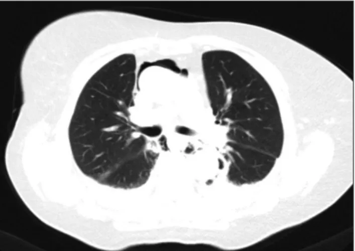
Esophageal perforation after endoscopic ultrasound-guided fine-needle aspiration for mediastinal staging is a rare but severe complication. We report 2 cases of patients with esophageal perforation who were treated using video-assisted thoracoscopic surgery in combination with esophageal stenting. Through these cases, the feasibility of minimally invasive thoracic surgery was evaluated.

Keywords: Endoscopic procedures, Video-assisted thoracic surgery, Injury, Esophageal perforation

Copyright

© The Korean Society for Thoracic and Cardiovascular Surgery. 2020. All right reserved.

This is an Open Access article distributed under the terms of the Creative Commons Attribution Non-Commercial License (http://creativecommons.org/licenses/

by-nc/4.0) which permits unrestricted non-commercial use, distribution, and reproduction in any medium, provided the original work is properly cited.

Case report

Endoscopic ultrasound-guided fine-needle aspiration (EUS-FNA) has emerged as a novel minimally invasive im- aging technique to obtain a histologic diagnosis in patients with mediastinal lymphadenopathies accompanying vari- ous benign or malignant conditions. It complements ad- vanced imaging methods such as positron emission to- mography-computed tomography (PET-CT), which have a high sensitivity for identifying enlarged lymph nodes, but limited specificity. In particular, differentiating between inflammatory processes and malignancy is especially diffi- cult and a pathological diagnosis is generally required. The overall sensitivity and specificity of EUS-FNA and/or en- dobronchial ultrasound (EBUS)-transbronchial needle as- piration were reported to be 97.4% and 100%, respectively [1]. Although severe complications are rare, we report 2 se- rious and life-threatening cases of esophageal wall rupture after EUS-FNA. Surgery via thoracotomy to clean the tho- racic cavity and close the esophageal defect is the gold standard treatment for esophageal perforations, but less in- vasive treatments are being explored and their role is

evolving. Hence, we report 2 cases of esophageal wall rup- ture after EUS-FNA in which minimally invasive surgery was successfully combined with endoscopic stenting.

This study was conducted according to the principles of the Declaration of Helsinki. Verbal informed consent was obtained from both patients for publication of their clinical details and images.

Case 1

Patient A was a 45-year-old man who underwent a work- up for recurrent right-sided chest pain and increased D-di- mer levels. Computed tomography (CT) was performed to exclude pulmonary embolism and showed mediastinal and hilar lymphadenopathy, which raised the suspicion of lym- phoma or sarcoidosis. An additional PET-CT examination showed axillary and mediastinal enlarged PET-positive lymph nodes, most prominently in the subcarinal region.

In addition, paraseptal emphysema and thickened peribronchial interstitium in the apex of the lung were present, with signs of active fibrosis. EUS-FNA of lymph node station 7 was performed with a 19G needle for patho-

https://doi.org/10.5090/kjtcs.19.064 pISSN: 2233-601X eISSN: 2093-6516

Korean J Thorac Cardiovasc Surg. 2020;53(5):313-316

(2)

314 www.kjtcvs.org

KJTCVS https://doi.org/10.5090/kjtcs.19.064

logical and microbiological evaluation.

The patient was readmitted after 12 days with excessive vomiting, followed by epigastric pain and fever. The patient was hemodynamically stable with a blood pressure of 130/80 mm Hg, a regular heart rate of 80 beats/min, a tem- perature of 37.2°C, and a respiratory rate of 15 breaths/

min. His C-reactive protein (CRP) level was 243 mg/L and white blood cell (WBC) count was 18×10

9

/L.

Chest CT showed posterior mediastinitis with possible abscess formation and pre-existing mediastinal and hilar lymphadenopathy without signs of extraluminal contrast leakage (Fig. 1). Gastroendoscopy was performed and showed a 5-mm tear in the esophagus with an inflow of pus at 33 cm from the incisor teeth. The lymph node cul- ture obtained earlier during EUS-FNA was positive for Streptococcus parasanguinis, Streptococcus salivarius, and Bacteroides fragilis, which were highly sensitive to clinda- mycin. These bacteria are normally present in the gastroin- testinal tract. The pathological report showed granuloma- tous inflammation.

Since there were no signs of sepsis, we decided to treat him with a combination of antibiotics, esophageal stenting, and minimally invasive video-assisted thoracoscopic sur- gery (VATS). Five days after re-admission, a 10-cm Ultraf- lex esophageal stent (Boston Scientific, Marlborough, MA, USA) was placed endoscopically and the posterior medias- tinum was drained surgically via right-sided uniportal VATS (u-VATS). The subcarinal lymph node package im- peded the view of the tear in the esophagus (Fig. 2). The pleural space was not involved in the abscess, most likely because the subcarinal nodes covered the tear in the esophagus. No further exploration of the subcarinal space

was performed to prevent contamination of the thoracic cage, as no abscess formation was observed on CT. Biopsy of the subcarinal lymph nodes was repeated. A 28F tube was placed in the posterior mediastinum. After the esoph- ageal stent was implanted, oral intake was gradually built up. On postoperative day 9, the chest tube was removed be- cause an upper gastrointestinal X-ray series showed no contrast dye leakage. In the following 7 days, no signs of sepsis were found, and the patient’s CRP level (70 mg/L) and WBC count (9.4×10

9

/L) decreased. Antibiotics were administered for 28 days. The patient was discharged 15 days after surgery. During further outpatient care (with a total follow-up of 6 months), the patient did not experience any related complications or secondary interventions.

Case 2

Patient B was a 74-year-old woman with a history of breast cancer that had been treated with curative intent 10 years earlier. She was referred with shortness of breath. A CT scan showed multiple lung nodules. In addition, a PET- CT scan showed 4 PET-positive lesions in the right lower lobe, as well as 2 nodules in the right middle lobe and an enlarged PET-positive lymph node in station 7, which were suspicious for metastatic breast cancer lesions. Mammog- raphy of the right breast showed no local recurrence of the tumor. EUS-FNA of station 7 was performed using a 19G needle. Directly after the procedure, the patient experi- enced pain and was admitted for observation. A chest X-ray showed subcutaneous emphysema on the left side of the neck. A non-contrast CT scan showed pneumatosis in

Fig. 1. Computed tomography scan showing mediastinitis.

Fig. 2. Uniportal video-assisted thoracoscopic surgery showing a

tear in the esophagus.

(3)

315

Anna C. M. Geraedts, et al. Esophageal Perforation after EUS-FNA

www.kjtcvs.org

KJTCVS

the mediastinum, and pleural effusion and atelectasis on both sides (Fig. 3). The laboratory results demonstrated a CRP of 600 mg/L and a WBC count of 22×10

9

/L. The pa- tient was placed on a fasting regimen and intravenous anti- biotics were started. The patient experienced clinical dete- rioration and was admitted to the intensive care unit (ICU) 2 days after the EUS-FNA. She was septic, with a blood pressure of 90/50 mm Hg, a regular heart rate of 110 beats/

min, a temperature of 37.7°C, and a respiratory rate of 21 breaths/min. Upper gastrointestinal endoscopy revealed a laceration of the esophagus at 27 cm from the incisor teeth.

Consequently an 8-cm colon stent was placed endoscopi- cally. The next day, a chest tube was placed in the left pleu- ral space, draining 400 mL of turbid fluid immediately.

Then, u-VATS debridement of a right-sided thoracic empy- ema was performed and 2 chest tubes were placed in the posterior mediastinum. Postoperatively the patient re- turned to the ICU. On the following days, the drains kept producing excessive turbid fluid. On day 4 after surgery, upper gastrointestinal endoscopy was repeated; the stent appeared to have been displaced and had to be replaced slightly more proximally. The chest tube in the left pleural space could be removed 6 days after surgery. The next day, another chest CT scan was performed due to an increase in production in the upper chest tube, and demonstrated a small area of fluid collection around the stent and pleural fluid around the lower lobes of both lungs, with atelectasis on the left side. The decision was made to debride the left pleural cavity in combination with opening and draining the posterior mediastinum via u-VATS. Two 28F chest tubes were placed. Because of the size of the esophageal defect and the continuing mediastinal and thoracic leak- age, it was decided to laparoscopically place a gastrostomy

for gastric decompression and a feeding jejunostomy.

Thereafter, a fast recovery and normalization of inflamma- tory markers was observed. At 14 days after the last surgi- cal procedure, a CT scan with contrast showed no leakage.

The gastrostomy could be removed, and the patient was admitted to the rehabilitation center. The esophageal stent was removed 2 months after initial placement. Upper gas- trointestinal endoscopy did not show a remaining esopha- geal defect and the patient returned to a normal diet. The pathology report of station 7 revealed metastatic breast cancer. The patient is currently receiving treatment for metastatic disease.

Discussion

EUS-FNA is an efficient and safe method for assessing mediastinal lymphadenopathies of unknown origin. In the revised guidelines of the European Society for Thoracic Surgeons, EUS or EBUS with FNA is recommended for mediastinal staging [2].

Post-procedural pain and mild hemorrhage are common complications of EUS-FNA. The risk of perforation is low, with a reported incidence of 0.02% [3]. The treatment of esophageal perforation after EUS is mainly based on the treatment principles of Boerhaave syndrome, but it remains unclear whether these are actually similar clinical entities that require similar treatment. Esophageal perforation in patients with Boerhaave syndrome has a poor prognosis and remains a life-threatening condition with a mortality rate of 14.8% [4]. It is a surgical emergency, in which early diagnosis is vital for achieving good outcomes and treat- ment should be started as early as possible.

The principles of definitive management of esophageal wall rupture are (1) control of the esophageal leak, (2) erad- ication of mediastinal and pleural contamination, (3) de- compression of the esophagus, and (4) nutritional support.

The gold standard surgical approach for esophageal perfo- rations is thoracotomy, laparotomy, or both [5]. The tradi- tional open surgical intervention to control the esophageal leak is primary closure with or without autogenous tissue reinforcement, esophageal resection, or exclusion and di- version. To clean the contaminated mediastinal and pleural spaces, tube drainage or simple drainage is performed during the surgical intervention. Anastomotic leakage can be a reason to delay the initiation of oral intake. Nasogas- tric tube intubation is then performed for enteral nutrition, medication administration, and for gastric decompression.

The purpose of surgery is to provide sufficient closure of the defect to allow esophageal healing by following the

Fig. 3. Computed tomography scan showing pneumomediastinum.

(4)

316 www.kjtcvs.org

KJTCVS https://doi.org/10.5090/kjtcs.19.064

aforementioned principles. The location of the injury in- fluences both the surgical approach and the surgical repair options that are chosen. The abdomen needs to be explored if the full extent of the perforation cannot be visualized and extends across the gastroesophageal junction. Howev- er, the majority of perforations after EUS are located in the cervical esophagus. Open repair is best performed via a right posterolateral approach. The esophagus is explored, and the defect is repaired by performing a longitudinal myotomy, mucosal repair, closure of the myotomy, and buttress reinforcement with intercostal muscle, pericardial fat, a diaphragm flap, or a pleural patch. Less invasive and endoscopic methods are currently being explored for steps 1 and 2. However, these methods have only been reported after esophageal rupture in patients with Boerhaave syn- drome [6], and not for patients with other causes of esoph- ageal perforation. Alternative less invasive treatments con- sist of a combination of thoracoscopic decortication, drainage, and esophageal stenting. The advantages of the thoracoscopic approach may be shorter thoracic drainage, less postoperative pain, a shorter hospital stay, and a more rapid return to normal function [4,7]. Another method to control the leak is endoscopically assisted mediastinal vac- uum therapy. However, nearly all recommendations made for the use of vacuum-assisted closure are based on expert opinions, rather than on scientific evidence.

This paper reports 2 cases of esophageal wall rupture due to EUS-FNA. In both cases, the 4 principles of defini- tive management of esophageal wall rupture were followed using a minimally invasive approach. In the first case, after eradication of mediastinal and pleural contamination the patient recovered to a considerable extent, and no sepsis occurred. The chest drain was removed on postoperative day 8 and the patient was discharged on postoperative day 15. In contrast, the esophageal leak in the second patient was more difficult to control, and she underwent 2 more operations for additional drainage and nutritional support.

The postoperative course was characterized by an oculogy- ric crisis, excessive production of both drains, and an over- all weakened condition.

In the second case, the patient was relatively older and also had distant metastases. These factors are associated with a higher risk of perforation according to the Ameri- can Society for Gastrointestinal Endoscopy guidelines [8].

In conclusion, to our knowledge, this is the first case re- port of esophageal wall rupture after EUS-FNA treated with minimally invasive surgery using a combination of

esophageal stenting and VATS. Both cases illustrate the need for early recognition and intervention. In conclusion, in selected cases, it is possible to perform minimally inva- sive surgery for esophageal wall rupture, even in patients with sepsis.

Conflict of interest

No potential conflict of interest relevant to this article was reported.

ORCID

Anna C. M. Geraedts: https://orcid.org/0000-0002-6476-6027 Pieter P. H. L. Broos: https://orcid.org/0000-0002-5274-2984 Michiel H. M. Gronenschild:

https://orcid.org/0000-0002-8864-3101

Frank L. J. Custers: https://orcid.org/0000-0001-5711-3303 Karel W. E. Hulsewe: https://orcid.org/0000-0001-8131-1895 Yvonne L. J. Vissers: https://orcid.org/0000-0002-2890-8390 Erik R. de Loos: https://orcid.org/0000-0001-6313-2658

References

1. Kang HK, Um SW, Jeong BH, et al. The utility of endobronchial ul- trasound-guided transbronchial needle aspiration in patients with small-cell lung cancer. Intern Med 2016;55:1061-6.

2. De Leyn P, Dooms C, Kuzdzal J, et al. Revised ESTS guidelines for preoperative mediastinal lymph node staging for non-small-cell lung cancer. Eur J Cardiothorac Surg 2014;45:787-98.

3. Wang KX, Ben QW, Jin ZD, et al. Assessment of morbidity and mor- tality associated with EUS-guided FNA: a systematic review. Gas- trointest Endosc 2011;73:283-90.

4. Kaman L, Iqbal J, Kundil B, Kochhar R. Management of esophageal perforation in adults. Gastroenterology Res 2010;3:235-44.

5. Mavroudis CD, Kucharczuk JC. Acute management of esophageal perforation. Curr Surg Rep 2014;2:34.

6. Cho JS, Kim YD, Kim JW, I HS, Kim MS. Thoracoscopic primary esophageal repair in patients with Boerhaave’s syndrome. Ann Tho- rac Surg 2011;91:1552-5.

7. Lee J, Lee JY, Choi JS, Sung SW. Comparison of uniportal versus multiportal video-assisted thoracoscopic surgery pulmonary segmen- tectomy. Korean J Thorac Cardiovasc Surg 2019;52:141-7.

8. ASGE Standards of Practice Committee, Early DS, Acosta RD, et al.

Adverse events associated with EUS and EUS with FNA. Gastroin-

test Endosc 2013;77:839-43.

수치

Fig. 2. Uniportal video-assisted thoracoscopic surgery showing a  tear in the esophagus.
Fig. 3. Computed tomography scan showing pneumomediastinum.

참조

관련 문서