• 검색 결과가 없습니다.

Unpredictable Spontaneous Fusion after Percutaneous Vertebroplasty and Kyphoplasty in Osteoporotic Compression Fracture

N/A
N/A
Protected

Academic year: 2021

Share "Unpredictable Spontaneous Fusion after Percutaneous Vertebroplasty and Kyphoplasty in Osteoporotic Compression Fracture"

Copied!
7
0
0

로드 중.... (전체 텍스트 보기)

전체 글

(1)

Received: November 6, 2015 Revised: November 16, 2015 Accepted: November 23, 2015

Corresponding Author: Jin Hwan Kim, Department of Orthopedic Surgery, Inje University Ilsan Paik Hospital, Inje University College of Medicine, 2240 Daehwa-dong, Ilsanseo-gu, Goyang 411-706, Korea

Tel: +82-31-910-7828, Fax: +82-31-910-7967, E-mail: oskim@paik.ac.kr

Unpredictable Spontaneous Fusion after Percutaneous Vertebroplasty and Kyphoplasty in Osteoporotic Compression Fracture

Jin Hwan Kim

Department of Orthopedic Surgery, Inje University, Ilsan Paik Hospital, Koyang-si, Gyeonggido, Korea

Introduction: We found a spontaneous fusion at adjacent vertebrae after percutaneous vertebroplasty (PVP) or kyphoplasty in osteoporotic compression fractures and analyze the radiologic & clinical characteristics.

Materials & Methods: Between January 2000 and December 2011, 555 patients were treated with PVP or kyphoplasty for osteoporotic compression fracture in our department. We classified the spontaneous fusion as two groups. One is solid spontaneous fusion group with at least three cortical continuity to adjacent vertebrae, the other is partially fusion group which progressed fusion compared to previous radiologic finding. We reviewed the plain film and analyzed the radiologic characteristics of those patients with duration of fusion, location and extent of fused segments. A clinical characteristic by visual analogue score (VAS) compared to our previous report was checked.

Results: Among them, 54 patients (9.7%) had an solid spontaneous fusion and 43 patients (7.7%) had partially fused on plane image. In solid fusion group, the average duration of fusion was 19 months ranged of 3 to 48 months. Forty six cases (85%) of solid fusion patients had occurred with proximal adjacent vertebrae and 7 cases (13%) had proximal with distal adjacent vertebrae. Forty one cases (76%) of spontaneous fusion occurred within 1 segment and 13 cases within multiple segments. The most cases of solid fusion group were occurred at thoracolumbar junction (40 patients, 74%). Mean VAS score of solid fusion group was 2.0 at final follow-up and were analyzed relatively low score compared to mean VAS of our previous report (2.0, 2.8 respectively).

Conclusion: After percutaneous vertebroplasty or kyphoplasty in osteoporotic compression fracture, unpredictable spontaneous fusion could develop more than 10% rate, especially with proximal vertebra within 1 segment at thoracolumbar junction in radiologic aspect. Clinically, patients with spontaneous fusion had a tendency of more reduced pain than others.

Key Words: Osteoporotic vertebral compression fracture, Percutaneous vertebroplasty, Spontaneous fusion, Kyphoplasty

The frequency of vertebral compression fractures caused by osteoporosis, metastatic disease, or trauma of the spine in the general population increases with age.

Among these various causes of vertebral compression fractures, osteoporosis is the most common. Recently, studies have shown an incidence of approximately 30%

prevalence of osteoporosis and 20% osteoporotic com-

pression fractures in postmenopausal women.

1

Osteo-

porotic compression fractures that increase the rate of

other osteoporotic compression fractures and kyphotic

deformities are associated with chronic back pain in

84% of symptomatic patients.

2

Mobility losses and

(2)

mortality rates due to medical complications of osteo- porotic compression fractures are also increasing.

3,4

There are two minimally invasive spine augmentation techniques for vertebral compression fractures, percu- taneous vertebroplasty (PVP) and kyphoplasty (KP).

PVP, introduced in France in 1984, involves the percutaneous injection of cement directly into the cancellous bone of a vertebral body. KP, introduced in 2001, is a modification of PVP in which a balloon device is inflated in the vertebral body to create a cavity for cement injection.

5

Both PVP and KP provide excellent short-term pain relief and functional resto- ration.

5-10

The area surrounding the osteoporotic com- pression fracture after PVP or KP goes through many healing-related changes in the long term.

11,12

Numerous reports have demonstrated the effective- ness of PVP and KP. Our recent report detailed the long-term follow-up results of PVP after osteoporotic compression fractures.

13

We observed the progression of bony bridging across the adjacent vertebrae after PVP or KP in osteoporotic compression fractures on serial radiographs, and named this phenomenon “spontaneous fusion.”

The purpose of this study was to analyze the clinical and radiological characteristics of spontaneous fusion.

MATERIALS AND METHODS

1. Demography

We examined 555 patients (233 men and 322 women) with a mean age of 72 years (39-94 years) who under- went PVP or KP for osteoporotic compression fractures of the thoracic or lumbar vertebrae between January 2000 and December 2011. All patients were suffering from persistent pain after post-traumatic conservative treatment or vertebral collapse after the progression of compression fracture. In terms of the injured area, compression fractures in the 1

st

lumbar vertebra accounted for 208 cases, followed by 173 cases in the 12

th

thoracic vertebra, 64 cases in the 2

nd

lumbar

vertebra, 46 cases in the 3

rd

lumbar vertebra, 46 cases in the 11

th

thoracic vertebra, and 18 cases in the 4

th

lumbar vertebra. Bone mineral densitometry showed a mean T-score of -2.9.

2. Preoperative patient evaluation, surgi- cal technique, and postoperative care Surgical intervention was performed in patients experiencing persistent pain or with progressive collapse after conservative treatment, which was followed by a reassessment after 3 weeks. Vertebrae were identified as symptomatic when they showed compression fractures on plain radiographs, focal tenderness on physical examination, and hot uptake on a bone scan. In addition, the degree and displacement of the fracture fragments were analyzed using pre- operative computed tomography, while the degree of osteoporosis was measured using bone mineral densito- metry. Patients who were suspected with metastatic cancer were analyzed using additional magnetic resonance imaging (MRI). Cases in which MRI was performed for other non-treatment reasons were also included.

The surgical procedure involved the administration of

fentanyl and midazolam as pre-medications after

preventive antibiotics 30 minutes prior to surgery,

administration of 1% lidocaine as a local anesthetic

agent on the surgical area with the patient in the prone

position, pinpoint skin incision, insertion of an 11-

gauge J-type bone marrow biopsy needle into the lesion

inside the vertebral body through both pedicles, and

confirmation of the needle location under an image

intensifier. Bone cement (Surgical Simplex P; How-

medica, Limerick, Ireland) was mixed with tungsten

powder and injected into the vertebral body, while

cement leakage was monitored by using a C-arm image

intensifier. Immediately after the surgery, activities on

the bed and early ambulation were allowed, and a

Juwett brace was provided for most of the patients with

thoracolumbar fractures.

(3)

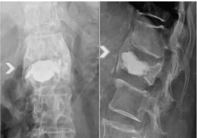
Fig. 1. Solid fusion group defined fractured vertebrae which continuity was achieved in at least 3 adjacent cortical vertebrae.

METHODS

To confirm objectified changes in the pre- and postoperative pain levels, anteroposterior and lateral radiographs were taken to measure the anterior body height and kyphotic angle of the vertebral bodies.

Changes in the injected cement were also retrospec- tively confirmed, and those in the symptomatic and adjacent vertebrae were observed on follow-up radio- graphs.

To evaluate the reproducibility of visual assessment, interpretation agreement among three independent orthopedic observers considering the plain radiographs of the 555 patients who underwent PVP or KP was evaluated. Serial radiographs showed progression of the bony bridging across the adjacent vertebrae.

The patients were distinguished into two groups, the solid spontaneous fusion group (Fig. 1) and the partial fusion group. The former included patients in whom continuity was achieved in at least 3 adjacent cortical vertebrae. The latter included patients in whom fusion had progressed compared to previous radiological findings.

We reviewed the plain radiographs and analyzed the radiological characteristics considering the fusion duration as well as the location and extent of fused segments.

We also compared the clinical characteristics with those of earlier reports using Visual Analogue Scale (VAS) scores of 0 (no pain) to 10 (most severe pain) to assess pain levels before the surgery and 2 days, 3 months, 1 year, and 2 years after the surgery.

13

RESULT

A total of 54 patients (9.7%) experienced spon- taneous fusion, while 43 patients (7.7%) experienced partial fusion as observed using plain radiography. In the solid fusion group, the average fusion duration was 19 months (range, 3 months to 48 years). A total of 46 cases (85%) of solid fusion occurred with the proximal adjacent vertebrae and 7 cases (13%) of the proximal with distal adjacent vertebrae. A total of 41 cases (76%) of spontaneous fusion occurred within a single segment, while the other 13 cases (24%) occurred within multiple segments. Most of the cases of solid fusion (40 patients; 74%) occurred at the thoracolumbar junction. The mean VAS score of the absolute fusion group was 2.0 at the final follow-up, which was rela- tively low compared to that of our previous report.

(2.0, 2.8 respectively)

CASES

1) A 73-year-old female patient underwent percu- taneous vertebroplasty due to an osteoporotic compression fracture of the 1

st

lumbar vertebra, which occurred when she fell. In the 3

rd

year of progression observation, the patient experienced another injury with a newly developed fracture of the 12

th

thoracic vertebra. Following that, findings of spontaneous fusion among vertebral segments were reported (Fig. 2).

2) A 69 years old female patient who underwent

percutaneous vertebroplasty due to an osteoporotic

compression fracture of the 3

rd

lumbar vertebra had

consecutive, multiple new fracture at adjacent level

(4)

Fig. 2. Simple lateral radiographs of a 73-year-old woman with L1,T12 compression fracture. (A) Preoperative radiograph showed L1 compression fracture, (B) Immediately postoperative radiograph of L1, (C) Post- operative 3 years follow-up radiograph show a new compression fracture on T12, (D) Immediately post- operative radiograph of T12 (3 years after L1 postoperatively), (E) Two years after vertebroplasty on T12, follow-up radiograph show a progressive bony bridging between T12 and L1, (F) Four years after vertebroplasty on T12, follow-up radiograph show a spontaneous fusion between T12 and L1.

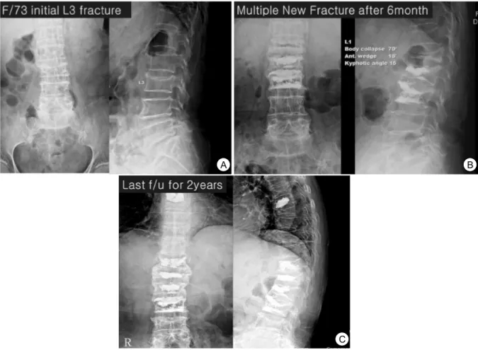
within 2 years. After initial L3 fracture, adjacent vertebrae had new fracture and progress solid spontaneous fusion (Fig. 3).

DISCUSSION

Numerous studies have successfully demonstrated the use of PVP and KP in the treatment of painful osteo- porotic compression fractures of vertebral bodies. Most patients who underwent PVP or KP experienced remarkable pain relief in the short-term follow-up period. Some of these patients also experienced spontaneous fusion during the healing process after

PVP or KP.

This study tried to analyze whether spontaneous fusion after PVP or KP in osteoporotic compression fractures is important to the healing process. We believe that spontaneous fusion at the level of the vertebral body can increase intervertebral stability, and is responsible for the better clinical characteristics observed in patients with bony bridging across adjacent vertebrae. Premik Nagad et al

14

reported that post- vertebroplasty instability at the vertebral body was associated with collapse and micro-motion after PVP in 5 cases after an average follow-up of 2 years. We believe that this is due to the collapse of soft

A B C

D E F

(5)

Fig. 3. (A) A 69 years old female had 3

rd

lumbar vertebrae fracture with minor trauma, (B) Six month later with L3 vertebroplasty, adjacent level fracture were occurred and repeated vertebroplasty, (C) Multiple level fracture with solid spontaneous fusion achieved within 2 years after initial fracture.

osteoporotic bones around the adjacent vertebral body that has been rendered stiff by the cement after PVP.

Spontaneous fusion occurs in some patients with osteoporotic compression fractures who undergo PVP or KP. However, it is unknown whether spontaneous fusion can occur without PVP or KP intervention. Why does spontaneous fusion occur? We have 2 hypotheses to explain this phenomenon. The first hypothesis is about the mechanical effect; polymethylmethacrylate does not have any kind of biological effect. This spontaneous fusion phenomenon came only from the mechanical stability of the fractured vertebral body.

15,16

In particular, the thoracolumbar junction is a transi- tional area of the relatively stiff thoracic spine to the flexible lumbar spine. It is vulnerable not only to

fracture but also to easy union due to kyphosis after the fracture.

Another hypothesis is that spontaneous fusion occurs most often in comminution of the upper end plate with slight kyphosis. Under the force of a kyphotic defor- mity, some comminuted fragments at the fractured vertebrae could be fused with the proximal adjacent vertebrae. The fact that spontaneous fusion after PVP or KP in osteoporotic compression fractures usually occurs in the anterior vertebral aspect supports both of the above hypotheses.

Nevertheless, the result of this study might be a better clinical outcome for all patients in the spon- taneous fusion group, which means that spontaneous fusion after PVP or KP in osteoporotic compression

A B

C

(6)

fractures acts as a mechanical stabilizer at the fracture site and provides structural support. This can eliminate interbody motion and expedite the healing process.

However, further studies are required to clarify the clinical relevance of our findings.

This study had a number of limitations. First, it was retrospective study without a control group. Second, comparisons with conservative treatments and other treatment modalities were not conducted. Finally, the natural course of spontaneous fusion is unclear.

CONCLUSION

After PVP or KP in osteoporotic compression fracture, unpredictable spontaneous fusion could develop at a rate ≥10%, especially with the proximal vertebrae within one segment at the thoracolumbar junction.

Patients who experienced spontaneous fusion reported a greater degree of pain relief than those who did not.

REFERENCES

1. Lee KS, Bae SH, Lee SH, Lee J, Lee DR. New Reference Data on Bone Mineral Density and the Prevalence of Osteoporosis in Korean Adults Aged 50 Years or Older: The Korea National Health and Nutrition Examination Survey 2008-2010. J Korean Med Sci 2014;29:1514-22.

2. Suk SI, Lee CK, Kang HS. Vertebral fracture in osteoporosis. J Korean Orthop Assoc 1993;28:

980-7.

3. Lee YK, Yoon BH, Koo KH. Epidemiology of Osteoporosis and Osteoporotic Fractures in South Korea. Endocrinol Metab 2013;28:90-93.

4. Lau E, Ong K, Kurtz S, Schmier J, Edidin A.

Mortality following the diagnosis of a vertebral compression fracture in the medicare population. J Bone Joint Surg Am 2008;90:1479-86.

5. Wang H, Sribastav SS, Ye F, Yang C, Wang J, Liu H, et al. Comparison of Percutaneous Verte-

broplasty and Balloon Kyphoplasty for the Treat- ment of Single Level Vertebral Compression Frac- tures: A Meta-analysis of the Literature. Pain Physician 2015;18:209-21.

6. Jensen ME, Evans AJ, Mathis JM, Kallmes DF, Cloft HJ, Dion JE. Percutaneous polymethyl- methacrylate vertebroplasty in the treatment of osteoporotic vertebral body compression fractures:

technical aspects. AJNR Am J Neuroradiol 1997;

18:1897-904.

7. Layton KF, Thielen KR, Koch CA. Vertebroplasty, first 1000 levels of a single center: evaluation of the outcomes and complications. AJNR Am J Neuroradiol 2007;28:683-9.

8. Barr JD, Barr MS, Lemley TJ, McCann RM.

Percutaneous vertebroplasty for pain relief and spinal stabilization. Spine (Phila Pa 1976) 2000;

25:923-8.

9. Grados F, Depriester C, Cayrolle G, Hardy N, Deramond H, Fardellone P. Long-term observations of vertebral osteoporotic fractures treated by percu- taneous vertebroplasty. Rheumatology (Oxford) 2000;39:1410-4.

10. Pérez-Higueras A, Alvarez L, Rossi RE, Quiñones D, Al-Assir I. Percutaneous ertebroplasty: longterm clinical and radiological outcome. Neuroradiology 2002;44:950-4.

11. Braunstein V, Sprecher CM, Gisep A. Long-term reaction to bone cement in osteoporotic bone: new bone formation in vertebral bodies after vertebro- plasty. J Anat 2008;212:697-70.

12. Cotten A, Duquesnoy B. Vertebroplasty: current data and future potential. Rev Rhum Engl Ed 1997;

64:645-9.

13. Kim JH, Yoo S, Kim J. Long-term Follow-up of Percutaneous Vertebroplasty in Osteoporotic Com- pression Fracture: Minimum of 5 Years Follow-up.

Asian Spine J 2012;6:1:6-14.

14. Nagad P, Rawall S, Kundnani V, Mohan K, Patil

SS, Nene A. Postvertebroplasty instability. J Neurosurg

(7)

Spine 2012;16:387-93.

15. Nagaraja S, Hassan K. Awada, BSa, Dreher ML, Gupta S, Miller SW. Vertebroplasty increases compression of adjacent IVDs and vertebrae in osteoporotic spines. The Spine Journal 2013;

13:1872-80.

16. Pneumaticos SG, Triantafyllopoulos GK, Evangelo-

poulos DS, Hipp JA, Heggeness MH. Effect of

vertebroplasty on the compressive strength of

vertebral bodies. The Spine Journal 2013;13:1921-

27.

수치

Fig.  1.  Solid  fusion  group  defined  fractured  vertebrae  which  continuity  was  achieved  in  at  least  3  adjacent  cortical  vertebrae.
Fig.  2.  Simple  lateral  radiographs  of  a  73-year-old  woman  with  L1,T12  compression  fracture
Fig.  3.  (A)  A  69  years  old  female  had  3 rd   lumbar  vertebrae  fracture  with  minor  trauma,  (B)  Six  month  later  with  L3  vertebroplasty,  adjacent  level  fracture  were  occurred  and  repeated  vertebroplasty,  (C)  Multiple  level  fra

참조

관련 문서

Although Korea has been playing a leading role in the development of fusion power generation and Korea is playing a leading role in the world stage,

discovered this was out with his girl friend the night after he realized that nuclear reactions must be going on in the stars in order to make them shine.. She said “Look

(3) Toroidally trapped particles tracing banana orbits reflected in the toroidal

The talent required by our educational status in Korea is ‘creative fusion talent’. Modern society is in the fourth industrial revolution. Future society

- We have seen that a perfectly conducting wall, placed in close proximity to the plasma can have a strong stabilizing effect on external kink modes. - In actual

- how magnetic, inertial, and pressure forces interact within an ideal perfectly conducting plasma in an arbitrary magnetic geometry.. - Any fusion reactor must

straight cylinder (no problems with toroidal force balance) - The 1-D radial pressure balance relation is valid for.. tokamaks, stellarators, RFP’s,

Magnetic field.. • Single particle motion of the plasma.