ISSN: 2233-601X (Print) ISSN: 2093-6516 (Online)
Received: August 30, 2016, Revised: December 18, 2016, Accepted: December 20, 2016, Published online: August 5, 2017
Corresponding author: Hyuk Ahn, Department of Thoracic and Cardiovascular Surgery, Seoul National University Hospital, Seoul National University College of Medicine, 101 Daehak-ro, Jongno-gu, Seoul 03080, Korea
(Tel) 82-2-2072-3349 (Fax) 82-2-762-3566 (E-mail) [email protected]
© The Korean Society for Thoracic and Cardiovascular Surgery. 2017. All right reserved.
This is an open access article distributed under the terms of the Creative Commons Attribution Non-Commercial License (http://creativecommons.org/
licenses/by-nc/4.0) which permits unrestricted non-commercial use, distribution, and reproduction in any medium, provided the original work is properly cited.
Extra-Anatomic Ascending Aorta to Abdominal Aorta Bypass in Takayasu Arteritis Patients with Mid-Aortic Syndrome
Hak Ju Kim, M.D., Jae-Woong Choi, M.D., Ho Young Hwang, M.D., Hyuk Ahn, M.D., Ph.D.
Department of Thoracic and Cardiovascular Surgery, Seoul National University Hospital, Seoul National University College of Medicine
Background: We evaluated the operative outcomes of an extra-anatomic bypass from the ascending aorta to the abdominal aorta in patients with type II or III Takayasu arteritis (TA) with mid-aortic syndrome.
Methods: From 1988 to 2014, 8 patients with type II (n=2) or III (n=6) TA underwent an ascending aorta to abdominal aorta bypass. The mean patient age was 43.5±12.2 years and the mean peak pressure gradient between the upper and lower extremities was 54.8±39.0 mm Hg. The median follow-up duration was 54.4 months (range, 17.8 to 177.4 months). Results: There were no cases of operative mortality. The mean peak pressure gradient significantly decreased to −2.4±32.3 mm Hg (p=0.017 compared to the preoperative value).
Late death occurred in 2 patients. The symptoms of upper extremity hypertension and claudication improved in all patients. The bypass grafts were patent at 47.1±58.9 months in 7 patients who underwent follow-up imaging studies. Conclusion: An extra-anatomic ascending aorta to abdominal aorta bypass could be an effec- tive treatment option for severe aortic steno-occlusive disease in patients with type II or III TA, with favor- able early and long-term outcomes.
Key words: 1. Extra-anatomic bypass 2. Takayasu arteritis 3. Mid-aortic syndrome
Introduction
Takayasu arteritis (TA) is a rare, nonspecific, sys- temic inflammatory arteriopathy that causes various types of steno-occlusion or aneurysmal dilatation in- volving the aorta, its branches, and pulmonary artery [1]. In patients with Ueno type II or III TA, narrow- ing of the thoracoabdominal aorta, which is known as mid-aortic syndrome, results in upper extremity hypertension and claudication that may require sur- gical correction. The standard anatomic repair con- sists of resection and anastomosis or the replacement of a diseased segment [2]. Even though extensive
aortic replacement is theoretically possible, complex vascular problems may call for extra-anatomic bypass grafts, especially in cases where long segments are narrowed or if multiple consecutive areas of stenosis are present [3]. The aim of this study was to eval- uate early and long-term outcomes after an ex- tra-anatomic bypass from the ascending to abdominal aorta in TA patients with mid-aortic syndrome.
Methods
1) Patient characteristics
From January 1998 to Decemb er 2014, 8 patients
https://doi.org/10.5090/kjtcs.2017.50.4.270
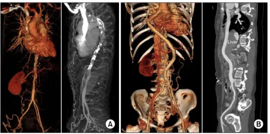
Fig. 1. (A) Preoperative computed tomography angiography. It shows diffuse calcification and narrowing of the descending thoracic and ab- dominal aorta. (B) Postoperative com- puted tomography angiography.
Table 1. Preoperative characteristics of the study patients (n=8)
Characteristic Value
Age (yr) 43.5±12.2
Male:female 2:6
Ueno type (II:III) 2:6
Smoking 0
Diabetes mellitus 2 (25.0)
Hypertension 5 (62.5)
History of stroke 1 (12.5)
Overweight (body mass index >25 kg/m
2) 1 (12.5)
Coronary artery disease 3 (37.5)
Chronic renal failure 1 (12.5)
Symptom 5 (62.5)
Claudication 4 (50.0)
Hypertension on upper extremity 4 (50.0)
Headache 2 (25.0)
Peak systolic blood pressure (mm Hg)
Upper extremity 148.6±41.2
Lower extremity 93.9±20.4
Peak pressure gradient 54.8±39.0
Left ventricle ejection fraction (%) 58.0±10.1 Left ventricle mass index (g/m
2) 164.2±65.8 Operative data
Diameter of ascending aorta (mm) 35.6±4.7 Diameter of narrowed aorta (mm) 11.3±1.5
Graft size (mm) 13.3±2.4
Cardiopulmonary bypass use 1 (12.5)
Operation time (min) 326.9±54.2
Values are presented as mean±standard deviation or number (%).
(2 males and 6 females) underwent an ascending aorta to abdominal aorta bypass for TA at Seoul National University Hospital. Two patients had Ueno type II TA and 6 patients had Ueno type III TA. The mean age at operation was 43.5±12.2 years. The most common symptoms were upper extremity hy- pertension (n=4, 50%) and claudication (n=4, 50%) (Table 1).
Seven patients had a preoperative computed to- mography (CT) scan (Fig. 1A) and 1 patient under- went aortography to evaluate the aorta and arch ves- sels, and transthoracic echocardiography was per- formed in all patients within 3 months before surgery. The mean diameters of the ascending and narrowed abdominal aorta were 35.6±4.7 mm and 11.3±1.5 mm, respectively (Table 1). The mean peak pressure gradient (PG) between the upper and lower extremities was 54.8±39.0 mm Hg. The left ven- tricular mass index was 164.2±65.8 g/m
2.
2) Operative strategy
All operations were performed via median sternot- omy and split midline laparotomy. After opening the pericardium, proximal anastomosis was done to the anterolateral aspect of the mid-ascending aorta in an end-to-side fashion. Seven patients underwent prox- imal anastomosis with a side-biting clamp without cardiopulmonary bypass. In the other patient, how- ever, cardiopulmonary bypass and total circulatory arrest were required due to a severely calcified as- cending aorta. After the completion of proximal anas- tomosis, the graft was passed down through the right pleural cavity and anterolateral border of the
right-side diaphragm to the peritoneal cavity. The
graft was then brought into the retroperitoneal cav-
ity, anteriorly to the liver and posteriorly to the
Fig. 2. (A) Operative photographs of the ascending aorta-to-infrarenal abdominal aorta bypass. The graft was anastomosed proximally to the ascending aorta and passed down through right pleural cavity and diaphragm via median sternotomy. (B) Then the graft was made to take ante-hepatic, retro-gastric course and brought into retroperitoneal cavity. (C) The distal anastomosis was performed to infrarenal abdominal aorta, just above the iliac bifurcation.
stomach, and was anastomosed to the infrarenal ab- dominal aorta, just above the iliac bifurcation under cross-clamps at the proximal and distal parts of the anastomosis site (Fig. 2). Commercially available vas- cular grafts were used with graft sizes ranging from 12 to 18 mm. The distal anastomosis site was wrap- ped with omentum and the posterior parietal peri- toneum was closed to prevent an aorto-intestinal fis- tula and pseudoaneurysmal changes.
3) Evaluation of early and long-term clinical out- comes
Patients underwent regular postoperative follow-up in our outpatient clinic at 3- to 4-month intervals.
The clinical follow-up period for this study ended on March 31, 2016. Follow-up was complete in all pa- tients, with a median follow-up duration of 54.4 months (range, 17.8 to 177.4 months). The peak sys- tolic PG between the upper extremity and lower ex- tremity (upper extremity systolic pressure minus lower extremity systolic pressure) was checked be- fore discharge in all patients. An early postoperative graft evaluation (within 2 months after surgery) was performed in 6 patients using CT (Fig. 1B), and a fol- low-up CT scan was carried out in 7 patients at 39.6 months (range, 0.4 to 170.9 months) after surgery.
Final follow-up echocardiography was done in 6 pa- tients at 45.8 months (range, 0.8 to 155.4 months) after surgery.
4) Statistical analysis
Statistical analyses were performed using IBM SPSS ver. 22.0 (IBM Corp., Armonk, NY, USA). Values were presented as mean±standard deviation, median with ranges, or proportions. The Wilcoxon signed- rank test was used to compare the changes in the peak PG and the left ventricular mass index. Survival analysis was performed using the Kaplan-Meier method. All p-values<0.05 were considered to in- dicate statistical significance.
Results
1) Early results
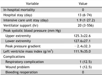
There were no cases of early mortality. The post- operative complications included respiratory compli- cations (n=1) and a wound problem (n=1) (Table 2).
The intensive care unit stay and hospital stay were 1.92 days (range, 1 to 27.2 days) and 12 days (range, 6 to 74 days), respectively. The peak systolic PG be- tween the upper and lower extremities decreased significantly immediately after surgery, from 54.8±
39.0 mm Hg to −2.4±32.3 mm Hg (p=0.017).
2) Follow-up results
Late death occurred in 2 patients. One patient died
suddenly without any notable cause 7 months after
surgery and the other patient died from multiple
myeloma 8 months after the operation. The actuarial
survival rates at 5 and 10 years were 75% and 75%,
Fig. 3. Kaplan-Meier overall survival estimates.
Table 2. Early clinical outcomes of the study patients(n=8)