pISSN 2288-9272 eISSN 2383-8493 J Oral Med Pain 2016;41(3):110-117 http://dx.doi.org/10.14476/jomp.2016.41.3.110
Factors Influencing the Duration of Occlusal Appliance Treatment for Patients with Temporomandibular Joint Internal Derangement
So-Youn Lee, Jin-Seok Byun, Jae-Kwang Jung, Jae-Kap Choi
Department of Oral Medicine, School of Dentistry, Kyungpook National University, Daegu, Korea
Received August 19, 2016 Revised September 12, 2016 Accepted September 12, 2016
Purpose: The purpose of this study is to determine factors influencing the duration of occlusal appliance (OA) treatment for patients with temporomandibular joint (TMJ) internal derange- ment.
Methods: Ninety patients were included for this study, who satisfied the following including criteria: (i) those who were diagnosed as disc displacement of TMJ by taking magnetic reso- nance imaging (MRI) and (ii) those who were finished OA treatment. The subjects were clas- sified into three groups according to the period of OA treatment: (i) early response group (<6 months), (ii) moderate response group (6 months-1 year), and (iii) delayed response group (>1 year). Demographic data, data from chief complaints and past history of temporomandibular disorder, data from clinical examination and diagnostic imaging including panoramic view and TMJ MRI were compared among groups. One-way ANOVA and chi-square analysis were used to test statistical significance.
Results: There were no significant differences in demographic data, data from chief complaints and TMJ imaging. However, only the prevalence of oral parafunctional habits including brux- ism, clenching, and unilateral chewing showed significant differences among groups.
Conclusions: Oral parafunctional habits could be factors to influence the duration of OA treat- ment in the patients with TMJ internal derangement.
Key Words: Conservative treatment; Occlusal appliance; Oral parafunction; Temporomandibu- lar joint internal derangement
Correspondence to:
Jae-Kap Choi
Department of Oral Medicine, School of Dentistry, Kyungpook National University, 2177 Dalgubeol-daero, Jung-gu, Daegu 41940, Korea Tel: +82-53-600-7321 Fax: +82-53-426-2195 E-mail: [email protected]
JOMP
Journal of Oral Medicine and PainCopyright Ⓒ 2016 Korean Academy of Orofacial Pain and Oral Medicine. All rights reserved.
CC This is an open-access article distributed under the terms of the Creative Commons Attribution Non-Commercial License (http://creativecommons.org/licenses/by-nc/4.0/),
INTRODUCTION
Temporomandibular joint internal derangement (TMJID) describes an abnormal positional relationship of the articu- lar disc to the mandibular condyle and the articular emi- nence. The disorder has been associated with characteristic clinical findings such as pain, joint sounds, and irregular or deviating jaw function.
It is generally accepted concept that temporomandibu- lar disorder (TMD) is self-limiting and the natural course of disc displacement of the TMJ is relatively favorable and the management goals for TMJID are similar to those of other orthopedic conditions, namely, reduction of pain and
adverse loading, improvement of function, and restoration of normal, daily activities. Several studies of conservatively treated patients showed that a series of fairly predictable adaptations were likely to occur in the majority of TMJID.
1)The emphasis should be on conservative treatment that fa- cilitates the musculoskeletal system’s natural healing ca- pacity and treatment that involves the patient in the physi- cal and behavioral management of their own problem. A multidisciplinary model that includes patient education and self-care, cognitive behavioral intervention, pharmacother- apy, physical therapy, and orthopedic appliance treatment is endorsed for the management of nearly all TMD patients.
2)Among several noninvasive and reversible treatments,
occlusal appliance (OA) has been predominantly recom- mended for the TMD treatment.
3)The use of OA relieves the amount of load placed on the condyle and protects the TMJ and articular disc from degeneration as well as the exces- sive tension placed on the joint.
4)In the research of Fricton et al.,
3)it has been supported that a group treated with OA shows the highest efficacy compared to the groups either treated with non-occluding appliance or with no OA in pa- tients with TMJ pain. Since the principles of mechanism and the efficiency of OAs have been proven in many re- searches, OAs are widely used as an initial and long-term treatment for TMD with pain.
Because clinical characteristics and other considerable contributing factors vary among patients, it is difficult to predict the outcome of conservative treatment, specifically OA treatment for TMD. There have been many studies on the comparisons on influential factors and predictive factors for the treatment outcome of TMD.
5)According to Ekberg and Nilner,
6)male patients showed more positive influence and severe pain at the initial visit showed more negative impact on the treatment outcome. Emshoff
7)had proposed the onset of pain is the most important factor determining the prognosis of OA treatment. In addition, Grossi et al.
8)concluded that neuropsychological and psychosocial prob- lems yield poor treatment outcome. As mentioned above, several studies have been suggested that the treatment out- come may be affected by various factors, such as demo- graphic factor, pain history, clinical findings and psycho- logical problems.
Most patients wonder about the period of treatment and the frequency of their visits to dental clinics to conserva- tively treat and alleviate their TMJ problems at their first visit to the clinic. Yet, because it is difficult to propose the exact period of treatment to the patients according to their varying symptoms, some patients quit receiving the treat- ment when the period of the treatment is prolonged than they expected. Moreover, 10% of TMD patients show no sign of improvement despite their efforts and time spent.
Surprisingly, this group of patients account for more than 40% of the expenses for the entire TMD patients.
5)Therefore, it is important for the clinicians to be able to dis- tinguish patients who would respond to certain treatment and who would not, so that the optimal treatment option
can be selected according to their varying symptoms to an- ticipate the best treatment results. However, there has been no attempt to predict the duration of the treatment based on patients’ predisposing factors by dividing the patients into groups of different treatment termination period and analyzing clinical characteristics of each group. Moreover, although the duration of treatment with OAs to relieve symptoms can be generally expected with clinical experi- ences, it is difficult to accurately estimate how the period of treatment would change depending on each symptom.
We expect that patients would show improved patient’s compliance without mid-treatment withdrawal if the treat- ment period of conservative treatment, specifically OA treatment, were predicted based on the information ac- quired at the patient’s initial visit. The purpose of this study was to classify the patients with TMJID, who had received conservative treatments including OAs, into three groups of different response rates and to analyze various clinical fac- tors observed at the initial visit of each group in order to predict the patient’s treatment period with the factors.
MATERIALS AND METHODS
1. Subjects and Data Collection
Subjects in this study were selected from patients who visited the Department of Oral Medicine at Kyungpook National University Hospital (Daegu, Korea) with disc dis- placement in TMJ from January 2011 to March 2014. Ninety patients were included for this study, who satisfied the fol- lowing including criteria: (i) those who were diagnosed as disc displacement of TMJ by taking magnetic resonance im- aging (MRI) and (ii) those who were finished OA treatment.
No subject had undergone invasive and surgical treatment.
The subjects were classified into three groups according to the period of OA treatment: (i) early response group (<6 months), (ii) moderate response group (6 months-1 year), and (iii) delayed response group (>1 year).
The criteria of the end of treatment were as follows:
1) The pain or discomfort during TMJ movement or func- tion is completely alleviated.
2) The normal activities of mandible are allowed.
3) Active range of motion (AROM) is greater than 38 mm.
4) No progressive sign of TMJ degenerative change.
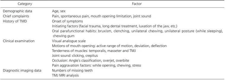
The data were recorded when patients visited the Depart- ment of Oral Medicine at Kyungpook National University Hospital for the first time. The data were based on modi- fied Research Diagnostic Criteria for Temporomandibular Disorders (RDC/TMD). Demographic data, data from chief complaints and past history of TMD, data from clinical ex- amination and diagnostic imaging including panoramic view and TMJ MRI were collected and compared among groups. Bruxism was exceptionally assessed by patient’s questionnaire and wear facets of the OA (Table 1). The study protocol was approved by the Institutional Review Board of Kyungpook National University Hospital (KNUH 2015-11-025-001).
2. Statistical Analysis
The one-way ANOVA and the chi-square analysis were used to test differences between the early response group, the moderate response group and the delayed response
group. The one-way ANOVA test was performed to compare the differences in age, onset of symptoms, visual analogue scale (VAS), AROM, overjet and overbite and the numbers of missing teeth between the three groups. The Pearson chi- square test was used to analyze the differences in extra fac- tors between the three groups. A difference of p<0.05 was considered statistically significant. Statistical evaluation of the data was performed using the IBM SPSS Statistics ver.
22.0 for Windows (IBM Co., Armonk, NY, USA), including the one-way ANOVA test and the Pearson chi-square test.
RESULTS
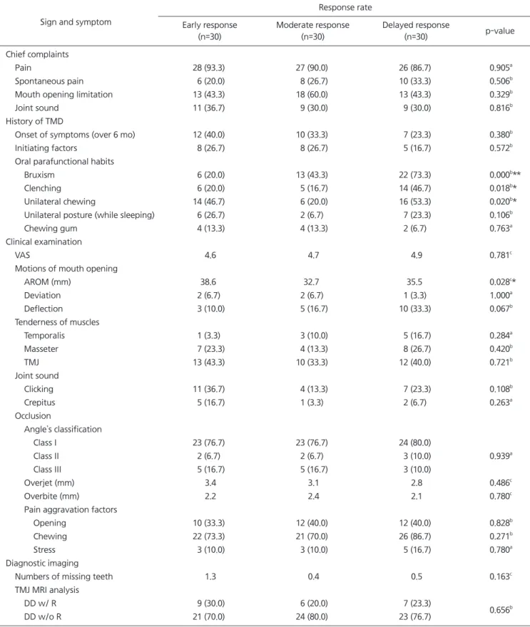
There were no significant differences in demograph- ic data, data from chief complaints and TMJ imaging.
However, only the prevalence of oral parafunctional hab- its including bruxism, clenching, and unilateral chewing showed significant differences among groups (Tables 2, 3).
Table 1. Summary of factors collected of patient’ s initial visit
Category Factor
Demographic data Age, sex
Chief complaints Pain, spontaneous pain, mouth opening limitation, joint sound
History of TMD Onset of symptoms
Initiating factors (facial trauma, long dental treatment, luxation of the jaw, etc.)
Oral parafunctional habits: bruxism, clenching, unilateral chewing, unilateral posture (while sleeping), chewing gum
Clinical examination Visual analogue scale
Motions of mouth opening: active range of motion, deviation, deflection Tenderness of muscles: temporalis, masseter and TMJ
Joint sound: clicking, crepitus
Occlusion: Angle’ s classification, overjet, overbite Pain aggravation factors: while opening, chewing, stress Diagnostic imaging data Numbers of missing teeth
TMJ MRI analysis
TMD, temporomandibular disorder; TMJ, temporomandibular joint; MRI, magnetic resonance imaging.
Table 2. Comparison of demographic data (n=90)
Demographic data Response rate
Early response Moderate response Delayed response p-value
Number of patients 30 (33.3) 30 (33.3) 30 (33.3)
Female 25 (83.3) 23 (76.7) 22 (73.3)
0.638
aMale 5 (16.7) 7 (23.3) 8 (26.7)
Mean age (y) 31.8 31.3 32.5 0.953
bValues are presented as number (%) or number only.
a
Chi-square.
bOne-way ANOVA.
Table 3. Comparison of signs and symptoms
Sign and symptom
Response rate Early response
(n=30)
Moderate response (n=30)
Delayed response
(n=30) p-value
Chief complaints
Pain 28 (93.3) 27 (90.0) 26 (86.7) 0.905
aSpontaneous pain 6 (20.0) 8 (26.7) 10 (33.3) 0.506
bMouth opening limitation 13 (43.3) 18 (60.0) 13 (43.3) 0.329
bJoint sound 11 (36.7) 9 (30.0) 9 (30.0) 0.816
bHistory of TMD
Onset of symptoms (over 6 mo) 12 (40.0) 10 (33.3) 7 (23.3) 0.380
bInitiating factors 8 (26.7) 8 (26.7) 5 (16.7) 0.572
bOral parafunctional habits
Bruxism 6 (20.0) 13 (43.3) 22 (73.3) 0.000
b**
Clenching 6 (20.0) 5 (16.7) 14 (46.7) 0.018
b*
Unilateral chewing 14 (46.7) 6 (20.0) 16 (53.3) 0.020
b*
Unilateral posture (while sleeping) 6 (26.7) 2 (6.7) 7 (23.3) 0.106
bChewing gum 4 (13.3) 4 (13.3) 2 (6.7) 0.763
aClinical examination
VAS 4.6 4.7 4.9 0.781
cMotions of mouth opening
AROM (mm) 38.6 32.7 35.5 0.028
c*
Deviation 2 (6.7) 2 (6.7) 1 (3.3) 1.000
aDeflection 3 (10.0) 5 (16.7) 10 (33.3) 0.067
bTenderness of muscles
Temporalis 1 (3.3) 3 (10.0) 5 (16.7) 0.284
aMasseter 7 (23.3) 4 (13.3) 8 (26.7) 0.420
bTMJ 13 (43.3) 10 (33.3) 12 (40.0) 0.721
bJoint sound
Clicking 11 (36.7) 4 (13.3) 7 (23.3) 0.108
bCrepitus 5 (16.7) 1 (3.3) 2 (6.7) 0.263
aOcclusion
Angle’ s classification
Class I 23 (76.7) 23 (76.7) 24 (80.0)
0.939
aClass II 2 (6.7) 2 (6.7) 3 (10.0)
Class III 5 (16.7) 5 (16.7) 3 (10.0)
Overjet (mm) 3.4 3.1 2.8 0.486
cOverbite (mm) 2.2 2.4 2.1 0.780
cPain aggravation factors
Opening 10 (33.3) 12 (40.0) 12 (40.0) 0.828
bChewing 22 (73.3) 21 (70.0) 26 (86.7) 0.271
bStress 3 (10.0) 3 (10.0) 5 (16.7) 0.780
aDiagnostic imaging
Numbers of missing teeth 1.3 0.4 0.5 0.163
cTMJ MRI analysis
DD w/ R 9 (30.0) 6 (20.0) 7 (23.3)
0.656
bDD w/o R 21 (70.0) 24 (80.0) 23 (76.7)
TMD, temporomandibular disorder; VAS, visual analogue scale; AROM, active range of motion; TMJ, temporomandibular joint; MRI, magnetic resonance imaging; DD w/ R, disc displacement with reduction; DD w/o R, disc displacement without reduction.
Values are presented as number (%) or mean number only.
a