체외배양 생쥐정소세포에서 합성에스트로젠이 P450 동위효소의 발 현 에 미치는 영 향
이호준1, 김묘경2, 고덕성2, 강희규3, 김동훈2
을지의과대학교 생리학교실1, 의과학연구소2, 서울보건대학교 임상병리과3
Effects o f xenoestrogens o n g e n e expression o f cytochrome P450 genes in in vitro cultured mice spermatogenic cells
Ho-Joon Lee1, Myo-Kyung Kim2, Duck-Sung Ko2, Hee-Kyoo Kang3, Dong-Hoon Kim2
Department of Physiology1, Eulji University School of Medicine, Medical Science Institute2, Eulji General Hospital, Seoul, 139-711, Korea, Department of Clinical
Pathology3, Seoul Health College, Sungnam-Si, Kyunggi-Do
* 교 신 저 자 이호준
서울시 노원구 하계동 280-1 을지병원 의과학연구소 을지의과대학교 생리학교실
전화번호: 02-970-8710 Fax: 02-970-8002 e-mail: [email protected]
* 본 연 구 는 2 0 0 0 년 식 약 청 내 분 비 계 장 애 물 질 용 역 사 업 (ED 2 0 0 0 - 4 0 )지 원 으 로 수 행 되 었 음 .
체 외 배 양 생 쥐 정 소 세 포 에 서 합 성 에 스 트 로 젠 이 P 4 5 0 동 위 효 소 의 발 현 에 미 치 는 영 향
이호준1, 김묘경2, 고덕성2, 강희규3, 김동훈2
을지의과대학교 생리학교실1, 의과학연구소2, 서울보건대학교 임상병리과3
Effects o f xenoestrogens o n g e n e expression o f cytochrome P450 genes in in vitro cultured mice spermatogenic cells
Ho-Joon Lee1, Myo-Kyung Kim2, Duck-Sung Ko2, Hee-Kyoo Kang3, Dong-Hoon Kim2
Department of Physiology1, Eulji University School of Medicine, Medical Science Institute2, Eulji General Hospital, Seoul, 139-711, Korea, Department of Clinical
Pathology3, Seoul Health College, Sungnam, Kyunggido
Objective : To know the effects of xenoestrogen on spermatogenesis, we investigated the expression of cytochrome P450s enzymes (CYPscc, CYP17α, CYP19) and 3β-HSD genes involved in steroidogenesis.
M e t h o d s : Mouse testicular cells were prepared from 15-day-old ICR mice which had only pre-meiotic germ cells by enzyme digestion using collagenase and trypsin.
Testicular cells were cultured in DMEM supplemented with FSH (0.1 IU/ml) and 10% FBS or medium with estrogen (E2), bisphenol-A (BPA), octylphenol (OP; 10-9, 10-7, 10-6, 10-5, 10-4 M, respectively) and aroclor 1254 (A1254) known as PCBs for 48 hours. The gene expression of cytochrome P450 enzymes were examined by semi-quantitive RT-PCR. The production of estrogen and testosterone was examined by RIA.
Results : As a results, expression of CYPscc mRNA was not significantly decreased, but 3β-HSD and CYP17α mRNA were significantly dose-dependent decreased. And production of testosterone and estrogen were not different except BPA and OP group (10-5 M).
Conclusion : BPA, OP and A1254 might inhibit steroidogenesis by decreasing the CYPscc, 3β-HDS and CYP17αmRNA expression in the mouse testis. These results suggest that BPA, OP and PCBs like as an endocrine disruptors inhibit the productions of steroidogenic enzymes and decrease the production of T and E by
P450
본 연구는 2000년 식약청 내분비계 장애물질 용역사업(ED 2000-40)지원으로 수행되었음.
인간을 포함한 포유동물의 생식계통은 시상하부-뇌하수체 전엽-생식소로 이어지는 기관에 서 분비되는 호르몬들에 의하여 조절되고 있으며, 특히 생식소 호르몬인 estrogen과 androgen은 난자형성과정, 여포형성과정과 정자형성과정을 조절할 뿐만 아니라 생식과 관 련된 여러가지 내분비 기능을 지니고 있다. 최근 급격한 산업화와 공업화에 따른 환경변화 는 여러가지 합성호르몬(xenoestrogen)들을 창출하여 식물뿐 아니라 인간을 비롯한 포유동 물에 내분비장애와 성분화억제 등 심각한 현상을 나타내고 있다.3 대부분 알려진 내분비계 장애물질들은 여성호르몬 또는 반여성호르몬적 작용능력을 가지고 있어 생식 호르몬의 표적 기관이나 표적세포에 작용하여 단계별로 발현되는 특정유전자들의 발현에 영향을 미치며, 소량으로도 생체내 생식기관의 기능을 교란시키는 것으로 보고되고 있다.20, 24 또한 생체 내 에서 쉽게 분해되지 않고 축적되는 특징이 있어 다음 세대까지 생식기관을 비롯한 신경계 및 면역계에 영향을 미치므로 그 심각성이 더욱 문제시되고 있다. 그 예로 1950년도에서 1970년 사이에 여성호르몬인 에스트로겐 기능을 가진 유산방지제 diethylstilbestrol (DES) 를 복용한 여성에 있어 자녀들의 생식기관에서 이상율의 증가가 보고 되었으며,14, 15 성인 남성들에 있어서도 요도하열, 잠복정소 정소외소증, 정자의 기형 증가 및 수 감소 등의 보 고가 있었다 .8, 21 내분비계 장애물질로 잘 알려진 bisphenol-A (BPA)와 octylphenol (OP)은 에스트로겐적인 활동성을 가진 화학물질들로 체내, 외에서 prolactin의 분비를 증가 시키고,1 뇌하수체에서 LH와 FSH의 분비를 억제시키는 것으로 알려져 있다 (Nikula 등, 1999).17 또한, 웅성 성체 흰쥐에 투여하였을 때, 혈청내 testosterone 양이 줄어드는 것을 확인하였으며,4, 5 임신한 흰쥐에 OP를 투여하게 되면 cytochrome P45017α
-hydroxylase/C17-20 lyase와 steroidogenic factor-1(SF-1)의 mRNA와 단백질 발현이 새끼 흰쥐의 정소에서 줄어드는 것을 확인하였다.14 이와 같이 내분비계 장애물질의 접촉 에 따른 생체 내 또는 체외에서 특히 생식기관에서 일어나는 변화에 관한 연구가 활발히 이루어지고 있으며, 그 작용기전을 밝히기 위한 많은 연구가 진행중이다. 따라서 본 연구 는 정자형성과정에 미치는 합성에스트로젠의 기작과 유해 효과를 확인하기 위해서 생쥐정자 세포의 체외배양법을 이용하여 페놀계 내분비계 장애물질로 알려진 bisphenol (BPA), octlyphenol (OP)와 polychlorinated biphenyl (PCBs)의 일종인 aroclor 1254 (A1254)를 체외배양 된 생쥐정자세포에 처리하여, 스테로이드 호르몬의 변화양상을 조사하고, 체내에 작용하는 정상적인 호르몬 기작에 어떤 영향을 미치는지를 알아보기 위해 steroidogenesis 를 조절하는 효소인 P450 동위효소군의 유전자발현양상을 조사함으로써 내분비계 장애물질 이 정자형성과정에 작용하는 기작을 규명하고자 실행하였다.
재 료 및 방 법
1. 미분화 정자세포 (감수분열이전의 정자세포)의 회수와 체외배양 1) 미분화 정자세포의 분리
본 연구에 사용된 실험동물은 ICR 계 (Charles's Rivers Korea Co.)의 생쥐를 사용하였 다. 미분화 정자세포의 분리는 Tres 등23 (1983)이 사용한 방법을 응용하여, 생후 14-15일 령의 ICR 웅성생쥐로 부터 정소를 회수하여 정소막을 제거한 후 erythrocyte-lysing buffer (NH4Cl 155 mM, KHCO3 10 mM, EDTA 2 mM, pH 7.2)를 처리하여 적혈구를 제 거하였다. 이들 정소조직을 PBS (Phosphate buffer saline) 용액에서 곡세정관을 아주 작 게 세절한 다음, 피펫으로 부드럽게 pipetting하여 세정관을 흐트러 주었다. 다시 세정관을 0.1% collagenase 와 20 ㎍/ml DNase가 함유된 PBS용액과 0.25% trypsin과 20 ㎍/ml DNase가 함유된 PBS용액에 각각 30 분, 15 분 동안 37℃ 에서 배양하여 세포 부유액을 만들어 70 ㎛ nylon mesh를 사용하여 여과한 후 1,200 rpm에서 5분간 원심분리를 실시하 였다. 원심분리 후, 상층액은 제거하고 제조된 배양액 (DMEM + 10% FBS)으로 2회 원심 분리하여 정자세포부유액을 준비하였다.
2) 공배양을 위한 생쥐 정소내 체세포 준비
생후 15일령 된 ICR 웅성생쥐로 부터 위의 정자생식세포 분리와 같은 방법으로 정자세 포부유액을 얻는다. 세포부유액 속의 Sertoli 세포는 32℃ 배양기에 이틀간 배양시켜 배양 접시 바닥에 부착할 수 있도록 유도한 후, 20 mM Tris (pH 7.3) 를 약 1 분간 처리하여 부유하고 있는 생식세포를 제거한 다음,13 새 배양액으로 교체하여 위에 준비한 정자세포와 공배양을 실시하였다.
3) 배양조건
기본배양액으로는 DMEM (Dulbecco's modified eagle's medium) 에 10% FBS, 1%
nonessential amino acid, 0.5% essential amino acid와 10 ㎍/ml gentamycin이 첨가된 배양액을 사용하였다. 첨가물질로 human FSH 0.1 IU/ml (Metrodin, Serono)을 기본배양 액에 첨가하였다. 배양조건은 35 mm dish에 세포농도는 5 × 10⁵/cm 으로 조정하여 32℃
배양기에서 6일 동안 배양하였다. 배양전후, 정자세포의 생존율은 0.4% trypan blue로 염 색하여 hemocytometer를 이용하여 판정하였다.
2 체외배양된 웅성생식세포에 합성에스트로젠(xenoestrogens)의 처리 1) 내분비계 장애물질의 처리
앞서 언급한 공배양을 통하여 정자세포를 체외배양하는 동안 합성에스트로젠인
bisphenol-A (BPA) (4,4'-isopropyl-idenediphenol; I-0635, Sigma), octylphenol (OP) (4-tert-octylphenol; Fluka), aroclor 1254와 생체내 호르몬인 estrogen (E2)을 각 군별로 농도별 (10-9, 10-7, 10-6, 10-5, 10-4 M)로 처리하여 이틀동안 배양하였다. 배양 후 얻은 정 소내 세포들의 생존율과 전체 세포수를 확인하였다.
3. Cytochrome P450 동위효소와 3β-HSD 유전자의 발현 1) 정자세포로부터 RNA 분리
체외배양된 미성숙 정자세포의 전체 RNA는 acid guanidinium phenol chloroform 방법 을6 응용하여 다음과 같이 분리하였다. Homogenizer를 이용하여 채취한 정소를 간 다음,
TRIZOL (Gibco BRL, USA)를 첨가하여 조직을 녹인 후, chloroform을 첨가하여 phenol층 과 분리한 다음 원심분리하였다. 상층액만을 분리하여 isopropyl alcohol을 동량 첨가하여 다시 원심분리하여 pellet으로 형성된 RNA를 얻었다.
2) 역전사중합효소 연쇄반응
(Reverse transcription-polymerase chian reaction, RT-PCR)
cDNA 합성을 위한 역전사 반응은 500 ng의 RNA에 10 mM Tris (pH 8.3), 50 mM KCl, 5 mM MgCl2, 1 mM dNTP mix, 2.5 uM Random primer p(dN)6, 2.5 U RNase inhibitor, 1 U 중합효소 연쇄반응AMV reverse transcripase (Boehringer Mannheim, Germany)를 혼합한 다음 42℃ 에서 1 시간 동안 수행하였다. 각 실험군들은 역전사에 의 해 만들어진 cDNA를 이용하여 cytochrome P450 (cholesterol side-chain cleavage enzyme: CYPscc, 17α-hydorxylase/C17-20 lyase: CYP17α, aromatase: CYP19) 동위효 소와 3β-hydroxysterodi dehydrogenase/Δ5,Δ4 isomerase: 3β-HSD 유전자의 발현양상을 조사하였는데, PCR에 사용된 primer들은 한국 Bioneer에서 제조하여 사용하였다25(Table 1). 각 반응은 최종 반응 부피 20 ㎕에 10 mM Tris (pH 8.3), 50 mM KCl, 1.5 mM MgCl2, 0.2 mM dNTP mix, 20 pmol primer쌍, 0.5 unit 의 Taq DNA polymerase (Boehringer Mannheim, Germany)을 혼합하여 DNA thermal cycler에서 94℃에서 45초;
54℃ 에서 45초; 72℃ 에서 1분 30초의 cycle을 40 회 수행하였다. 반응이 끝난 증폭 산 물은 2% agarose gel 전기영동법으로 분석하였다.
4. estrogen과 testosterone의 양 분석
체외배양동안 정소세포로부터 분비된 스테로이드 호르몬의 분석을 위해서 배양된 배양액 으로부터 estrogen과 testosterone 양을 RIA 방법으로 조사하였다.
5. 통 계
각 군간의 유의성은 χ2- test로 검정하였으며, P 값이 0.05 이하일 경우에 통계적으로 유 의하다고 보았다.
결 과
1. 웅성생식세포에 합성에스트로젠의 처리
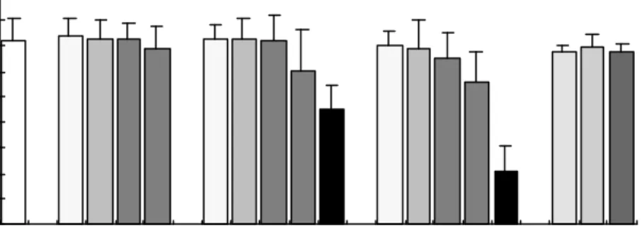
실험군으로 합성에스트로젠 중 phenol계인 bisphenol-A (BPA), octylphenol (OP)를 10-9, 10-7, 10-6, 10-5, 10-4 M 농도별로 aroclor 1254(A1254)를 0.02, 0.2, 1 ㎍/ml로 처 리한 후 2일간 정자세포를 배양한 후 세포를 회수하여 trypan blue (0.4%) 로 생존율을 확 인하였다. BPA와 OP를 1 mM 첨가하였을때는 첨가시 바로 모든 세포가 죽는 것을 관찰하 였으며, 10-4 M 처리하였을 때 대조군이나 다른군에 비해 유의하게 세포의 생존율이 감소 하였다 (Figure 1).
2. Xenoestrogens 처리를 통한 유전자 발현
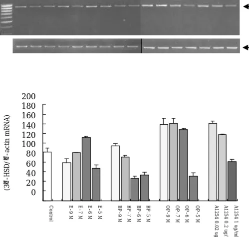
1) Cytochrome P450 동위효소 (CYPscc, 3β-HSD, CYP17α, CYP19) 의 유전자 발현
BPA, OP (10-9, 10-7, 10-6, 10-5, 10-4 M)와 PCB (0.02, 0.2, 1 ㎍/ml)를 처리한 세포 군에서 2일간 배양 후 각 군의 모든 세포를 회수하여 RNA를 분리하여 RT-PCR방법으로 Leydig 세포에서 조절되는 호르몬의 작용을 확인하기 위해서 cholesterol에서 androgen 그리고 estrogen을 형성하기 위해 작용하는 전이효소인 P450 동위효소군의 작용을 조사하 기 위해 이들 효소의 유전자 발현변위를 semi-quantitative PCR방법으로 조사하였다. 합성 에스트로젠이 첨가된 군에서 CYPscc의 유전자 발현이 대조군에 비해 낮은 경향을 나타내 었고 (Figure 2), 3β-HSD의 유전자 발현은 BPA 처리군에서는 농도 의존적으로 그 발현양 이 감소하였으며 (Figure 3), OP와 PCB처리군에서는 저농도에서는 대조군보다 다소 높은 발현을 보였지만 10-5 M의 처리군에서는 대조군에 비해 낮은 발현양상을 보였다. 그러나, CYP17α의 유전자 발현은 모든 합성에스트로젠 처리군에서 농도가 증가할 수록 낮게 나타났 다 (Figure 4). 특히 OP와 PCB의 모든 처리군에서 유의하게 낮은 발현율을 나타내었다.
CYP19 유전자의 발현은 발현양이 적어서 인지 관찰되지 않았다.
3. estrogen 과 testosterone 양 분석
체외에서 2일간 배양된 정자세포로부터 분비된 호르몬의 분석을 위해서 배양된 배양액 으로부터 estrogen (E) 과 teststerone (T)을 검사하였다. 배양액내 E와 T의 양은 대조군 과 비교하여 처리군에서 유의한 차이가 없었으나, 고농도인 BPA와 OP (10-4 M) 처리군에 서 대조군보다 낮은 수치가 측정되었다 (Figure 5, 6).
0 10 20 30 40 50 60 70 80 90
Control E-9 M E-7 M E-6 M E-5 M BP-9 M BP-7 M BP-6 M BP-5 M BP-4 M OP-9 M OP-7 M OP-6 M OP-5 M OP-4 M PCB0.02g/ml PCB0.2g/ml PCB1g/ml
T r e a t m e n t
Viability (%)
*
*
Table 1. Primers used for RT-PCR
Primer Sequence Size (bp)
5' β-actin 5'-GTGGGCCGCTCTAGGCACCAA-3'
3‘β-actin 5'-CTCTTTGATGTCACGCACGATTTC-3' 540 5' CYPscc 5'-AGTGGCAGTCGTGGGGACAGT-3'
3' CYPscc 5'-TAATACTGGTGATAGGCCACC-3' 411 5' CYP
17α5'-CCCATCTATTCTCTTCGCCTGGGTA-3'
3' CYP
17α5'-GCCCCAAAGATGTCTCCCACCGTG-3' 743 5' CYP19 5'-ATAATGTCACCATCATGGTCCCGG-3'
3' CYP19 5'-GCATGATGTGTCTCATGAGGGTCA-3' 579 5' 3β-HSD 5'-TGGTGACAGGAGCAGGA-3'
3' 3β-HSD 5'-AGGAAGCTCACAGTTTCCA-3' 890
Figure 1. The viability of mouse testicular cells treated E
2, BPA, OP and
A1254 (10
- 9, 10
- 7, 10
- 6, 10
- 5, 10
-4M) and A1254 (0.02, 0.2, 1
ug/ml) for 48 hr. (* P<0.05).
CYPscc
β−actin
M 1 2 3 4 5 6 7 8 9 10 11 12 13 14 15 16
411 bp
540 bp
0 20 40 60 80 100 120 140 160 180
Control E-9 M E-7 M E-6 M E-5 M BP-9 M BP-7 M BP-6 M BP-5 M OP-9 M OP-7 M OP-6 M OP-5 M A1254 0.02 ug/ml A1254 0.2 ug/ml A1254 1 ug/ml
Treatment Relative value (CYPscc/-actin mRNA)
A
B
Figure 2. The relative mRNA levels of CYPscc in E2, BPA, OP and A1254 treated pre-pubertal mouse testicular cells for 48 hr.
A. RT-PCR amplification of CYPscc and β-actin mRNA. M : 100 bp ladder, lane 1: control, 2-5: E
2, 6-9 : BP, 10-13: OP (10
-9, 10
-7, 10
-6, 10
-5M, repectively) and 14-16 : A1254 (0.02, 0.2, 1
㎍/ml).
B. Relative changes in the amount of CYPscc mRNA
(relevant to β-actin).
890 bp
540 bp 3 β -HSD
β−actin
M 1 2 3 4 5 6 7 8 9 10 11 12 13 14 15 16
0 20 40 60 80 100 120 140 160 180 200
Control E-9 M E-7 M E-6 M E-5 M BP-9 M BP-7 M BP-6 M BP-5 M OP-9 M OP-7 M OP-6 M OP-5 M A1254 0.02 ug/ml A1254 0.2 ug/ml A1254 1 ug/ml
Treatment
Relative value (3β-HSD/β-actin mRNA)* * *
A
B
Figure 3. The relative mRNA levels of 3β-HSD in E2, BPA, OP and A1254 treated pre-pubertal mouse testicular cells for 48 hr. (* P<0.05).
A. RT-PCR amplification of 3β-HSD and β-actin mRNA. M : 100 bp ladder, lane 1: control, 2-5: E
2, 6-9 : BP, 10-13: OP (10
-9, 10
-7, 10
-6, 10
-5M, repectively) and 14-16 : A1254 (0.02, 0.2, 1
㎍/ml).
B. Relative changes in the amount of 3β-HSD mRNA
(relevant to β-actin).
M 1 2 3 4 5 6 7 8 9 10 11 12 13 14 15 16 CYP17α
β−actin
743 bp
540 bp
0 20 40 60 80 100 120 140 160 180
Control E-9 M E-7 M E-6 M E-5 M BP-9 M BP-7 M BP-6 M BP-5 M OP-9 M OP-7 M OP-6 M OP-5 M A1254 0.02 ug/ml A1254 0.2 ug/ml A1254 1 ug/ml
Treatment Relative value (CYP17αactin mRNA)
* * *
* * *
*
A
B
Figure 4. The relative mRNA levels of CYP
1 7αin E2, BPA, OP and A1254 treated pre-pubertal mouse testicular cells for 48 hr. (* P<0.05).
A. RT-PCR amplification of CYP
17αand β-actin mRNA. M : 100 bp ladder, lane 1: control, 2-5: E
2, 6-9 : BP, 10-13: OP (10
-9, 10
-7, 10
-6, 10
-5M, repectively) and 14-16 : A1254 (0.02, 0.2, 1
㎍/ml).
B. Relative changes in the amount of CYP
17αmRNA
(relevant to β-actin).
0 0.5 1 1.5 2 2.5 3 3.5 4
Control E-9 M E-7 M E-6 M E-5 M BP-9 M BP-7 M BP-6 M BP-5 M BP-4 M OP-9 M OP-7 M OP-6 M OP-5 M OP-4 M A1254 0.02 ug/ml A1254 0.2 ug/ml A1254 1 ug/ml
Treatment
Testosterone (ng/ml)
0 20 40 60 80 100 120 140 160
Control BP-9 M BP-7 M BP-6 M BP-5 M BP-4 M OP-9 M OP-7 M OP-6 M OP-5 M OP-4 M A1254 0.02 ug/ml A1254 0.2 ug/ml A1254 1 ug/ml
Treatment
Estrogen (ng/ml)
Figure 5. The effects of E2, BP, OP and A1254 on testosterone production
of
in vitro cultured pre-pubertal mouse testicular cells.고 찰
환경호르몬으로 알려진 내분비계 장애물질들은 흔히 일상생활에서 사용하는 캔, 플라스 틱이나 일회용품, 살충제, 농약류등에서 검출될 뿐만 아니라, 공기중이나 물과 토양에 오염 되어 동 식물은 물론 먹이사슬을 통해 축적된 형태로 인간에게 돌아오게 된다.7 최근 들어 내분비계 장애물질에 대한 연구가 활발히 이루어지고 있으며, 그 대부분이 스테로이드계 호르 몬과 유사작용이나, 방해, 억제작용을 통해 여러가지 생식기 발달 이상, 신경계 및 면역계에 영향을 일으키는 것으로 알려지고 있다.12, 22
웅성생식기인 정소에서 이루어지는 주요기능 중의 하나는 Leydig 세포에서 남성호르몬 인 testosterone (T)을 만드는 것으로, 정자세포의 분화와 정자형성과정을 유지하고 남성적 성징을 나타내는 중요한 기능을 하고 있다.9, 16 이 호르몬의 합성은 cholesterol이 4가지 효소, cholesterol side-chain cleavage enzyme (CYPscc), 3β-hydroxysterodi dehydrogenase/Δ5,Δ4 isomerase (3βHSD), 17α-hydorxylase/C17-20 lyase (CYP17α) 그 리고, 17-ketosteroid reductase의 작용을 받아 합성된다.18 또한 이렇게 만들어진 T는 CYP19 에 의해 정소내 E2를 형성하게된다. 정소내 E2의 기능은 아직 완전히 알려져 있지 는 않지만 주로 웅성 생식관의 유류, 재흡수. 정자의 성숙 및 뇌하수체호르몬의 조절에도 관여하는 것으로 알려지고 있다.10 따라서 내분비계 장애물질이 이러한 일련의 과정을 방해 한다면, 정상적인 steroidogenesis가 이루어지지 않아, 정자형성이나 정소의 다른 세포의 기능에도 영향을 미칠 것으로 보여진다. Johnson 등 (1992)11은 Dioxin (2,3,7,8- tetrachlorodibenzo-ρ-dioxin)을 성체 흰쥐에 처리하였을 때는 농도 의존적으로 Leydig 세 포의 부피와 기능이 저하되었다고 보고 하고 있으며, Akinbemi 등 (2000)2은 HPTE를 Leydig 세포에 처리하였을 때, 스테로이드 합성의 주원료인 콜레스테롤을 pregnenolone 으로 전환시키는 효소인 CYPscc의 유전자발현이 감소되고, 특히 Leydig 세포에 대한 민감 성이 출생직직후에서부터 미성숙 그리고 성체순으로 나타난다고 보고하고 있다. Andric 등 (2000)3도 polychlorinated biphenyls (PCBs)의 일종인 aroclor 1248을 흰쥐에 투여하였 을 때, 투여 농도에 따른 반응은 조금 차이가 있었으나 3β-HSD, 17α-hydroxylase/lyase 와 17β-hydroxysteroid dehydrogenase의 활성이 저하되어 정소 내 androgenesis가 방해 를 받는다고 확인하였다.
본 연구에서는 정소내 정자세포들의 분화가 완전히 일어나기 전인 생후 15일령의 생쥐 정 자세포를 이용하여 에스트로겐성격을 가지고 있는 것으로 알려진 BPA, OP와 A1254가 정자 형성과정에 미치는 영향을 알아보기 위해 체외배양법을 이용하여 스테로이드 호르몬 합성에 관여하는 효소들의 발현과 성호르몬의 생성량을 확인하였다. 또한 내분비계 장애물질들이 에 스트로겐과 같은 결과를 정소 세포에서 유발시키는 가를 확인하기 위해 E2도 농도별로 처리 하였다. 본 실험결과에서는 합성호르몬인 BPA, OP와 A1254가 10-5M 이하의 농도에서는 세포의 생존에는 크게 영향을 미치지 않아 Raychoudhury 등 (1999)19의 실험결과와 유사 하게 나타났으며, CYPscc, 3β-HSD와 CYP17α유전자의 발현은 억제되어 생쥐 정소내 steroidogenesis를 방해하는 것을 확인할 수 있었다. 또한 E2는 이들과 같은 농도에서는 이 와 같은 억제작용은 일어나지 않는 것으로 보아 이들 내분비계 장애물질이 생체내 에스트론 겐과는 다른 대사를 하는 것으로 여겨진다. 이는 이들 합성에스로젠들이 생체내 E2와 유사
하게 작용함으로써 negative feedback 기작에 의해 뇌하수체 분비호르몬의 분비를 억제하 고 시상하부-뇌하수체 전엽-생식소 축의 호르몬 균형의 교란을 유발, 뇌하수체 분비호르몬 인 FSH나 LH의 분비를 줄임으로써 정상적인 정소의 기능을 방해하여 결과적으로 정소의 분화와 정자형성과정에 이상을 초래할 것으로 사료된다.
이러한 결과는 내분비계장애물질이 포유동물의 성성숙 및 생식세포의 발생을 조절하는 스테로이드 호르몬의 생성 및 발현에 영향을 미치게 된다는 것을 의미하며 따라서, 정자형 성과정 뿐만 아니라 정상적인 생식기관의 분화 및 정소의 발생을 저해할 것으로 사료된다.
그리고, steroidogenesis의 억제가 태아기나 사춘기의 성성숙시기에 일어난다면, 성인보다 더욱 치명적인 해를 입게 될 것으로 예측할 수 있으며 내분비계 장애물질의 규정에 대한 screening방법과 내분비계 장애물질이 미치는 영향에 대하여 체내 및 체외 실험을 통한 정 확한 기전에 관한 연구와 대책이 필요할 것이다. 본 연구는 환경오염물질인 합성에스트로젠 에 대한 동물실험을 통해 이들 물질이 웅성생식계에 작용하는 영향을 연구함으로서 인간에 게도 나타날 여러가지 현상을 규명하고 그 기작을 밝혀내는 기초자료로 이용될 수 있을 것 이다.
참 고 문 헌
1. Abraham E.J., Frawley, S.L. Octylphenol, an environmental estrogen, stimulates prolactin gene expression. Life sciences 1997;60(17):1457-1465.
2. Akinbemi, B.T., Ge, R., Klinefelter, G.R., Gunsalus, G.L., Hardy, M.P. A metabolite of methoxychlor, 2,2-bis(ρ-hydroxyphenyl)-1,1,1-trichloroethane, reduces testosterone biosynthesis in rat leydig cells through suppression of steady-state messenger ribonucleic acid levels of the cholesterol side-chain cleavage enzyme. Biol. Reprod. 2000;62:571-578.
3. Andric, S.A., Kostic, T.S., Stojilkovic, S.S., Kovacevic, R.Z. Inhibition of rat testicular androgenesis by a polychlorinated biphenyl mixture aroclor 1248. Biol.
Reprod 2000;62:1882-1888.
4. Blake, C.A., Boockfor, F.R. Chronic administration of the environmental pollutant 4-tert-octylphenol to adult male rats interferes with the secretion of luteinizing hormone, follicle-stimulation hormone, prolactin and testosterone. Biol. Reprod.
1997;57:255-266.
5. Boockfor, F.R., Blake, C.A. Chronic administration of 4-tert-octylphenol to adult male rats causes shrinkage of the testis and male accessory sex organs, disrupts spermatogenesis and increases the incidence of sperm deformities. Biol.
Reprod. 1997;57:267-277.
6. Chomzynski, P., Sacchi, N. Singly-step method of RNA isolation by acid guanidinium thiocyanate-phenol-chloroform extraction. Annual Biochem.
1998;162,: 156-159.
7. Colborn, T., Dumanoski, D., Myers, J.P. Our stolen Future. Putton, New York, 1996. Colborn, T., vom Saal, F.S. and Solto, A.M.: Developmental effects of endocrine-disruption chemicals in wildlife and humans. Envion. Health Perspect.
1993;101:378-384.
8. Gill, W., Schumacher, G., Bibbo, M., Straus, F., Schoenberg, H. Association of diethylstibestrol exposure in utero with cryptorchidism, testicular hypoplasia and semen abnormalities. J. Urol. 1979;122:36-39.
9. George, F.W., Wilson, J.D. Sex determination and differentiation. IN:Knobil E, NeillJD (eds). The Physiology of Reproduction. Raven Press, New York, 1994;3-28.
10. Hess, R.A., Bunick, D., Lee, K. Bahr, J. Taylor, J.A., Korach, K.S., Lubahn, D.B.
A role for oestrogens in the male reproductive system. Nature 1997;390:509-512.
11. Johnson, L., Dickerson, R. Safe, S.H., Nyberg, C.L., Lewis, R.P., Welsh, T.H. Jr.
Reduced leydig cell volume and function in adult rats exposed to 2,3,7,8-tetrachlorodibenzo-ρ-dioxin without a significant effect on
spermatogenesis. Toxicology 1992;76:103-118.
12. Kuiper, G.J.M., Lemmen, J.G., Carlsson, B. Interaction of oestrogenic chemicals and phytoestrogens with oestrogen receptor β. Endocrinology 1998;139:4252-4263.
13. Le Magueresse-Battistoni, B., G rard, N., J gou, B. Pachytene spermatocytes can achieve meiotic process in vitro. Biochem. Biophys. Res. Commun.
1991;179; 1115-1121.
14. Majdic, G., Sharpe, R.M., O'S haughnessy, P.J., Saunders, P.T.K. Expression of cytochrome P450 17α-hydroxylase/C17-20 lyase in the fatal rat testis is reduced by maternal exposure to xenogenous estrogens. Endocrinology 1996;137:1063-1070.
15. McLachalan, J., Newbold, R., Bullock, B. Reproductive tract lesions in male mice exposed prenatally to diethylstilbestrol. Science 1975;190:991-992.
16. Miller, W.R. Molecular biology of steroid hormone synthesis. Endocri. Rev.
1988;9: 295-318.
17. Nikula, H., Talonpoika, T., Kaleva, M., Toppari, J. Inhibition of hCG-stimulated steroidogenesis in cultured mouse leydig tumor cells by bisphenol A and octylphenols. Toxicol. Appl. Pharmacol 1999;157:166-173.
18. Payne, A.H., Youngblood, G.L. Regulation of expression of steroidogenic enzymes in leydig cells. Biol. Reprod. 1995;52:217-225.
19. Raychoudhury, S.S., Blake, C.A., Millette, C.F. Toxic effects of octylphenol on cultured rat spermatogenic cells and sertoli cells, Toxicol. Appl. Pharmacol 1999;157:192-202.
20. Reinhart, K.C., Dubey, R.K., Keller, P.J., Lauper, U., Rosselli, M.
Xeno-oestrogens and phyto-oestrogens induce the synthesis of leukaemia inhibitory factor by human and bovine oviduct cells. Mol. Hum. Reprod.
1999;5(10):899-907.
21. Saunders, P.T.K., Majdic, G., Parte, P., Millar, M.R., Fisher, J.S., Turner, K.J., Sharpe, R.M. Fetal and perinatal influence of xenoeostrogens on tesits gene expression. Adv. Exp. Med. Biol.1997;424:99-110.
22. Spearow, J.L., Doemeny, P., Sera, R., Leffler, R., Barkley, M. Genetic variation in susceptibility to endocrine disruption by estrogen in mice. Science 1999;285:1259-1261.
23. Tres, L.L., Kierszenbaum, A.L.: Viability of rat spermatogenic cells in vitro is facilitated by their co-culture with sertoli cells in serum-free
J.C. Refinement of the differentiated phenotype of the spermatogenic cell line GC-2spd(ts). Biol. Reprod. 1996;55:923-932.