장관기종은 장관벽의 점막하층이나 장막하층에 다수의 공기 를 포함한 낭종으로 나타나는 특징을 가지고 있다(1). 이는 특 발성으로 발생하거나 이차원인에 의해 나타나며, 기계적장폐 쇄, 감염, 폐쇄성폐질환, 수술, 외상 등의 다양한 원인에 의해 야기된다. 장관기종은 복부일반촬영에서도 보이지만 전산화단 층촬영(Computed tomography, CT)가 진단에 좀 더 민감하 며, CT 이용증가로 장관기종의 발생률이 높아졌다. 또한, 새로 운 시술과 약물과 관련된 장관기종도 발생률 증가에 기여하였 다. CT에서 장관기종의 양상과 동반된 문맥 및 장간막정맥 내 공기여부, 장관의 허혈변화가 환자 예후에 영향을 미친다고 되 어 있다. 이에 저자들은 CT에서 장관기종을 보인 환자의 영상 소견과 임상양상을 알아보고자 하였다.
대상과 방법
2000년 1월에서 2007년 10월까지 CT 소견 상 장관기종으
로 진단된 15명의 환자를 대상으로 하였으며, 남자는 10명, 여 자는 5명이었고 평균 나이는 57세였다.
CT는 13명 의 환 자 에 서 Somatom Sensation 16 scanner(Siemens Medical Systems, Erlangen, Germany) 기 종을 이용하여 140 mL의 조영제를 정맥을 통하여 초당 3 mL 를 주입하였고, 5 mm 폭 조절과 5 mm 두께, 1.5 pitch로 조 영전기, 동맥기, 문맥기, 지연기 축상면영상과 재구성문맥기관 상면영상을 얻었고, 나머지 2명의 환자에서 Lightspeed spiral CT(GE medical systems, Milwaukee, Wis) 기종을 이용하여 140 mL의 조영제를 정맥을 통하여 초당 3 mL를 주입하였고, 7 mm 폭 조절과 7 mm 두께, 1 pitch로 조영전기, 동맥기, 문 맥기 영상을 얻었다.
CT 상 장관기종을 공기방울양상과 선상으로 분류하였으며, 침범부위, 문맥과 장간막정맥 내 공기음영여부, 동반된 장관변 화를 후향분석하였고, 환자의 동반 질환과 주소 및 임상경과를 비교하였다.
장관기종환자: 영상소견 및 임상양상1
김혜린・이혜경・박성진・이범하・고봉민2・홍현숙・백상현
목적: 본원에서 경험한 장관기종환자의 영상소견과 임상양상을 알아보고자 하였다.
대상과 방법: 2001년 1월에서 2007년 10월까지 전산화단층촬영(CT)상 장관기종으로 진단된
15명의 환자를 대상으로 하였으며, CT 상 장관기종의 양상, 침범부위, 문맥 및 장간막정맥 내 공기 여부, 동반된 허혈변화의 유무와 임상양상을 분석하였다.
결과: 15명의 환자는 말기 신질환(5예), 위전절제술(2예) 척추후궁절제술(1예), 결핵성장염(1 예), 폐암(1예), 폐렴(1예)의 병력이 있었으나, 나머지 4예는 특별한 병력이 없었다. CT에서 장관기종은 공기방울양상 6예, 선상 1예, 공기방울 및 선상은 8예에서 보였다. 침범부위는 소 장 6예, 대장 6예, 소장과 대장을 모두 침범한 예는 3예이었다. 문맥 및 장간막정맥 내 공기가 동반된 경우는 6예였으며, 장관의 교액 및 허혈변화는 5예에서 관찰하였다. 또한, 복강수기종 2예에서, 복강기종과 충양돌기 천공이 1예에서 동반되었다. 수술은 CT에서 허혈병변과 복강기 종 및 수기종이 동반된 7예, 충양돌기 천공에 의한 1예와 범복막염 의심하에 1예에서 시행되 었으며, 수술하지 않는 6예 중 3예에서 보존치료 후 호전되어 퇴원하였고, 진단 후 치료경과 중 3예는 사망하였다.
결론: 장관기종환자에서 동반질환은 말기 신질환이 가장 많았으며, CT에서 문맥 및 장간막정 맥 공기, 동반된 허혈변화, 선상장관기종이 있으면 예후가 나빴다.
1순천향대학교 부천병원 영상의학과
2순천향대학교 부천병원 소화기내과
이 논문은 2007년 11월 22일 접수하여 2007년 12월 28일에 채택되었음.
결 과
15명의 환자는 말기신질환(5예), 위암으로 위전절제술(2예)
척추후궁절제술(1예), 결핵성 장염(1예), 폐암(1예), 폐렴(1 예)의 병력이 있었으나, 나머지 4예는 특별한 병력이 없었다.
주소로는 복통(12예) 및 복부팽만감(2예)을 호소하였으나, 1 예에서는 증상이 없었다(Table 1).
Table 1. Clinical Profile and CT Findings of Pneumatosis Intestinalis Patients
Sex/ Underlying CT Finding
Age Disease Involvement Gas PV / MV Associated Progress
Site Pattern Gas Finding
01 F/28 N-C TI, AC B - / - - Conservative treatment
02 M/56 N-C I B + / + - Conservative treatment
03 F/80 N-C R B - /- - Conservative treatment
04 M/5 N-C AC B - / - Appendicitis Appendectomy
05 F/44 ESRD DC, SC B / L - / - Strangulation of colon Total colectomy
06 F/49 ESRD J B - / - Strangulation Segmental resection
of jejunum of jejunum
07 M/58 ESRD I B / L + / + Strangulation of ileum Segmental resection
of ileum
08 M/53 ESRD DC, SC B / L - / - - Conservative treatment
09 M/70 ESRD J, I, B / L - / - Ischemic change and Diagnostic laparoscopy
TC, SC hydropneumoperitoneum without perforation
of bowel
10 M/57 Gastrectomy I B / L + / - - Adhesiolysis &
due to AGC Serosa repair of ileum
11 M/75 Gastrectomy J, I B / L - / - Ischemic change and Primary repair &
due to AGC hydropneumoperitoneum segmental resection
of ileum
12 F/64 Laminectomy AC L - / - Pneumoperitoneum Primary repair
due to SDH of peforated ileum
13 M/69 Tuberculosis enteritis I B / L + / - - Expired
14 M/60 Lung cancer AC B + / - - Expired
15 M/86 Pneumonia Small & large bowel B / L + / - - Expired
N-C, nonspecific; ESRD, end state of renal disease; AGC, advanced gastric cancer; SDH, subdural hematoma; J, jejunum; I, ileum; TI, ter- minal ileum; AC, ascending colon; TC, transverse colon; DC, descending colon; SC, sigmoid colon; R, rectum; B, bubble; L, linear; PV, portal vein; MV, mesenteric vein
A B
Fig. 1. 44-years-old woman with end stage renal disease
Contrast enhanced CT scan shows (Fig. 1A, B) markedly dilated descending colon with large amount of hematoma filling the thick- ened wall (arrows) represent extensive mucosal necrosis. Linear and bubble like pneumatosis intestinalis (arrow heads) is noted within the wall and hematoma. There is no evidence of wall enhancement.
A B
C D
E F
Fig. 2. 55-years-old man with abdominal pain
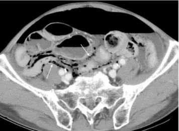
Contrast enhanced CT scan shows bubble like pneumatosis intestinalis in the ileum (Fig. 2A) and branching pattern of air in portal (Fig. 2B) and ileal vein branch of superior mesenteric vein (Fig. 2C, arrow).
Follow up image after 8 days shows air in portal and mesenteric veins are absorbed (Fig. 2D). Air in bowel loops are also subsided but diffuse enhancing wall thickening are developed (Fig. 2E) in ileum (arrow heads) and descending colon(arrow). Sigmoidoscopic findings show well demarcated erythematous and friable mucosal changes at distal descending colon (Fig. 2F).
CT에서 장관기종은 공기방울양상과 선상으로 8예에서 보였 고(Fig. 1A), 공기방울양상만으로 보인 예는 6예이었으며(Fig.
2A), 선상만으로 보인 예는 1예이었다. 침범부위는 소장에 국 한된 예가 6예이었고, 대장에 국한된 예는 6예이었으며 소장 과 대장에 모두 있었던 예는 3예이었다. 또한, 문맥 및 장간막 정맥 내 공기가 있었던 경우는 6예였으며(Fig. 2B), 동반된 장 관의 교액변화(strangulation)는 3예에서 있었고(Fig. 1B) 허 혈변화는 2예에서 관찰하였다. 복강수기종(Fig 3A)은 2예, 복 강기종 및 천공충수염(Fig. 4)은 각각 1예에서 동반되었다.
수술은 총 9예에서 시행하였으며, CT에서 교액변화를 보였 던 3예 중 1예는 수술시 상행 결장을 제외한 전 대장에 허혈 변화가 있어 전결장절제술을 시행하였으며, 병리소견에서 허혈 변화가 있었다. 다른 1예에서는 공장구역절제술을 시행하였고,
병리소견에서 장관전층을 침범한 출혈괴사가 있었으며, 나머지 1예에서는 회장구역절제술을 하였고, 병리소견에서 장관 일부 에만 괴사가 있었다. 또한, CT에서 허혈변화와 복강수기종이 있는 2예 중 1예에서는 회장천공이 있어 천공부위의 복원과 회장구역절제술을 하였으나 병리소견에서 급성화농염증이 국 소적으로 있었다. 다른 1예에서는 수술시 소량의 복수만 있고 장관의 허혈변화가 없었으며, 복강기종의 원인을 찾지 못하였 다. CT에서 허혈변화가 없었으나 천공충수염 1예, 장관 유착 1예, 복강기종 1예, 임상적으로 범복막염이 의심되었던 1예등 의 4예에서 수술을 시행하였으나 모두 장관의 이상소견은 없 었다. 4예 중 천공된 충수돌기절제술을 1예에서 시행하였으며, 장관 유착으로 인한 부종 및 확장과 장막파열이 1예에서 보여 유착분리술과 장막복원을 시행하였고, 복강기종이 있는 1예에 서는 회장천공이 있어 복원하였다. 나머지 1예에서는 수술시 장관이상소견은 없었으나 회장에 메켈게실이 있어 제거하였다.
수술 후 계속적인 복부 통증과 소화불량이 해소되지 않았으며 수술 8일 후 시행한 CT에서 문맥 및 장간맥 정맥 내 공기는 소실되었으나(Fig. 2D), 골반강 내에 위치한 회장 및 S-결장, 하행결장 말단부의 벽이 전반적으로 두꺼워졌으며 조영증강이 되었고 골반강 내 복수가 생겨(Fig. 2E) 허혈변화가 생겼음을 알 수 있었다. 이후 시행한 직장 내시경검사에서 상부하행결장 에 경계가 좋은 홍반과 쉽게 망가지는 점막이 보였으며 그 이 하 부위는 홍반과 함께 전반적인 궤양이 있었고 S자 결장은 주 변 점막홍반과 함께 협착이 있었으나 보존치료로 호전되었다 (Fig. 2F).
수술하지 않는 6예 중 3예에서 보존치료 후 호전되어 퇴원 하였고, 나머지 3예는 진단 후 치료경과 중 사망하였다. 보존 치료를 시행한 3예 중 1예는 퇴원 후 5개월 후 다시 재발하였 으나 보존치료 후 호전되었다.
A B
Fig. 4. 5-years-old boy with perforated appendicitis
Contrast enhanced CT scan shows bubble like pneumatosis intestinalis in the ascending colon (Fig. 4A). Appendix reveals dilata- tion, diffuse wall thickening and appendicoliths (Fig. 4B, arrow).
Fig. 3. 75-years-old man with total gastrectomy due to AGC Contrast enhanced CT scan shows linear and bubble like pneumatosis intestinalis in the ileum (arrows) and hydropneu- moperitoneum.
고 찰
장관기종은 장벽 내에 공기 낭종으로 나타나며 이는 여러 상 태에서 발생하며 특발성과 이차성 등의 두 가지 유형으로 나 누며, 약 15%는 원인이 없는 일차성으로 주로 성인에서 발병 하며 하부결장을 침범하는 경우가 많고 예후가 좋다. 일차성 장관기종은 드물게 발생하며 병리학적으로 다수의 서로 연결 되지 않은 공기 낭종이 점막하 및 장막하층에 위치하며 점막 이나 근육층은 정상소견을 보인다.
장관기종은 질환이 아니고 영상학적 소견이다(2, 3). Lisa 등(3)은 장관기종을 양성과 생명을 위협하는 원인에 의한 두 분류로 나누면서 장관기종이 질환이 아니고 영상학적 소견이 라는 것을 이해하는 것이 중요하며, 환자의 전반적인 임상상태 를 숙지하는 것이 장관기종을 일으키는 양성과 생명을 위협하 는 원인을 판단하는데 중요한 단서가 된다고 하였다. 양성 원 인으로는 다양한 위장관 질환, 폐기종과 천식 등의 폐질환, 의 인성, 장기이식과 원인이 없는 일차성 장관기종이 있다. 생명 을 위협하는 원인으로는 장허혈, 장간막혈관질환, 장폐쇄, 장 염, 부식제섭취, 독성거대결장, 외상, 교원성혈관질환이 있다 (3). 이번 연구에서 동반질환은 말기신질환이 많았으며(5/15, 33.3%) 이는 만성신질환이 있는 경우 나타날 수 있는 점막궤 양이나 장관 내 세균증식(4)이 야기되거나, 구토나 설사 등(5) 에 의한 장관 내 압력증가에 의한 것으로 여겨진다.
장관기종의 정확한 발병기전은 알려지지 않았다. 여러 가지 가설(6-10)이 있으며, 세균이론은 공기를 형성시키는 세균들 이 점막틈새를 통해 장막하층에 도달하거나 장관 벽 내에서 공 기를 형성하여 점막의 투과성을 증가시켜 일으키게 되며, 기계 이론은 외상이나 장관폐쇄, 연동운동 등에 의해 증가한 압력으 로 장관 내 공기가 장벽을 뚫고 들어가 점막하나 장관 벽에 다 양한 크기의 공기 방울들이 장벽 내에 보이게 되는 것을 말한 다. 점막손상이론은 장염 및 장허혈, 크론병 등에 의한 점막분 열로 의해 발생한다고 하였으며, 폐질환이론은 폐쇄성폐질환에 서 증가한 압력에 의해 일부 막힌 기관지와 기침이 폐포를 파 열시켜 공기가 기관지 및 혈관을 따라 벗겨져 종격동으로 이 동하여 식도공을 따라 후복막강에 간 후 장간막 사이를 벗겨 내 장막과 점막하 위치에 도착함으로써 야기된다고 하였다. 그 외 화학적, 영양학적, 종양학적 이론이 있는데, 영양상태에 따 라 정상세균총이 변화하여 야기된다고 보고 있으며, 암환자에 있어서는 장관의 허혈변화보다는 화학요법제에 의한 점막변화 나 세균증식에 의한다고 보고 있다.
장관기종의 진단에 대부분 복부단순촬영과 CT가 이용되며 (11), CT는 단순촬영보다 장벽내기종과 문맥 및 장간막 정맥 내 공기발견에 좀 더 민감하다고 보고하고 있다(12, 13). 또한, 이전 나선 CT와 비교하여 16채널 CT에서는 재구성영상을 얻 을 수 있기 때문에 축상면에서 선상장관기종이 공기방울양상 으로 확인되어 좀 더 공기방울양상의 공기 낭종 발견이 높아졌 다. 장관기종이 선상을 보이는 경우가 공기방울양상보다 장벽 전층을 침범한 괴사가 있을 때 좀 더 많이 관찰된다고 보고하
였다(14, 15). 이번 연구에서도 선상장관기종이 있었고 사망한 두 예를 제외하고 7예 모두 수술을 시행하였으며(7/9, 77.8%) 이 중 4예에서는 장관 벽 괴사가 동반되었다. 공기방울양상으 로만 있는 경우는 보존치료(3/6, 50%)로 치료되었으며, 수술 을 시행한 두 명 중 한 명에서 장관 벽 괴사가 동반되었다. 다 른 1예는 폐암에 의한 전신상태불량으로 사망하였으며 장관기 종에 의한 결과로 보기는 어렵다. 문맥 공기는 간 주변부까지 확대되는 관상으로 가지치는 양상의 저음영으로 보이며, 장간 막 공기와 함께 장벽 전층괴사가 있는 경우에 초래된다(14, 15). Wiesner 등(14)은 CT에서 장관기종이 문맥-장간맥 정 맥공기와 동반된 경우는 장관 전층 벽에 의한 괴사와 동반되었 을 가능성이 매우 큰 반면 방울모양의 장관기종이나 단지 문 맥-장간맥 정맥 공기만 있는 경우에는 약 1/3에서 일부 근육 층허혈증이 동반된다고 하였다. 이번 연구에서 문맥-장간막 정 맥 공기가 있는 6예 중 2예에서 수술을 시행하였고 3예는 치 료경과 중 사망하였다. 다른 1예는 수술시 장관 이상은 없었으 나, 수술 후 증상악화와 함께 허혈장염소견이 나타났다.
장관 벽의 비후, 점막 조영증강감소, 장관확장, 동맥 또는 정 맥 폐색, 복수 등이 보일 수 있다(12, 16). 장관 허혈에 의한 경우에는 특정혈관분포에 따라 국한될 수 있으며, 장막 및 장 막하 기종파열에 의해 특발기복증이 야기될 수 있다(17).
이번 연구의 제한점으로는 연구대상이 너무 적어 장관기종 의 영상소견빈도를 알아내는 것에 무리가 있었으나 다양한 영 상소견과 동반질환 및 예후를 갖고 있어 결론을 내리는데 문 제가 없을 것으로 생각하였다.
결론적으로 장관기종환자에서 동반질환은 말기신질환이 가 장 많았으며, CT에서 선상장관기종, 동반된 문맥 및 장간막 정 맥 공기 및 허혈변화가 있으면 예후가 나빴다.
참 고 문 헌
1. SzucS Ra, Wolf EL, Gramm HF, Scholz FJ, Eisenberg RL, Hall DA, et al. Miscellaneous abnormalities of the colon. In Gore RM, Levine MS. Textbook of gastrointestinal radiology. 2nd ed. Philadelphia: W.
B. Saunders, 2000:1114-1115
2. Pear BL. Pneumatosis intestinalis: a review. Radiology 1998;207:
13-19
3. Ho LM, Paulson EK, Thompson WM. Pneumatosis intestinalis in the adult: benign to life-threatening causes. AJR Am J Roentgenol 2007;188:1604-1613
4. Strid H, Simre′n M, Stotzer PO, Ringstro¨m G, Abrahamsson H, Bjo¨rnsson ES. Patients with chronic renal failure have abnormal small intestinal motility and a high prevalence of small intestinal bacterial overgrowth. Digestion 2003;67:129-37
5. Skorecki K, Green J, Brenner BM. Chronic renal failure. In Braunwald F, Fauci AS, Kasper DL, Hauser SL, Longo DL, Jameson JL. Harrison’s Principles of internal medicine. 15th ed. New York: McGraw-Hill, Medical Pub. Division, 2001:1551-1562 6. Galandiuk S, Fazio VW. Pneumatosis cystoides intestinalis: a re-
view of the literature. Dis Colon Rectum 1986;29:358-363
7. Pieterse AS, Leong AS, Rowland R. The mucosal changes and pathogenesis of pneumatosis cystoides intestinalis. Hum Pathol 1985;16:683-688
8. Priest RJ, Goldstein F. Pneumatosis cystoides intestinalis. In Bockus HL, Berk JE Bockus’ gastroenterology. 4th ed. Philadelphia:
Saunders, 1985:2474-2483
9. Yale CE, Balish E, Wu JP. The bacterial etiology of pneumatosis cystoides intestinalis. Arch Surg 1974;109:89-94
10. Feczko PJ, Mezwa DG, Farah MC, White BD. Clinical significance of pneumatosis of the bowel wall. Radiographics 1992;12:1069- 1078
11. Caudill JL, Rose BS. The role of computed tomography in the eval- uation of pneumatosis intestinalis. J Clin Gastroenterol 1987;9:223- 226
12. Schindera ST, Triller J, Vock P, Hoppe H. Detection of hepatic por- tal venous gas: its clinical impact and outcome. Emerg Radiol 2006;12:164-170
13. Fisher JK. Computed tomography of colonic pneumatosis intesti-
nalis with mesenteric and portal venous air. J Comput Assist Tomogr 1984;8:573-574
14. Wiesner W, Mortele KJ, Glickman JN, Ji H, Ros PR. Pneumatosis intestinalis and portomesenteric venous gas in intestinal ischemia:
correlation of CT findings with severity of ischemia and clinical outcome. AJR Am J Roentgenol 2001;177:1319-1323
15. Kernagis LY, Levine MS, Jacobs JE. Pneumatosis intestinalis in pa- tients with ischemia: correlation of CT findings with viability of the bowel. AJR Am J Roentgenol 2003;180:733-736
16. Smerud MJ, Johnson CD, Stephens DH. Diagnosis of bowel infarc- tion: a comparison of plain films and CT scans in 23 cases. AJR Am J Roentgenol 1990;154:99-103
17. Koss LG. Abdominal gas cysts (pneumatosis cystoides intestino- rum hominis): an analysis with a report of a case and a critical re- view of the literature. Arch Pathol 1952;53:523-549
J Korean Radiol Soc 2008;58:149-154
Address reprint requests to : Hae Kyung Lee, M.D., Department of Radiology, Soonchunhyang University Hospital Bucheon 1174, Jung-dong, Wonmi-gu, Bucheon-si, Gyeonggi-do 420-767, South Korea.
Tel. 82-32-621-5851 Fax. 82-32-621-5874 E-mail: [email protected]
Pneumatosis Intestinalis: CT Findings and Clinical Features1
Hye Lin Kim, M.D., Hae Kyung Lee, M.D., Seong Jin Park, M.D., Boem Ha Yi, M.D., Bong Min Ko, M.D.2, Hyun Sook Hong, M.D., Sang Hyun Paik, M.D.
1Department of Radiology, Soonchunhyang University Hospital Bucheon
2Department of Gastroenterology, Soonchunhyang University Hospital Bucheon
Purpose: The purpose of this study is to evaluate the CT findings and clinical features of patients with pneu- matosis intestinalis.
Materials and Methods: From January 2001 to October 2007, 15 patients with pneumatosis intestinalis were diagnosed by the use of CT. We analyzed the clinical features and CT findings to assess the involvement site, the presence of portal and mesenteric vein gas, and the existence of accompanied ischemic change.
Results: Of the 15 patients, five patients had end stage renal disease (33.3%), two patients underwent a gas- trectomy, one patient underwent a laminectomy, one patient had tuberculous enteritis, one patient had lung cancer and one patient had pneumonia. Four patients presented with no specific disease. There was portal or mesenteric venous gas in six cases, and strangulation or an ischemic change of the bowel in five cases.
Otherwise, pneumatosis intestinalis was associated with hydropneumoperitoneum in two cases, pneumoperi- toneum in one case and a single case of perforated appendicitis. Nine patients underwent surgery for ischemic change of the bowel, hydropneumoperitoneum, appendicitis, and a clinical sign of panperitonitis. Among the remaining six patients, three patients recovered and were discharged, and three patients expired during pro- gression of the disease.
Conclusion: End stage renal disease is the most common condition associated with pneumatosis intestinalis.
The presence of portomesenteric venous gas, ischemic change of the bowel, and linear pneumatosis intesti- nalis are indicative of a poor prognosis.
Index words :Pneumatosis cystoides intestinalis Portal vein
Kidney failure
Computed tomography (CT)