34 Copyright © 2016 Korean Neurotraumatology Society
Introduction
Traumatic vertebral artery dissection (VAD) is rare af- ter a cervical spine injury.1,3,5) Bilateral VAD occasionally causes vertebrobasilar stroke; however, unilateral VAD does not usually cause symptomatic brain ischemia and is treated conservatively. Nevertheless, physicians should carefully suspect a silent vertebral artery injury because dissection can cause symptomatic cerebral infarction after the latency period or due to an unexpected triggering fac- tor. Here, we report a case of unilateral VAD presenting
with symptomatic, multiple cerebral infarctions and dis- cuss the clinical course.
Case Report
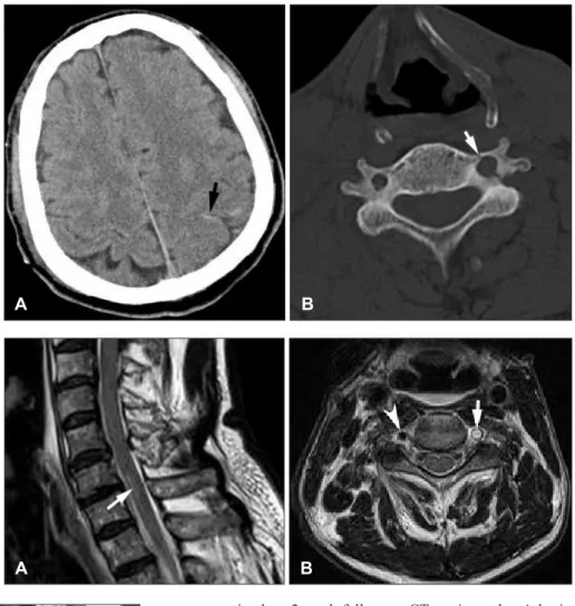
A 73-year-old man was referred from a local hospital with impaired consciousness and left hemiparesis after a motor car accident. He was diagnosed with traumatic sub- arachnoid hemorrhage on the left parietal lobe and left lat- eral mass fractures on C2, C5, and C6, involving the transverse foramen (Figure 1). A neurological examina- tion revealed stupor with a Glasgow Coma Scale score of 8. Severe grade 2 hemiparesis of the left arm and leg was detected. High signal was seen at the C6-7 levels of the spi- nal cord on a sagittal cervical spine magnetic resonance imaging (MRI). The signal void on the left vertebral ar- tery was lost on the C6 axial image (Figure 2). He was di- agnosed with left VAD and was managed conservatively.
Neck computed tomography (CT) angiography showed obliteration of the left second segment of the vertebral ar- tery (V2) (Figure 3). The obliteration of the V2 segment
Multiple Cerebral Infarctions due to Unilateral Traumatic Vertebral Artery Dissection after Cervical Fractures
Sang-Youl Yoon, MD, Seong-Hyun Park, MD, PhD, Jeong-Hyun Hwang, MD, PhD, and Sung-Kyoo Hwang, MD, PhD
Department of Neurosurgery, Kyungpook National University Hospital, Daegu, Korea
We report a case of multiple symptomatic cerebral infarctions from a traumatic vertebral artery dissection (VAD) after cervical fractures. A 73-year-old man was admitted with stuporous mentality and left hemiparesis after a motor-vehicle accident. A brain computed tomography (CT) scan at admission showed a traumatic subarachnoid hemorrhage on the left parietal lobe. A cervical CT scan showed left lateral mass fractures on C2, C5, and C6, involving the transverse foramen.
Cervical spine magnetic resonance imaging (MRI) revealed loss of signal void on the left vertebral artery. Neck CT angi- ography showed left VAD starting at the C5 level. Brain MRI revealed acute, multiple cerebral infarctions involving the pons, midbrain, thalamus, corpus callosum, and parietal and frontal lobes on diffusion weighted images. The patient was treated conservatively at the intensive care unit in the acute stage to prevent extent of stroke. Aspirin was started for anti- platelet therapy in the chronic stage. The possibility of symptomatic cerebral infarctions due to traumatic VAD following cervical fracture should be considered.
(Korean J Neurotrauma 2016;12(1):34-37) KEY WORDS: Cerebral infarction ㆍCervical vertebrae ㆍSpinal fractures ㆍVertebral artery dissection.
CASE REPORT
Korean J Neurotrauma 2016;12(1):34-37
pISSN 2234-8999 / eISSN 2288-2243 http://dx.doi.org/10.13004/kjnt.2016.12.1.34
Received: October 10, 2015 / Revised: December 2, 2015 Accepted: December 16, 2015
Address for correspondence: Seong-Hyun Park, MD, PhD Department of Neurosurgery, Kyungpook National University Hos- pital, 130 Dongdeok-ro, Jung-gu, Daegu 41944, Korea
Tel: +82-53-200-5652, Fax: +82-53-423-0504 E-mail: [email protected]
cc This is an Open Access article distributed under the terms of Cre- ative Attributions Non-Commercial License (http://creativecommons.
org/licenses/by-nc/3.0/) which permits unrestricted noncommercial use, distribution, and reproduction in any medium, provided the original work is properly cited.
Sang-Youl Yoon, et al.
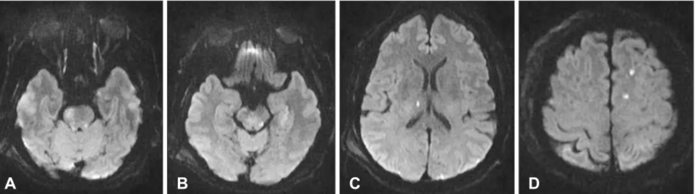
http://www.kjnt.org 35 remained on 2-week follow-up CT angiography. A brain MRI was performed to evaluate the cause for impaired consciousness and motor weakness. Diffusion weighted im- ages revealed multiple area of abnormally high signals in the right side of the pons, left side of the midbrain, right thalamus, corpus callosum splenium, both parietal lobes, and the left frontal lobe, indicating multiple acute cerebral infarctions (Figure 4). Ischemic areas were diffusely dis- tributed to the anterior and posterior circulation of the Circle of Willis. Ischemic stroke occurred immediately after cervical spine trauma and neurological deficits after trauma resulted from brain ischemia. Aspirin was started for antiplatelet therapy 1 week after the trauma after con- firming disappearance of the acute traumatic hemor- rhage. He was discharged from the hospital on aspirin to receive neurological follow-up and rehabilitation therapy.
A brain CT scan at discharge showed subdural fluid col- lection in both frontal lobes. His motor weakness and consciousness state were improving gradually to a grade 4 weakness and alert state on follow-up examination. Bi- lateral chronic subdural hematomas (CSDHs) were ob- served with headache 4 months after the trauma (Figure 5).
FIGURE 1. A: Brain computed tomography (CT) scan showing traumatic subarachnoid hemor- rhage (arrow) on the left parietal lobe after trauma. B: Cervical CT scan demonstrating fracture of the left transverse process of C6 (arrow) with encroachment on
the neural foramen. A B
FIGURE 2. Cervical spine mag- netic resonance imaging. A: Sag- ittal T2 weighted image show- ing a high signal (arrow) on the C6-7 cord on a sagittal image. B:
Loss of the signal void was de- tected on the left vertebral ar- tery (arrow) on the C6 axial T2 weighted image. See the normal signal void of the right vertebral
artery (arrowhead). A B
FIGURE 3. Neck computed tomography angiography showing obliteration of the V2 segment on the left vertebral artery (ar- row).
36 Korean J Neurotrauma 2016;12(1):34-37 Vertebral Artery Dissection
Burr hole trephinations were performed on both sides for the CSDHs.
Discussion
The incidence of vertebral artery injury after blunt trau- ma of the cervical spine is unknown, as it is rarely symp- tomatic and therefore easily overlooked.3) Vertebral artery injury may be more prevalent than commonly believed af- ter cervical spine fracture. Traumatic VAD following se- vere neck trauma was first reported in 1955, and most cas- es result from chiropractic maneuvers of the neck or traffic accidents.5) The possibility of vertebral artery injury should be considered when establishing the clinical management scheme for blunt trauma of the cervical spine.10)
Although the mechanisms are not absolutely clear, there seems to be an important relationship between arterial dissection and cervical trauma. Giacobetti et al.1) reported that flexion distraction-type injuries are the most common
mechanism of non-penetrating vertebral artery injuries, followed by a flexion compression mechanism. Because of their unique course through four or five transverse fo- ramen, the vertebral arteries are particularly prone to di- rect traumatic damage.5) Fractures involving the lateral masses, particularly fractures of the transverse foramen, may damage the artery within its bony confines.The ini- tial injury to the artery most likely involves intimal dis- ruption through excessive distraction and stretching of the artery between two adjacent transverse foramens.10) Uni- lateral occlusion of the vertebral artery seldom results in symptomatic ischemic stroke. Our case with normal con- tralateral vertebral artery rarely developed neurological deficits due to embolic infarction, considering thrombosis had not extended distally to the basilar trunk. A delay in clinical presentation has been attributed to propagation of distal extension of the dissection, mural thrombus into the basilar system, and embolization to the brain.11) Blunt trau- ma on the vertebral artery can result in occlusion, which is not necessarily confined to the level of the bony injury. The site of dissection can serve as a site of progressive throm- bosis or artery-to-artery emboli, the latter causing occlu- sion of the distal branch artery.6,8)
Clinical symptoms of VAD after neck trauma consist of neck and/or head pain, often localized to the site of intimal disruption, which are common warning symptoms of dis- section. Symptoms are often only minor and there may be no neurologic deficits. However, patients who develop ver- tebrobasilar ischemia often die or are severely impaired.5) Vertebrobasilar ischemia after cervical spine injury was first described in 1955.7) In this report, the posterior infe- rior cerebellar artery (PICA) syndrome developed several days after a C5 fracture. Unilateral VAD with subsequent thrombosis may not lead to clinical symptoms due to ade- quate blood supply through PICA from the contralateral side and/or reconstitution through the intramuscular col- lateral vessels of the thyrocervical trunk.4) Willis et al.10) FIGURE 4. Brain magnetic resonance imaging showing multiple high signals on diffusion weighted images of the pons (A), mid- brain (B), thalamus (C), and frontal lobe (D).
A B C D
FIGURE 5. Brain computed tomography scan showing chronic subdural hematomas on both frontal lobes (*) 4 months after the trauma.
Sang-Youl Yoon, et al.
http://www.kjnt.org 37 reported on a prospective clinical study in which none of
26 patients with a vertebral artery injury clearly devel- oped neurological dysfunction or other sequelae. Our pa- tient is rare, as he presented with ischemic brain stroke at anterior and posterior circulation of the Circle of Willis.
Cerebral infarction sequelae remained, such as limb weak- ness resulting from the pons infarction. Parent et al.4) re- ported a retrospective review of 640 patients with cervical spine fractures in which five patients had a lateral disloca- tion of the cervical spine and injuries to the vertebral ar- teries. Three of these patients developed symptoms of vertebrobasilar ischemia, which led to a VAD diagnosis based on angiographic or postmortem findings. Conven- tional angiography remains the gold standard but Doppler ultrasound, duplex sonography, MRI, and MR angiogra- phy demonstrate vessel stenosis or occlusion, and a hema- toma may be seen in the vessel wall.2) It is important to detect VAD before ischemic sequelae occur. Recent prog- ress in neuroradiology and neurosonography allows a noninvasive approach to diagnose VAD.
Treatment for traumatic VAD is controversial. No guide- lines have been established for the timing or means of an- ticoagulation or interventional therapy. However, the ther- apeutic goal for acute stage VAD remains preventing ischemic stroke. Antiplatelet or anticoagulation therapy is generally recommended for intimal disruption.9) However, anticoagulation in a patient with trauma causes a signifi- cant hemorrhagic risk. Interventional therapy is an alter- native in patients with severe vertebral artery injuries or symptoms of a vertebrobasilar embolism.12)
The prognosis for VAD is highly variable, but usually remains good, depending on the extent of the damage and the presence of adequate collateral circulation. VAD does not usually cause a critical manifestation. The rate of VAD- associated ischemia in patients with cervical spine trauma is low because the opposite vertebral artery fills the intra- cranial portion of the occluded vessel. However, if collat- eral blood flow is inadequate, an occlusion may present severe vertebrobasilar stroke. In addition, a thrombus in an occluded segment of the vertebral artery can create an embolic shower, as shown here. Early recognition of VAD
following cervical injury and considering the natural his- tory of VAD may lead to proper treatment and improve survival.
Conclusion
We report a rare case of symptomatic, multiple cerebral infarctions from unilateral VAD after cervical fractures.
Physicians should consider the possibility of ischemic brain stroke, even though the incidence of cerebral infarction due to unilateral VAD following cervical injury is low.
■ The authors have no financial conflicts of interest.
REFERENCES
1) Giacobetti FB, Vaccaro AR, Bos-Giacobetti MA, Deeley DM, Albert TJ, Farmer JC, et al. Vertebral artery occlusion associated with cervical spine trauma. A prospective analysis. Spine (Phila Pa 1976) 22:188-192, 1997
2) Hoffmann M, Sacco RL, Chan S, Mohr JP. Noninvasive detec- tion of vertebral artery dissection. Stroke 24:815-819, 1993 3) Miyachi S, Okamura K, Watanabe M, Inoue N, Nagatani T, Takagi
T. Cerebellar stroke due to vertebral artery occlusion after cervical spine trauma. Two case reports. Spine (Phila Pa 1976) 19:83-88, 1994 4) Parent AD, Harkey HL, Touchstone DA, Smith EE, Smith RR. Lat- eral cervical spine dislocation and vertebral artery injury. Neuro- surgery 31:501-509, 1992
5) Schellinger PD, Schwab S, Krieger D, Fiebach JB, Steiner T, Hund EF, et al. Masking of vertebral artery dissection by severe trauma to the cervical spine. Spine (Phila Pa 1976) 26:314-319, 2001 6) Schwarz N, Buchinger W, Gaudernak T, Russe F, Zechner W. In-
juries to the cervical spine causing vertebral artery trauma: case reports. J Trauma 31:127-133, 1991
7) Suechting RL, French LA. Posterior inferior cerebellar artery syndrome; following a fracture of the cervical vertebra. J Neu- rosurg 12:187-189, 1955
8) Tekin S, Aykut-Bingöl C, Aktan S. Case of intracranial vertebral artery dissection in young age. Pediatr Neurol 16:67-70, 1997 9) Watridge CB, Muhlbauer MS, Lowery RD. Traumatic carotid
artery dissection: diagnosis and treatment. J Neurosurg 71:854- 857, 1989
10) Willis BK, Greiner F, Orrison WW, Benzel EC. The incidence of vertebral artery injury after midcervical spine fracture or sublux- ation. Neurosurgery 34:435-441; discussion 441-442, 1994 11) Wirbel R, Pistorius G, Braun C, Eichler A, Mutschler W. Bilateral
vertebral artery lesion after dislocating cervical spine trauma. A case report. Spine (Phila Pa 1976) 21:1375-1379; discussion 1380, 12) Yamaura A, Watanabe Y, Saeki N. Dissecting aneurysms of the 1996
intracranial vertebral artery. J Neurosurg 72:183-188, 1990