CLINICAL ARTICLE
J Kor Neurotraumatol Soc 2011;7:24-28 ISSN 1738-8708
Received: December 2, 2010 / Revised: February 14, 2011 Accepted: April 11, 2011
Address for correspondence: Sung Bae Park, MD
Department of Neurosurgery, Inje University College of Medicine, Seoul Paik Hospital, 85 Jeo-dong 2-ga, Jung-gu, Seoul 100-032, Korea
Tel: +82-2-2270-0029, Fax: +82-2-2270-0573 E-mail: [email protected]
흉요추부 분쇄골절의 흉강경 척추 수술에 대한 임상적 및 방사선적 분석
인제대학교 의과대학 서울백병원 신경외과교실
정 종 락 .박 성 배
Clinical and Radiological Analysis of Thoracoscopic Spinal Surgery in Thoracolumbar Burst Fracture
Jong Rak Jung, MD and Sung Bae Park, MD
Department of Neurosurgery, Inje University College of Medicine, Seoul Paik Hospital, Seoul, Korea
Objective: To evaluate the effectiveness and reasonability of thoracoscopic spinal surgery in patients with thoracolumbar burst fractures. Methods: The authors reviewed the patients who had undergone thoracoscopic spine surgery for thoraco- lumbar burst fracture between January 2008 and October 2010. Spine lateral radiographs were taken at preoperative and follow-up periods and thoracolumbar kyphosis (T11-L2 Cobb angle) was measured. Oswestry disability index (ODI) and Visual analogue scale (VAS) scores were also measured preoperatively and follow-up. Results: There were three male patients and 2 female patients and a mean age of patients was 51.6±14.38 years. Mean follow-up duration was 14.0±7.8 months. The mean ODI score in follow-up period was significantly improved (40±1.5 and 13±11.4, p=0.043). The differ- ence between preoperative and follow-up mean VAS scores was significant (8±1.1 and 1±1.7, p=0.042). Mean preopera- tive kyphotic angle was 21.4±6.48 degree and mean follow-up kyphotic angle was 10.3±7.41 degree. There was significant difference (p=0.036). One patient complained a transient intercostal neuralgia and there was postoperative chylothorax in other patient. Conclusion: In this study, there are good clinical and radiological results after thoracoscopic spine surgery in short-term period. Although thoracoscopic spine surgery requires a steep learning curve, it could be a promising tech- nique with good results in thoracolumbar burst fracture. (J Kor Neurotraumatol Soc 2011;7:24-28)
KEY WORDS: Thoracoscopy ㆍThoracolumbar ㆍSpine ㆍFracture.
서
론흉강경을 이용한 수술은 1865년 Dublin의 Cruise에 의해서 처음 사용되었으며 그 후 1990년대 초반부터 vid- eo-assisted thoracoscopic surgery (VATS)를 사용 하여 독일과 미국 등을 중심으로 많은 발전을 이루게 된
다.6,11,12) 현재에는 이러한 VATS를 이용하여 흉막 및 폐
의 여러 병변에 대한 진단 및 치료가 널리 이용되고 있
다.2) 주로 흉부외과 의사들에 의해서 사용되어 왔던 흉강 경은 Mack에 의해서 처음 사용된 이후로 현재는 염증, 외상, 전이성 종양, 측만증 교정과 같은 다양한 흉추 병변 의 치료에 이용되고 있다.3,13,16)
개흉술을 통해서 치료하였던 여러 흉추 병변들이 흉강 경의 등장으로 큰 혁신을 가져왔으나 가파른 숙련곡선, 기 술적인 요구도, 익숙하지 않은 수술환경 등으로 인하여 아 직 흉강경의 사용이 많이 제한적이다.1,10) 그래서 흉추 병 변에 대한 치료는 후방접근법이 더욱더 보편적이다.7) 외상 성 척추 골절은 주로 흉요추에 발생하며 척추체의 심한 손상의 경우 흉추부의 전방 접근은 전방 병변의 직접적 접 근, 즉각적인 전방 부하 지탱력 복원 및 단분절 고정과 같 은 생역학적 장점을 제공할 수 있다.4,9) 이러한 전방 접근 법에 의한 수술적 치료가 흉강경을 통해서 이루어진다면
아주 유용한 최소 침습 수술이 될 수 있다. 이에 본 교실 에서는 흉강경을 이용하여 치료한 흉요추 분쇄 골절의 환 자를 방사선학적 및 임상적으로 분석하여 흉강경 수술에 대한 효과를 평가하고자 한다.
대상 및 방법
2008년 3월부터 2010년 10월까지 흉추11번부터 요추 2 번까지의 흉요추 분쇄골절 환자로 내원한 환자 20명 중에 서 흉강경을 이용하여 척추 수술을 시행 받은 환자 5명을 기준으로 후향적으로 분석하였다. 흉강경을 이용한 수술에 대한 적응증은 흉요추부 손상에서 손상에 대한 생역학적 기전, 후방구조의 손상 유무 및 신경학적 상태를 모두 반 영한 Thoracolumbar injury severity score system (TLISS)(Table 1)의 점수가 5점 이상이며 척추체 손상 정도 를 반영하는 McCormck classification (Table 2)의 점수 가 7점 이상인 환자는 20명 중 15명이었으며 이중 환자와 보호자에게 수술 방법에 대해서 설명 후, 동의한 경우에 흉강경을 이용하여 척추체 제거 및 전방 고정 후, 척추경 을 통한 후방 고정술을 시행하였다.5,14)
흉강경 척추 수술
수술자세는 좌측 측면와위를 취하고 다리를 굽힌 자세
에서 무릎에 욕창이 발생하지 않도록 주의한다. 병변 부위 에 견인기, 흉강경, 작업창을 위하여 세개의 삽입구를 위 치하며 이 삽입구는 삼각형 모양이 되게 하며 흉강경의 삽 입구는 중앙 겨드랑이 선과 앞쪽 겨드랑이 선의 사이에 위 치하게 한다 (Figure 1). 척추체 제거 전에 C형 투시기를 이용하면서 위, 아래 척추체에 고정판을 위치한 후 척추체 를 제거한다 (Figure 2, 3). 척추체 제거 후 이식골편이나 확장 케이지(expandable cage)(SynexTM, Synthes, USA) 를 삽입하여 전방한다 (Figure 4).
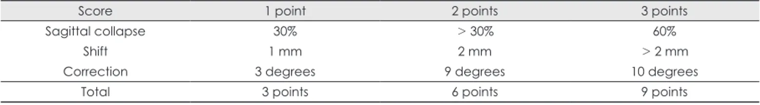
TABLE 2. McCormck’s classification
Score 1 point 2 points 3 points
Sagittal collapse 30% >30% 60%
Shift 1 mm 2 mm >2 mm
Correction 3 degrees 9 degrees 10 degrees
Total 3 points 6 points 9 points
TABLE 1. Thoracolumbar injury severity score (TLISS) system
1. Injury mechanism Points
a. Compression Simple compression 1
Lateral angulation > 10° 1
Burst 1
b. Translation/Rotation 3
c. Distraction 4
2. PLC disrupted in tension, rotation, or translation
a. Intact 0
b. Suspected/interminate 2
c. Injured 3
3. Neurologic status
Nerve root involvement 2
Cord, conus medullaris involvement Incomplete 3
Complete 2
Cauda equine involvement 3
The score is the total of 3 components: injury mechanism, neurologic status, and PLC distruption. A score of ≤3 suggests nonop- erative treatment, 4, operative or nonoperative treatment, and ≥5 suggests operative treatment
FIGURE 1. Portal placement. The working portals are located between anterior and middle axillary line and the triangulation should be considered.
방사선학적 및 임상적 평가
방사선학적 평가는 수술 전 및 마지막 경과관찰 시 촬 영한 일반 측면 방사선 사진에서 T11과 L2 사이의 Cobb angle을 측정하여 비교하였으며 임상적 평가는 수술전 및 마지막 경과관찰 시의 Oswestry disability index (ODI)와 Visual analogue scale (VAS)로 비교 평가하였 다. 수술 전 후 ODI, VAS 및 Cobb angle의 통계학적 비 교 평가는 비모수 통계방법인 윌콧슨 검정 (Wilcoxon signed rank test)을 이용해 분석하였으며, 이는 SPSS 12.0.1 (SPSS Institue, Inc., Chicago, Illinois, USA)라 는 통계프로그램을 이용하여 시행되었다.
결
과총 5명의 환자가 이번 연구의 적응증에 해당되었으며 L1 burst fracture 환자가 80%이었고 평균 경과 관찰 기간은 14개월이었다. 모든 환자는 TLISS 점수가 5점 이상 이며 McCormck classification 점수가 7점 이상인 환자였고 외상의 원인은 모두 추락이었다 (Table 3). 임상적 평가에 서는 경과관찰 시에 측정한 ODI와 VAS의 평균 값은 각각 13±11.4과 1±1.7이며, 이는 ODI와 VAS의 수술 전 평균 값인 40±1.5과 8±1.1에 비해서 의미 있게 향상되었다 (p=0.043, p=0.042)(Table 4).
방사선학적 평가에서는 경과관찰시 측정한 평균 후만 각 (10.3±7.41°)과 수술 전 측정한 평균 후만각 (21.4±
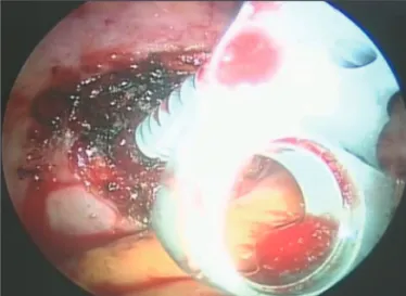
6.48°)에 비해서 의미 있는 차이를 보이며 (p=0.036), 수술 후 정상 후만각으로 복원되고 있음을 알 수 있다 (Figure 5). 두 명의 환자에서 수술과 연관된 합병증이 발생하였 으며 그 중 한 명의 환자는 수술 후 유비흉이 발생되었으 나 지방제거식을 복용하면서 치료되었으며 또 다른 환자 는 수술 후 1개월까지 늑간 신경통을 호소하였으나 그 후 에 통증을 호소하지 않았다. 그 이외에 폐렴이나 무기폐와 같은 합병증은 없었으며, 평균 수술 시간은 361±64.1 min 이고 평균 출혈량은 1,440±642.7 cc였다 (Table 4). 경과 관찰 기간 동안 모든 환자에게서 흉,요추 굴곡 및 신전 방 사선 사진을 촬영하였고, 이 방사선 사진에서 모두 3도 FIGURE 2. The first screw of MACS TL plate system (Aesculap)
is inserted into the caudal vertebral body.
FIGURE 3. This picture shows the partial corpectomy site of
thoracic vertebra. FIGURE 4. This picture shows the intraoperative view of cage
and bone segment insertions.
TABLE 3. The characteristics of patients
Case Sex/Age Level Margel classification McCormck TLISS F/U (months)
1 M/57 L1 A3.3 9 5 21
2 F/67 T12 A3.3 8 5 17
3 M/53 L1 A3.2 7 6 18
4 F/28 L1 A3.1 7 5 13
5 M/53 L1 A3.2 7 5 01
TLISS: thoracolumbar injury severity score
미만의 움직임만 있어서, 융합 부위의 불안정성을 일반 방 사선 촬영에서 보이는 환자는 없었다. 하지만 융합 여부의 가장 중요한 판단 근거인 척추체간의 골교(bridging bone) 여부는 오직 1명의 환자에서 수술 후 1년 뒤 촬영한 흉, 요 추부 CT에서 확인되었다.
고
찰전방 및 전측방 척추체의 복원은 전방 부하 지탱에 매 우 효과적이다.12) 이러한 수술에 흉강경을 이용할 경우 많 은 장점이 있다. 장점들로는 늑골제거나 불필요한 늑골견 인이 필요가 없으며 적은 크기의 피부 절개로 전방 병변 부위를 직접 보면서 수술이 가능하여 수술 후 정상 활동 수준으로의 복귀 시간이 단축된다.3,8) 이 연구에서는 밝 히지 않았지만 다 분절 후방 고정술만을 시행한 환자의 경
우 수술 후 상당기간 등 통증 및 활동의 제한에 대한 불편 감을 호소하나 흉강경을 이용한 단분절 고정술을 시행한 환자들은 등 통증보다는 등 이물감을 호소하며 활동의 제 한에 대한 호소 역시 미비하였고, 환자 모두 수술 상처의 크기에 대해 만족해하였다. 하지만, 흉강경 척추 수술은 의사가 숙달하는데도 시간이 오래 걸리며, 모니터의 평면 영상에 대한 입체감이 없어 해부학적 구조의 파악이 쉽지 않으며 사용되는 기구 사용이 익숙지 않아서 갑작스런 출 혈에 대한 조절이 용이하지 않다.15,17) 이러한 제함점들을 극복하기 위해서는 동물이나 사체 실습 등으로 충분한 연 습을 한 뒤에 시행해야 해야 하겠다.
향후 광학기술이 발전하고 수술기구들이 좀 더 개선이 되 면 영상 유도 장치를 흉강경 수술에 사용될 수도 있으며, 좀 더 정확한 병소의 접근이나 나사못 삽입 등에 용이할 수 있고, 로봇을 이용한 흉강경 수술도 가능하리라 생각된다.
TABLE 4. The clinical and radiological outcomes
CASE ODI VAS Khyphotic angle
Preop Postop Preop Postop Preop Postop Cx.
1 40 26 08 4 14.5 07.10 -
2 42 24 08 2 31.4 09.00 +
3 39 10 08 1 18.5 16.08 -
4 39 02 07 0 18.9 08.00 ++
5 42 03 10 0 23.8 15.20 -
40±1.5 13±11.4* 8±1.1 1±1.7* 21.4±6.48 10.3±7.41
*p<0.05. +: Chylothroax, ++: Transient intercostal neuralgia, ODI: Oswestry disability index, VAS: Visual analogue scale
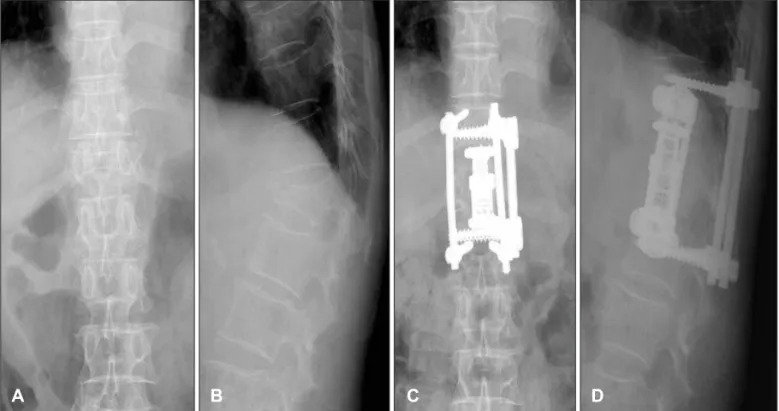
A B C D
FIGURE 5. Postoperatively anteroposterior (A) and lateral (B) plain films show T12 burst fracture and postoperatively anteroposte- rior (C) and lateral (D) plain films show partial corpectomy and circumferential fixation at thoracolumbar vertebrae.
결
론외상에 의한 흉, 요추부 전방 척추체의 골절로 인하여 전 방 부하의 지탱이 생역학적으로 불안정한 경우에는 전방 접근법에 의한 수술이 필요하다. 비록 흉강경을 이용한 수 술이 척추 외과의사에게서 익숙한 수술은 아니지만, 흉강 경을 이용하여 추체간 고유합술 및 내고정술을 시행하는 것은 만족할 만한 임상적 방사선학적 결과를 가질 수 있 는 유용한 최소침습수술로 생각된다.
중심 단어: 흉강경·흉요추·척추·골절.
■ The authors have no financial conflicts of interest.
REFERENCES
1) Bartels RH, Peul WC. Mini-thoracotomy or thoracoscopic treat- ment for medially located thoracic herniated disc? Spine (Phila Pa 1976) 32:E581-E584, 2007
2) Colthap WH, Arnold JH, Alford WC Jr, Burrus GR, Glassford DM Jr, Lea JW 4th, et al. Videothoracoscopy: improved technique and expanded indications. Ann Thorac Surg 53:776-778, 1922 3) Dickman CA, Rosenthal D, Karahalios DG, Paramore CG, Mican
CA, Apostolides PJ, et al. Thoracic vertebrectomy and reconstruc- tion using a microsurgical thoracoscopic approach. Neurosur- gery 38:279-293, 1996
4) Han PP, Kenny K, Dickman CA. Thoracoscopic approaches to the thoracic spine: experience with 241 surgical procedures. Neu- rosurgery 51:S88-S95, 2002
5) Harrop JS, Vaccaro AR, Hurlbert RJ, Wilsey JT, Baron EM, Shaf- frey CI, et al. Intrarater and interrater reliability and validity in the assessment of the mechanism of injury and integrity of the posterior ligamentous complex: a novel injury severity scoring system for thoracolumbar injuries. Invited submission from the Joint Section Meeting On Disorders of the Spine and Peripheral
Nerves, March 2005. J Neurosurg Spine 4:118-122, 2006 6) Horowitz MB, Moossy JJ, Julian T, Ferson PF, Huneke K. Thorac-
ic discectomy using video assisted thoracoscopy. Spine (Phila Pa 1976) 19:1082-1086, 1994
7) Huang TJ, Hsu RW, Liu HP, Hsu KY, Liao YS, Shih HN, et al.
Video-assisted thoracoscopic treatment of spinal lesions in the thoracolumbar junction. Surg Endosc 11:1189-1193, 1997 8) Khoo LT, Beisse R, Potulski M. Thoracoscopic-assisted treatment
of thoracic and lumbar fractures: a series of 371 consecutive cases.
Neurosurgery 51:S104-S117, 2002
9) Kim SJ, Sohn MJ, Ryoo JY, Kim YS, Whang CJ. Clinical Analysis of Video-assisted Thoracoscopic Spinal Surgery in the Thoracic or Thoracolumbar Spinal Pathologies. J Korean Neurosurg Soc 42:293-299, 2007
10) Landreneau RJ, Mack MJ, Hazelrigg SR, Dowling RD, Acuff TE, Magee MJ, et al. Video-assisted thoracic surgery: basic technical concepts and intercostal approach strategies. Ann Thorac Surg 54:800-807, 1992
11) Lewis RJ, Caccavale RJ, Sisler GE. Imaged thoracoscopic sur- gery: a new thoracic technique for resection of mediastinal cysts.
Ann Thorac Surg 53:318-320, 1992
12) Longo UG, Papatietro N, Maffulli N, Denaro V, Thoracoscopy for minimally invasive thoracic spine surgery. Orthop Clin North Am 40:459-464, 2009
13) Mack MJ, Regan JJ, Bobechko WP, Acuff TE. Application of tho- racoscopy for diseases of the spine. Ann Thorac Surg 56:736-738, 14) McCormack T, Karaikovic E, Gaines RW. The load sharing clas-1993
sification of spine fractures. Spine (Phila Pa 1976) 19:1741-1744, 15) Niemeyer T, Freeman BJ, Grevitt MP, Webb JK. Anterior thora-1994 coscopic surgery followed by posterior instrumentation and fu- sion in spinal deformity. Eur Spine J 9:499-504, 2000
16) Rosenthal D, Dickman CA. Thoracoscopic microsurgical excision of herniated thoracic discs. J Neurosurg 89:224-235, 1998 17) Rosenthal D, Rosenthal R, de Simone A. Removal of a protruded
thoracic disc using microsurgical endoscopy. A new technique.
Spine (Phila Pa 1976) 19:1087-1091, 1994