Treatment of Disseminated Superficial Actinic Porokeratosis
Vol. 23, Suppl. 2, 2011 S211 Ann Dermatol Vol. 23, Suppl. 2, 2011 http://dx.doi.org/10.5021/ad.2011.23.S2.S211
CASE REPORT
Received January 7, 2011, Revised February 13, 2011, Accepted for publication April 25, 2011
Corresponding author: Jun Young Lee, M.D., Department of Dermato- logy, Seoul St. Mary’s Hostpial, The Catholic University of Korea, 505 Banpo-dong, Seocho-gu, Seoul 137-701, Korea. Tel: 82-2-2258-6222, Fax: 82-2-594-3255, E-mail: jylee@catholic.ac.kr
This is an Open Access article distributed under the terms of the Creative Commons Attribution Non-Commercial License (http://
creativecommons.org/licenses/by-nc/3.0) which permits unrestricted non-commercial use, distribution, and reproduction in any medium, provided the original work is properly cited.
Photodynamic Therapy Combined with CO 2 Laser Vaporization on Disseminated Superficial Actinic Porokeratosis: A Report of 2 Cases on the Face
Hei Sung Kim, M.D., Ji Hye Baek, M.D., Young Min Park, M.D., Hyung Ok Kim, M.D., Jun Young Lee, M.D.
Department of Dermatology, Seoul St. Mary’s Hospital, The Catholic University of Korea College of Medicine, Seoul, Korea
Disseminated superficial actinic porokeratosis (DSAP) is a skin condition that usually shows a poor response to different modalities of treatment. Herein we describe 2 patients with DSAP on the face, each treated with 3 to 4 sessions of photodynamic therapy combined with laser vaporization.
(Ann Dermatol 23(S2) S211∼S213, 2011) -Keywords-
Disseminated superficial actinic porokeratosis (DSAP), Face, Laser vaporization, Photodynamic therapy (PDT)
INTRODUCTION
Photodynamic therapy (PDT) is a treatment modality which involves the sequential administration of a photo- sensitizer drug and light. PDT has been reported to be useful in treating nonmelanoma skin cancers and a variety of benign skin conditions. Here, we examined whether PDT might be effective in the treatment of disseminated superficial actinic porokeratosis.
CASE REPORT
Case 1
A 61-year-old woman presented with a 10-year history of asymptomatic lesions on the distal part of the upper limb and face which were slowly increasing in size and number. She was distressed by the appearance of the lesions, especially those on the face. Clinical examination revealed multiple, small, circular scaly lesions on the arms and face, each surrounded by a fine, raised hyperkeratotic ridge, and with a clinical appearance typical of dissemi- nated superficial actinic porokeratosis (DSAP).
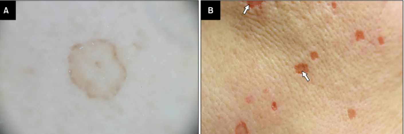
Dermoscopic findings aided the diagnosis (Fig. 1A). Three sessions of topical methyl aminolevulinate (MAL)-photo- dynamic therapy (PDT) treatment was done on the face (1 week and 20 days apart). Cream containing 160 mg/g MAL (MetvixⓇ, Galderma, France) was applied in an occlusive manner following CO2 laser ablation of the individual lesions (Sharplan 40C, Sharplan Lasers Inc., Tel Aviv, Israel) with a continuous output power of 0.7 W in the superpulse mode. Multiple passes of the CO2 laser were applied to the rim, whereas a single pass was given to the central area. Despite vigorous CO2 laser ablation, the rims remained intact in many cases (Fig. 1B). After 3 hours of incubation, the lesions were irradiated with a commercial broadband red light source (Waldmann PDT 1200LⓇ, Waldmann Medizintechnik, villingen-Schwen- ningen, Germany, wave length: 570∼730 nm, light dose of 37 J/cm2 at a fluence rate of 70 mW/cm2, Lamp-detector distance of 35 cm). The red light was repeatedly shined at 3 angles (the frontal face and right and left sides of the face) for even coverage. CO2 laser ablation was repeatedly performed in lesions with visible rims before the 2nd and 3rd MAL-PDT treatments. The patient experienced mild
HS Kim, et al
S212 Ann Dermatol
Fig. 1. Dermoscopic findings of a disseminated superficial actinic porokeratosis lesion. The peripheral rim is prominent (A), despite vigorous CO2 laser ablation the rims remain intact (arrows) (B).
Fig. 2. Initial lesions of disseminated superficial actinic porokeratosis on the face (A), after 3 sessions of methyl aminolevulinate (MAL)-photodynamic therapy (PDT) (B), & diffuse scaling and aggravation of the underlying melasma following MAL-PDT (C).
The pigmentation was prominent at her follow-up visit (which occurred 1 week after the MAL-PDT) and it gradually faded with time, lasting for about a month.
burning sensations during red light illumination and post- inflammatory hyperpigmentation following treatment.
Marked clinical improvement was observed with slight residual hyperpigmentation at a follow-up visit 6 months after the 3rd MAL-PDT treatment.
Case 2
A 62-year-old woman had a 7-year history of DSAP lesions first appearing on the lower legs and spreading to involve the arms and face (Fig. 2A). The lesions were increasing in size and she found the appearance em- barrassing. There was no family history of the condition.
On examination, she had multiple, small, scaly lesions with a raised hyperkeratotic ridge on her legs, arms and face. A punch biopsy taken from a representative lesion on the face showed a keratin-filled epidermal invagination with a characteristic cornoid lamella, and confirmed the diagnosis of DSAP. Subsequently, treatment with MAL- PDT was performed on the face (she was given 4 treat- ments). The 2nd and 3rd MAL-PDT treatment was given after an interval of 3 weeks and a 4th MAL-PDT after 4 months. The treatment protocol was the same as in case 1.
A majority of the lesions disappeared after 3 sessions of MAL-PDT (Fig. 2B) and the 4th session was performed to
Treatment of Disseminated Superficial Actinic Porokeratosis
Vol. 23, Suppl. 2, 2011 S213 remove the few remaining lesions. The patient compl-
ained of diffuse scaling of the face and aggravation of the underlying melasma after MAL-PDT treatment (Fig. 2C).
The pigmentation was prominent at her follow-up visit which was made 1 week after the 1st MAL-PDT and gra- dually faded with time, lasting for about a month.
DISCUSSION
DSAP, a clinical variant of porokeratosis, is an inherited, autosomal dominant cutaneous disorder of keratinization.
DSAP skin lesions appear predominantly on the sun exposed areas of the extremities with facial involvement in about 15% of all DSAP cases1. Dermoscopy aids the diagnosis where a “white track” structure is identified at the periphery of the lesion with a brownish pigmentation in the inner side and “double white track” in some parts of the lesion. The single or double “white track” structure at the margin corresponds to the cornoid lamella and is characteristic of porokeratosis2. Although different therapeutic options have been proposed, including cryotherapy, topical 5-fluorouracil and tacalcitol, the therapy of DSAP is still a challenge, mostly because of the multiplicity of the skin lesions and the frequent relapse of the disease.
PDT is a relatively new treatment modality that is currently used for various skin cancers, actinic keratosis and acne. Lesions in DSAP are thought to arise from proliferation of localized clones of abnormal epidermal cells. PDT is potentially a useful treatment option for DSAP as it can selectively target highly active, atypical cells and cause destruction by the creation of toxic intermediates3. So far, 3 studies have reported the use of PDT with red light for the treatment of DSAP with varying photo-sensitizers, light sources and parameters4-6. Striking clinical improvement was observed in a DSAP patient treated with MAL and AnkiliteⓇ 635 nm at a fluence of 37 J/cm2, whereas the same treatment was not so successful in a group of 6 patients with DSAP. In another study, DSAP of the arms and legs were treated with 20%
aminolevulinic acid (ALA) and Waldmann PDT 1200LⓇ 570∼730 nm at a fluence of 100 J/cm2 with disappointing results. Based on previous reports, we considered MAL to be a better choice over ALA with its higher penetration depth, although we do not have experience with the counterpart agent in DSAP. Two different red light sources are mainly used for MAL-PDT: Ankilite (635 nm) and Waldman 1200 (broad band, 570∼730 nm). Since Waldman 1200 has a broader wavelength, a higher fluence is required than with Ankilite. As previously mentioned, common energy settings in previous studies were 37 J/cm2 with Ankilite and 100 J/cm2 with Waldman 1200, and
there has not been a study comparing the effects of a single light source at different energy settings. In my case, I chose to use Waldman 1200 at a low fluence setting because post-inflammatory hyperpigmentation was reported to be severe in a case treated with Waldman 1200 at a setting of 100 J/cm2. A light dose of 37 J/cm2 with a broadband light source resulted in an adequate response in our cases. A low irradiance of 70 mW/cm2 was used because it correlates with an increase in photobiological damage. It also causes less pain and theoretically covers a wider area. As a pretreatment modality, light curettage was chosen in previous studies whereas in our cases we pretreated the individual lesions with CO2 laser ablation to enhance the effects of MAL-PDT.
CO2 laser vaporization itself has been a treatment option for DSAP, but as seen in our patients, it is not sufficient by itself for removing the rims. We wanted to see whether PDT can supplement the effects of a CO2 laser. Overall, PDT was found to remove some of the remnant rims of DSAP following CO2 laser ablation, but the degree of improvement was not striking. Even with the addition of MAL-PDT to CO2 laser vaporization, multiple sessions of treatment were required and complications (ex. aggra- vation of melasma, residual pigmentation) associated with PDT raised some concern. However, there is a need for further controlled studies before drawing a conclusion on the effects of PDT on DSAP. In the meantime, we should also continue searching for a better treatment option for patients with DSAP.
REFERENCES
1. Sawyer R, Picou KA. Facial presentation of disseminated superficial actinic porokeratosis. Ear Nose Throat J 1989;68:
57-59.
2. Zaballos P, Puig S, Malvehy J. Dermoscopy of disseminated superficial actinic porokeratosis. Arch Dermatol 2004;140:
1410.
3. Levitt J, Emer JJ, Emanuel PO. Treatment of porokeratosis of mibelli with combined use of photodynamic therapy and Fluorouracil cream. Arch Dermatol 2010;146:371-373.
4. Nayeemuddin FA, Wong M, Yell J, Rhodes LE. Topical photodynamic therapy in disseminated superficial actinic porokeratosis. Clin Exp Dermatol 2002;27:703-706.
5. Cavicchini S, Tourlaki A. Successful treatment of dissemi- nated superficial actinic porokeratosis with methyl aminole- vulinate-photodynamic therapy. J Dermatolog Treat 2006;
17:190-191.
6. Fernández-Guarino M, Harto A, Pérez-Garcia B, Martin- González M, Urrutia S, Jaén P. Photodynamic therapy in disseminated superficial actinic porokeratosis. J Eur Acad Dermatol Venereol 2009;23:176-177.