Ann Hepatobiliary Pancreat Surg 2016;20:201-203
https://doi.org/10.14701/ahbps.2016.20.4.201
Case Report
Cholangiocarcinoma in choledochal cyst after cystoenterostomy:
how a mistreated choledochal cyst can progress to malignancy
HyungJoo Baik, Yo-Han Park, Sang Hyuk Seo, Min Sung An, Kwang Hee Kim, Ki Beom Bae, Chang Soo Choi, Sang Hoon Oh, and Young Kil Choi
Department of Surgery, Busan Paik Hospital, College of Medicine, Inje University, Busan, Korea
This case report presents an unusual case of cholangiocarcinoma arising nearly 35 years after cystoduodenostomy for choledochal cyst. The patient visited our hospital with dyspepsia and studies revealed bezoar within the choledochal cyst caused by bile and food reflux. The patient underwent pancreaticoduodenectomy and a biopsy revealed ad- enocarcinoma, stage IIB. After 19 months, the patient has no recurrence to date and has recovered well. This case shows that proper surgical management and meticulous, long-term follow-up is imperative for patients with congenital choledochal cyst. (Ann Hepatobiliary Pancreat Surg 2016;20:201-203)
Key Words: Choledochal cyst; Cholangiocarcinoma; Pancreaticoduodenectomy
Received: May 9, 2016; Revised: July 15, 2016; Accepted: July 28, 2016 Corresponding author: Chang Soo Choi
Division of Hepatobiliary Surgery, Department of Surgery, Busan Paik Hospital, College of Medicine, Inje University, Bokji-ro 75, Busangjin-gu, Busan 47392, Korea
Tel: +82-51-890-6352, Fax: +82-51-898-9427, E-mail: [email protected]
Copyright Ⓒ 2016 by The Korean Association of Hepato-Biliary-Pancreatic Surgery
This is an Open Access article distributed under the terms of the Creative Commons Attribution Non-Commercial License (http://creativecommons.org/
licenses/by-nc/4.0) which permits unrestricted non-commercial use, distribution, and reproduction in any medium, provided the original work is properly cited.
Annals of Hepato-Biliary-Pancreatic Surgery ∙ pISSN: 2508-5778ㆍeISSN: 2508-5859
INTRODUCTION
A choledochal cyst is a cystic dilatation of the biliary tree and a well-known cause of biliary tract malig- nancies.1-3 It is a relatively rare disease; incidence rates ranging from 0.32% in Asia and 1 in 13,000 to 1 in 2 million births in western countries.3,4 Most cases are diag- nosed during early childhood, but between 20% to 30%
of cases are first detected in adults as choledochal cyst either causing symptoms or found incidentally during imaging for an unrelated cause.4 It has been reported that only a small number of patients who underwent surgical management for choledochal cyst had progression to bili- ary malignancy.5 The incidence of cancer in patients with primary choledochal cyst is 9.9%, whereas the incidence of cancer development after cyst excision is 0.6%.6 Herein, we present a case of a patient who had been treat- ed for choledochal cyst in an alternative manner with no follow-up, which then progressed to cholangiocarcinoma.
CASE
A 46-year-old woman visited Busan Paik Hospital Hepatobiliary clinic with dyspepsia and right, upper quad- rant, abdominal pain. The patient had a history of a cys- toduodenostomy nearly 35 years ago. There was a pal- pable mass in her abdomen in the right, upper quadrant.
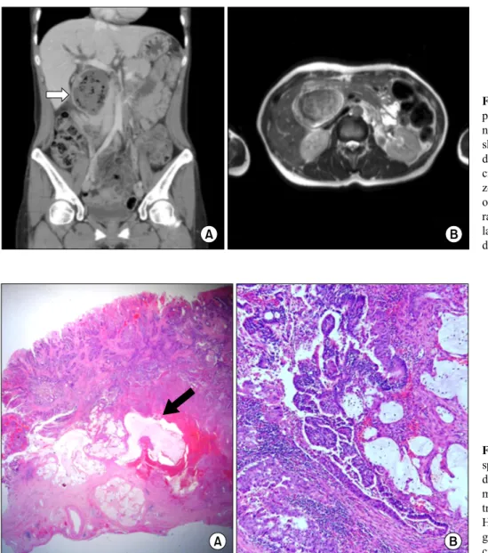
Initial laboratory findings were as follows: total bilirubin 0.5 mg/dl, aspartate transaminase/alanine transaminase 16/14 IU/L, alkaline phosphatase 276 U/L, alpha-fetopro- tein 2.01 ng/ml, carcinoembryonic antigen 0.756 ng/ml, carbohydrate antigen 19-9 11.70 U/ml, and hepatitis B surface antibody-positive. Abdomino-pelvic computed to- mogram (APCT) scan and magnetic resonance chol- angiopancreaticography (MRCP) showed moderate central dilatation of the intrahepatic duct with pneumobilia and a large, cystic, mass-like lesion with internal echogenicity and probable bezoar formation within a choledochal cyst of 6.8 cm×8.5 cm×5.0 cm in size (Fig. 1). There was also mild proximal pancreatic duct dilatation with lobulated mass suggestive of intraductal papillary mucinous neoplasm. Her esophagogastroduodenoscopy showed an
202 Ann Hepatobiliary Pancreat Surg Vol. 20, No. 4, November 2016
Fig. 1. Imaging studies of the patient. (A) Patient’s abdomi- no-pelvic computed tomogram shows dilatation of intrahepatic duct, pneumobilia, and a 6.8 cm×8.5 cm×5.0 cm-sized be- zoar (arrow). (B) Magnetic res- onance cholangiopancreaticog- raphy also represents cystic di- latation of the extrahepatic bile duct.
Fig. 2. Microphotographs of the specimen. (A) The common bile duct wall is thickened with tu- mor invasion and shows ex- tracellular mucin pools (arrow, HE, ×10). (B) Tumor reveals glandular architecture and mu- cin formation (HE, ×100).
opening, at the second portion of the duodenum,with gastritis.
The initial plan for the operation was to undergo re- vision of the cystoduodenostomy and/or cyst excision with a hepaticojejunostomy. Because of the bezoar-like mass in the cyst, the patient was in a fasting state for 2 days prior to surgery, hoping that the mass would be dismantled. An incision was made via the previous right paramedian scar. Underneath the scar there was a severe adhesion so adhesiolysis was performed. The previously performed cystoduodenostomy was identified with multi- ple lymph node enlargement near the superior mesenteric vein and stomach. A small portion of the common bile duct was excised for frozen biopsy, which revealed ad- enocarcinoma; moreover, cystic dilatation had extended to the pancreas because of an anomalous pancreaticobiliary
ductal union (APBDU). Thus, a pancreaticojejunostomy was inevitable for complete excision of the cyst and prop- er management for cholangiocarcinoma. Within the cyst, there was a mass: a conglomeration of food material with bile. A cholecystectomy, subtotal gastrectomy, pancreati- cojejunostomy, and gastrojejunostomy were performed.
The patient’s extrahepatic bile ducts were unusually low-lying, so the surgeon performed a ductoplasty, con- joining the left and right extrahepatic bile ducts side to side, making it easier to carry out hepaticojejunostomy.
The final pathology report proved the presence of ad- enocarcinoma: moderate differentiation arising in the choledochal cyst, invading to surrounding adipose tissue (pT2a), accompanying severe inflammatory infiltration, and one metastatic lymph node out of seven excised lymph nodes, stage IIB (Fig. 2). It has been nineteen
HyungJoo Baik, et al. Cholangiocarcinoma in choledochal cyst 203
months since the patient had her surgery and she is going through oral doxifluridine chemotherapy without any complications or recurrences to date.
DISCUSSION
Choledochal cysts are congenital anomalies of the bile ducts, which are subdivided into five different categories.
The most common types are I and IVa, the dilatation of the extrahepatic bile duct and both the extrahepatic and intrahepatic bile ducts, consecutively. Asians and women tend to have a higher incidence than Caucasians and men, though clear reasons for these tendencies are yet to be elucidated.2 In many cases, diagnosis is made early during childhood in 80%; however, due to the advance of imag- ing techniques, some diagnoses are incidentally made in adulthood.4 Symptom triads are abdominal pain, jaundice, and an abdominal mass, but only rarely do these symp- toms coexist. Treatment of choice is a total cystectomy and Roux-en-Y hepaticojejunostomy; less frequently, cys- toenterostomy has been used as an alternative method.1 The standard form of surgery prevents subsequent compli- cations such as pancreatitis, cholangitis, portal hyper- tension and malignancy. The risk of cholangiocarcinoma in a choledochal cyst is as high as 20-30% in early adult- hood if no surgical intervention is performed. On the other hand, cholangiocarcinoma following a choledochal cyst resection is relatively rare, with an incidence rate of 0.7-6%.5
The exact pathophysiology of cholangiocarcinoma in a choledochal cyst is still unknown, but it is suspected that the reflux of pancreatic juice is the main cause, promoting chronic inflammatory changes and carcinogenesis.3 This patient had a Type I choledochal cyst and, due to a pre- vious cystoduodenostomy, not only pancreatic juice but also food material had refluxed into the biliary tract, caus- ing bezoar formation and eventually malignancy. Previous studies have emphasized the necessity of long-term fol-
low-up for patients who have had a cyst excision, since it is known that the risk of biliary malignancy in the rem- nant bile duct increases more than 15 years after surgery, as in the case of our patient.5 For those who had a cys- toenterostomy,7,8 it is mandatory that they have regular check-ups since the incidence rate of malignancy or com- plications such as cholangitis, hepatolithiasis, and pan- creatitis could be as high as 70%.1
In conclusion, it should be emphasized that the standard treatment of choledochal cyst is complete cyst excision and hepaticojejunostomy. If for some reason this is not possible, a cystoenterostomy can be an alternative treat- ment, but meticulous life-long follow-up is essential for early detection and management of complications.
REFERENCES
1. Khandelwal C, Anand U, Kumar B, Priyadarshi RN. Diagnosis and management of choledochal cysts. Indian J Surg 2012;74:
401-406.
2. Sallahu F, Hasani A, Limani D, Shabani S, Beka F, Zatriqi S, et al. Choledochal cyst - presentation and treatment in an adult.
Acta Inform Med 2013;21:138-139.
3. Kim SH, Kim HW, Kang DH, Kim MD, Lee JH, Lee JH, et al. A case of intrahepatic cholangiocarcinoma associated with type iv choledochal cyst. Korean J Gastroenterol 2012;60:
123-127.
4. Franko J, Nussbaum ML, Morris JB. Choledochal cyst chol- angiocarcinoma arising from adenoma: case report and a review of the literature. Curr Surg 2006;63:281-284.
5. Ohashi T, Wakai T, Kubota M, Matsuda Y, Arai Y, Ohyama T, et al. Risk of subsequent biliary malignancy in patients under- going cyst excision for congenital choledochal cysts. J Gastroenterol Hepatol 2013;28:243-247.
6. Lee SE, Jang JY, Lee YJ, Choi DW, Lee WJ, Cho BH, et al.
Choledochal cyst and associated malignant tumors in adults: a multicenter survey in South Korea. Arch Surg 2011;146:
1178-1184.
7. Ong J, Campbell W, Taylor MA. Metastatic cholangiocarcinoma following choledochal cyst excision: an unusual cause of ab- dominal pain in a 35-year-old female. Ulster Med J 2013;82:
21-22.
8. Ono S, Fumino S, Shimadera S, Iwai N. Long-term outcomes after hepaticojejunostomy for choledochal cyst: a 10- to 27-year follow-up. J Pediatr Surg 2010;45:376-378.