© 2019 The Korean Ophthalmological Society
This is an Open Access article distributed under the terms of the Creative Commons Attribution Non-Commercial License (http://creativecommons.org/licenses /by-nc/3.0/) which permits unrestricted non-commercial use, distribution, and reproduction in any medium, provided the original work is properly cited.
Original Article
Comparison of Laser In Situ Keratomileusis Flap Morphology and Predictability by WaveLight FS200 Femtosecond Laser and Moria
Microkeratome: An Anterior Segment Optical Coherence Tomography Study
Zeiad H. Eldaly1, Mahmoud A. Abdelsalam1, Mohamed S. Hussein1, Mohamed A. Nassr1,2
1Department of Ophthalmology, Assiut University, Assiut, Egypt
2University Hospitals of Leicester NHS Trust, Leicester, UK
Purpose: To evaluate laser in situ keratomileusis (LASIK) flap thickness predictability and morphology by femto- second (FS) laser and microkeratome (MK) using anterior segment optical coherence tomography.
Methods: Fifty-two candidates for the LASIK procedure were stratified into two groups: FS laser-assisted (Al- legretto FS-200) and MK flap creation (Moria 2). Flap thickness was determined at five points. The side-cut angle was measured in three directions at the margin interface. LASIK flap assessment was performed one month postoperatively by Spectralis anterior segment optical coherence tomography.
Results: Fifty-two patients (93 eyes) were recruited; 49 eyes were stratified to the FS group and 44 eyes to the MK group. The FS group had relatively even flap configurations, and the MK group had meniscus-shaped flaps. Mean differences between planned and actual flap thickness were 12.93 ± 8.89 and 19.91 ± 5.77 μm in the FS and MK groups, respectively. In thin flaps (100 to 110 μm), there was a significant disparity between the two groups (7.80 ± 4.71 and 19.44 ± 4.46 μm in the FS and MK groups, respectively). However, in thicker flaps (130 μm), comparable flap thickness disparity was achieved (18.54 ± 9.52 and 20.83 ± 5.99 μm in the FS and MK groups, respectively). Mean side-cut angle was 74.29 ± 5.79 degrees and 32.34 ± 4.94 degrees in the FS and MK groups, respectively.
Conclusions: Comparable flap thickness predictability was achieved in thicker flaps (130 μm), while the FS la- ser technique yielded a more predictable result in thinner flaps (100 to 110 μm). Different flap morphology was observed in meniscus flaps in MK-LASIK and flap morphology in FS-LASIK.
Key Words: Anterior segment optical coherence tomography, Flap morphology, Laser in situ keratomileusis
Corneal refractive surgery has shown remarkable prog- ress during the last decade, with fast-growing updates in
operative techniques, devices, and instrumentation. Howev- er, laser in situ keratomileusis (LASIK) remains the most common corneal refractive procedure [1]. Flap creation is the most critical step during LASIK eye surgery, so the con- sistency and predictability of the corneal flap thickness are crucial for a successful LASIK outcome. Thus, improving the predictability of LASIK flap thickness and morphology
Received: April 2, 2018 Accepted: June 28, 2018
Corresponding Author: Zeiad H. Eldaly, MD. Department of Ophthal- mology, Assiut University Hospital, 6th floor, Assiut 71516, Egypt. Tel:
20-88-230-9970, Fax: 20-88-241-3643, E-mail: [email protected]
is worthy of attention [2].
In mechanical microkeratome (MK)-assisted flap cre- ation, an oscillating blade is used to create corneal flaps.
Despite the advances in MK designs, flap-related complica- tions, such as free caps, button holes, incomplete cuts, and torn flaps, remain a challenge for most refractive surgeons [3,4]. In femtosecond (FS)-assisted flap creation, a cleavage line is created through the cornea at a predetermined depth by photo-ionization of optically transparent tissues with a resultant acoustic shock wave and gas bubble formation, disrupting treated tissues [5].
Many FS laser systems are available now, including In- traLase (Abbott Medical Optics, Santa Ana, CA, USA), Vi- suMax (Carl Zeiss Meditec AG, Dublin, CA, USA), Femto LDV (Zeimer Group, Port, Switzerland), and FS 200 Wave- Light (Alcon Laboratories, Fort Worth, TX, USA) [6].Dif- ferent devices are available for measuring postoperative flap thickness depending on subtraction from the preoperative planned residual stromal bed; however, these measurements are rough and mostly inaccurate [7,8]. Using real-time im- ages, anterior segment optical coherence tomography (AS- OCT) is now the most widely-used corneal imaging system not only to assess post-LASIK flap thickness, but also to evaluate flap morphology and determine the side cut angle at the flap margin interface [9,10]. The objective of our study was to assess LASIK flap thickness predictability created by a FS laser versus a mechanical MK, in addition to flap morphology and side cut angle using AS-OCT.
Materials and Methods
Setting
A prospective, comparative, open-label study was con- ducted from January 2016 to April 2016 after approval of the ethical committee of the Faculty of Medicine, Assiut University, Egypt (0032016) and under the tenets of Helsin- ki declaration. A written informed consent was signed by all patients after a thorough explanation of the procedure and its possible complications.
Participants and selection criteria
The inclusion criteria were myopic LASIK correction up to -12 diopters with or without myopic astigmatism up to -6
diopters, a corneal thickness at the thinnest location ≥500 μm, and a residual stromal bed ≥280 μm. Exclusion criteria were hyperopic refraction, mixed astigmatism, systemic disease that contraindicates LASIK, and intraoperative or postoperative complications.
Pre-LASIK assessment
All candidates underwent detailed history-taking and complete ophthalmic examination, including uncorrected visual acuity (UCVA) and best-corrected visual acuity by Landolt’s C-chart, slitlamp biomicroscopy (Haag-Streit, Mason, OH, USA), intraocular pressure measurements (Goldmann Applanation tonometer mounted on a slitlamp), Schirmer I test, and Pentacam evaluation (Oculus Penta- cam, Oculus GmbH, Wetzlar, Germany).
Flap creation
Candidates were stratified into two groups according to flap creation technique: FS or MK. In the FS group, patients underwent FS-assisted flap creation using Allegretto Wave- Light FS-200 FS laser (Alcon Laboratories). The device used a 200-kHz repetition rate, 1,030-nm wavelength, and 5-µm spot size. The settings of the flap creation procedure were set so that the hinge was superior with a fixed flap di- ameter of 9 mm and a side cut angle of 70 degrees in all pa- tients. The planned flap thickness was subdivided into 100 to 110 μm and 130 μm groups according to the patient’s cor- neal and refractive profile and according to surgeon’s pref- erence.
In the MK group, patients underwent mechanical flap cre- ation using a Moria 2 Microkeratome (Moria SA, Antony, France), where flaps with superior hinges were created with variable diameters according to keratometric readings.
Planned flap thickness was also subdivided into 100 to 110 μm and 130 μm groups according to the surgeon’s preference after choice of a suitable suction ring.
Myopic laser ablation was performed using EX-500 exci- mer laser (Alcon Laboratories) with a planned full correc- tion and post-operative emmetropia. All cases were done by the same experienced surgeon (MA) in both groups. Post- LASIK follow-up was scheduled 1-day, 1-week, 2-week, and 1-month post-LASIK. A combination of Dexamethasone-To- bramycin eye drops (Tobradex, Alcon Laboratories) four times daily for 1 week and topical lubricant eye drops (Sys-
tane Ultra, Alcon Laboratories) four times daily for 3 months were prescribed.
Flap assessment
All patients underwent anterior segment optical coher- ence tomography (AS-OCT) evaluation of flap morpholo- gy, thickness, and side-cut by Spectralis spectral domain optical coherence tomography (Heidelberg Engineering, Heidelberg, Germany) 1-month post-LASIK. The Spectra- lis AS-OCT had an acquisition speed of 40,000 A-scans per second, with an axial resolution of 3.9 to 7 μm and a transverse resolution of 14 μm. Flap assessment by AS- OCT was carried out by a single experienced ophthalmolo- gist (ZE). Flap thickness was measured at five points along the horizontal meridian passing through the corneal center.
The corneal center was determined by the presence of high reflective artifacts while scanning for corneal apex. The flap was evaluated with a horizontal line scan measuring 20 degrees and averaged to 10 frames only to avoid over- exposure and loss of LASIK interface details. The LASIK flap thickness was measured at five points: center, 1 mm nasal and temporal, and 2.5 mm nasal and temporal. Flap thickness was defined as the distance between the tear film-epithelial interface and the LASIK flap interface and perpendicular to tear film-epithelial interface. It was mea- sured three times, and the average was calculated. The ad- justments were done manually, and readings were calculat-
ed accordingly based on the measuring tool of the device.
For side-cut angles, a 15-degree line scan averaged to 10 frames was aligned perpendicularly across nasal, tempo- ral, and inferior edges of the flap to acquire scans across the three flap edges. Images were then transferred to Im- ageJ software ver. 1.8.0 (National Institutes of Health, Bethesda, MD, USA) [11]. The angle measuring tool in the software was utilized to measure the side-cut angles three times, and the mean reading was recorded by a single ex- perienced ophthalmologist (MN).
Statistical analysis
The statistical analysis was done by IBM SPSS Statistics ver. 20.0 (IBM Corp., Armonk, NY, USA). Descriptive sta- tistics were evaluated to compare patient characteristics between groups. Mann-Whitney test was used to compare means among groups. A p-value less than 0.05 was consid- ered significant.
Results
Patient baseline characteristics
This study included 93 eyes of 52 patients. FS-assisted LASIK surgery was done in 49 eyes of 30 patients (14 males and 16 females), while 44 eyes of 22 patients (6
Table 1. Patient characteristics in both femtosecond laser-assisted LASIK and microkeratome-assisted LASIK
FS-LASIK MK-LASIK p-value*
Patients 30 (49 eyes) 22 (44 eyes) -
Age (yr) 28.83 ± 8.09 29.55 ± 6.32 0.943
Male : female 14 : 16 6 : 16 -
Pre-LASIK MRSE - 6.18 ± 4.06 - 6.05 ± 3.96 0.543
Pre-LASIK BCVA 0.91 ± 0.11 0.89 ± 0.13 0.338
Post-LASIK UCVA 0.94 ± 0.74 0.92 ± 0.61 0.899
Pre-LASIK CCT 548.83 ± 38.09 539.83 ± 29.91 0.124
Pre-LASIK K value 44.46 ± 2.31 43.72 ± 2.87 0.078
Pre-LASIK Schirmer value 15.57 ± 1.71 15.8 ± 1.89 0.921
Values presented as mean ± standard deviation.
LASIK = laser in situ keratomileusis; FS-LASIK = femtosecond laser-assisted LASIK; MK-LASIK = microkeratome-assisted LASIK;
MRSE = manifest refraction spherical equivalent; BCVA = best-corrected visual acuity; UCVA = uncorrected visual acuity; CCT = cen- tral corneal thickness; K value = keratometric readings.
*Mann-Whitney test.
males and 16 females) underwent LASIK surgery with MK-assisted flap creation. Table 1 demonstrates the base- line patient characteristics.
Flap thickness and predictability
Mean flap thickness was 127.37 ± 16.83 μm in FS- LASIK patients and 136.34 ± 14.86 μm in MK-LASIK pa- tients. There was a significant difference between planned and actual flap thickness between the two groups regard- ing the overall mean flap thickness measurement (p <
0.001) (Table 2).
Furthermore, planned flap thickness was categorized in each group into two sub-groups: 100 to 110 μm and 130 μm planned flap thickness. In the FS-LASIK group, planned flap thickness of 100 to 110 μm was significantly more pre-
dictable than 130 μm (p < 0.001), with planned-actual flap thickness of 7.80 ± 4.71 and 18.54 ± 9.52 μm, respectively.
Meanwhile, in MK-LASIK patients, there was no signifi- cant difference between 100 to 110 μm and 130 μm planned flap thickness (p = 0.330), with planned-actual flap thickness of 19.44 ± 4.46 μm and 20.83 ± 5.99 μm, re- spectively.
FS-LASIK yielded a significantly more predictable flap thickness when comparing planned 100 to 110 μm flap thickness in both treatment groups (planned-actual flap thickness, 7.80 ± 4.71 and 19.44 ± 4.46 μm in FS-LASIK and MK-LASIK groups, respectively, p < 0.001). On the other hand, the two treatment groups yielded a comparable flap thickness when planned flap was adjusted for 130 μm with insignificant difference (18.54 ± 9.52 and 20.83 ± 5.99 μm in FS-LASIK and MK-LASIK groups, respectively, p = 0.296).
Table 2. Planned and actual flap thickness in both femtosecond laser-assisted LASIK and microkeratome-assisted LASIK
FS-LASIK MK-LASIK p-value*
Mean flap thickness (μm) 127.37 ± 16.83 136.34 ± 14.86 <0.001
Planned-actual flap thickness (μm) 12.93 ± 8.89 19.91 ± 5.77 <0.001
Sub-group Sub-group
Planned flap thickness (μm) 100–110 130 p-value* 100–110 130 p-value*
Eyes 25 24 22 22
Actual post-LASIK flap thickness (μm) 114.58 ± 6.12 140.82 ± 13.78 125.80 ± 5.79 146.88 ± 13.63 Planned-actual flap thickness (μm) 7.80 ± 4.71 18.54 ± 9.52 0.000 19.44± 4.46 20.83 ± 5.99 0.330
18.54 ± 9.52 20.83 ± 5.99 0.296
7.80 ± 4.71 19.44 ± 4.46 <0.001
Values are presented as mean ± standard deviation.
LASIK = laser in situ keratomileusis; FS-LASIK = femtosecond laser-assisted LASIK; MK-LASIK = microkeratome-assisted LASIK.
*Mann-Whitney test.
Table 3. Actual flap thickness in five locations across the horizontal meridian in FS-LASIK and MK-LASIK groups
100–110 μm 130 μm
FS-LASIK MK-LASIK p-value* FS-LASIK MK-LASIK p-value*
Eyes 25 22 24 22
Actual post- LASIK flap thickness (μm)
Central 113.6 ± 6.87 124.59 ± 6.32 <0.001 140.77 ± 13.88 144.32 ± 15.95 0.438 Nasal 1 mm 114.64 ± 6.49 124.50 ± 7.90 <0.001 139.23 ± 15 145.5 ± 15.37 0.196 Temporal 1 mm 115.16 ± 7.18 125.86 ± 5.13 <0.001 141.68 ± 14.02 145.05 ± 17.78 0.492 Nasal 2.5 mm 115.56 ± 6.55 126.27 ± 6.58 <0.001 140.27 ± 14.54 149.86 ± 11.18 0.078 Temporal 2.5 mm 113.96 ± 7.32 127.77 ± 4.95 <0.001 142.36 ± 13.96 149.68 ± 10.77 0.059 Values are presented as mean ± standard deviation.
LASIK = laser in situ keratomileusis; FS-LASIK = femtosecond laser-assisted LASIK; MK-LASIK = microkeratome-assisted LASIK.
*Mann-Whitney test.
Fig. 2. Average flap thickness in femtosecond laser-assisted la- ser in situ keratomileusis (LASIK) and microkeratome-assisted LASIK groups as measured in five different locations (centrally, 1 mm nasally and temporally, 2.5 mm nasally and temporally across horizontal meridian). The difference between two groups is more evident in planned flap thickness of 100 to 110 μm. (A) Planned flap thickness (pooled date), (B) planned LASIK flap (130 μm), and (C) planned LASIK flap (100 to 110 μm). N = nasal; T = temporal.
140 135 130 125 120 115
N 2.5 mm N 1 mm Central T 1 mm T 2.5 mm Femtosecond laser Microkeratome
A
155 150 145 140 135 130
N 2.5 mm N 1 mm Central T 1 mm T 2.5 mm Femtosecond laser Microkeratome
B
130 125 120 115 110 105
N 2.5 mm N 1 mm Central T 1 mm T 2.5 mm Femtosecond laser Microkeratome
C
Fig. 1. Laser in situ keratomileusis (LASIK) flap side-cut angle and edge outline in (A) microkeratome-assisted LASIK and (B) fem- tosecond laser-assisted LASIK groups by anterior segment optical coherence tomography.
A
B
Mean flap thicknesses at five different points (center, 2.5 mm nasally, 1 mm nasally, centrally, 1 mm temporally, and 2.5 mm temporally) are demonstrated in Table 3. The point-to-point difference with regard to treatment groups and planned flap thickness agreed with mean flap thick- ness difference between the two groups.
Flap configuration
In the FS-LASIK groups, there was a relatively even flap configuration where mean flap thickness was nearly equal when measured at five different points across the horizon- tal meridian, while in the MK-LASIK group, the LASIK flap had a meniscus configuration, being thinnest at the center and gradually increasing in thickness toward the periphery (Fig. 1A, 1B).
This meniscus configuration in MK-LASIK was best demonstrated when planned flap thickness was 130 μm, while a less prominent meniscus configuration was found 100 to 110 μm planned flap thickness.
The overall center-to-periphery difference was not sta- tistically significant between FS-LASIK and MK-LASIK.
Despite the disparity in flap configuration between the two treatment groups (Fig. 1), there was no significant cen- ter-to-periphery difference in sub-group analysis (Table 4).
Side-cut angle of LASIK flap
LASIK flaps in the FS group had a mean side-cut angle of 74.29 ± 5.79degrees (range, 59 to 84 degrees; 95% confi- dence interval [CI], 72.62 to 75.95 degree,) while the MK group had a mean side-cut angle of 32.34 ± 4.94 degrees (range, 23 to 41 degrees; 95% CI, 30.84 to 33.85 degree). As FS-LASIK flap side-cut angle was pre-operatively adjusted to 70 degrees, mean difference between planned and actu- al post-LASIK flap side-cut was 6.12 ± 3.73 degrees (range, 0 to 14 degrees; 95% CI, 5.05 to 7.2 degree).
In AS-OCT evaluation of side-cut angle, FS-LASIK side-cut edges were well-demarcated and regularly out- lined, while MK-LASIK side-cut edges showed a ragged outline in many cases (Fig. 2A-2C).
Refractive Outcomes
Mean post-LASIK manifest refraction spherical equiva- lent (MRSE) were -0.43 ± 0.62 and -0.49 ± 0.46 diopters in FS-LASIK and MK-LASIK groups, respectively, with sig- nificant difference from pre-LASIK MRSE (p < 0.001).
There was a tendency for MK-LASIK to induce slightly
Table 4. Center-to-periphery difference in FS-LASIK and MK-LASIK groups
FS-LASIK MK-LASIK p-value*
Center-periphery difference (μm) 3.59 ± 2.76 4.51 ± 5.71 0.449
Sub-group Sub-group
Planned flap thickness (μm) 100–110 130 p-value* 100–110 130 p-value*
Center-periphery difference (μm) 3.72 ± 2.49 3.45 ± 3.09 0.494 3.06 ± 1.94 5.95 ± 7.65 0.113
3.72 ± 2.49 3.06 ± 1.94 0.528
3.45 ± 3.09 5.95 ± 7.65 0.787
Values are presented as mean ± standard deviation.
LASIK = laser in situ keratomileusis; FS-LASIK = femtosecond laser-assisted LASIK; MK-LASIK = microkeratome-assisted LASIK.
*Mann-Whitney test.
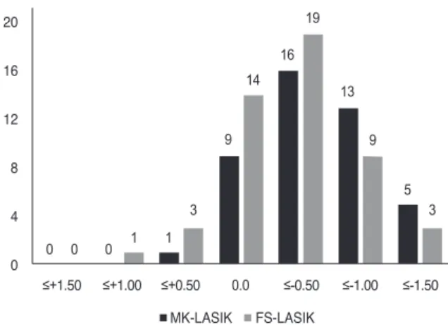
Fig. 3. Frequency of distribution of postoperative (microker- atome-assisted laser in situ keratomileusis [MK-LASIK] and femtosecond laser-assisted laser in situ keratomileusis [FS- LASIK]) manifest spherical equivalent.
20 16 12 8 4 0 ≤+1.50
0 0 0 1
≤+1.00 ≤+0.50 0.0 ≤-0.50 ≤-1.00 ≤-1.50 FS-LASIK
MK-LASIK 1
3 5
3 9
14 13
9 16
19
more myopic post-LASIK refraction than FS-LASIK after one month; however, there was no significant impact on post-LASIK UCVA (Fig. 3).
Discussion
In this prospective study, we investigated the predict- ability of LASIK flap thickness and its configuration by two different methods, FS- and MK-assisted flap creation.
We analyzed LASIK flap characteristics and measure- ments by AS-OCT. Many clinical studies have highlighted the utility of AS-OCT in the evaluation of LASIK flaps created by either MK or FS laser [10,12].
We evaluated the LASIK flap at five different points along the horizontal meridian of the cornea rather than a single central corneal point, as used in other studies [13], to provide a comprehensive overview of the LASIK flap and its configuration both qualitatively and quantitatively. In addition, flap configuration and peripheral architecture should not be overlooked. Planned LASIK flap thickness was stratified into two subgroups: 100 to 110 μm and 130 μm. Our study highlighted the predictability of flap thick- ness by MK and FS laser in both thin flaps (110 to 110 μm) and thicker flaps (130 μm) to spotlight the advantage of FS-assisted LASIK. Other studies have overlooked such stratification, and different planned flap thicknesses were pooled together, neglecting the possible difference in flap thickness. Thereafter, a separate comparison was carried out between planned and actual post-LASIK flap thickness in both groups. Such a detailed comparison clarifies the different performances of FS- and ML-assisted flap cre- ation in different planned flap thickness. Regarding mean flap thickness in pooled data, there was a significant planned-actual flap thickness difference between FS- and MK-assisted groups (12.93 ± 8.89 and 19.91 ± 5.77 μm, re- spectively, p < 0.001), with less difference in the FS-assist- ed group. However, after stratifying both groups into 100 to 110 μm and 130 μm subgroups, different results were produced. In the 100 to 110 μm subgroup, there was a sig- nificant planned-actual flap thickness difference (7.80 ± 4.71 and 19.44 ± 4.46 μm, respectively, p < 0.001). The 100 to 110 μm actual FS-assisted flap was less than 10 μm than planned, reflecting high reproducibility of FS in this flap thickness category. On the contrary, there was no signifi- cant planned-actual flap thickness difference in the 130
μm subgroup between FS- and MK-assisted flap creation (18.54 ± 9.52 and 20.83 ± 5.99 μm, respectively, p = 0.296).
Therefore, if a surgeon plans a 130 μm LASIK flap, there is no preference of FS- over MK-assisted flap regarding flap thickness reproducibility. Moreover, point-to-point comparison revealed a significant difference in all points between FS- and MK-assisted flap creation in the 100 to 110 μm subgroup and a non-significant difference in all points in the 130 μm subgroup (Table 3).
Flap configuration varied between the two groups. A meniscus flap configuration was obtained in the MK group and was more pronounced at 130 μm flap thickness. The FS group yielded a more uniform flap regardless of flap thickness (Fig. 1). This could be explained by the course of the MK through the cornea during flap creation, while in the FS group, flattening produced by a suction cup over the cornea results in uniform FS laser application and flap configuration. Despite the difference in flap configuration, center-periphery difference was not significant in any sub- group analysis, reflecting the reproducibility of both tech- niques in flap creation. Though flap configuration was a key predictor of flap stability, side-cut angles remain one of the most important factors in flap stability. One of the advantages of FS-assisted flap creation is the ability to ad- just the side-cut angle to ensure greater flap stability. In the FS group, planned-actual side-cut angle difference (70-degree planned side-cut angle) was 6.12 ± 3.73 degrees, highlighting the accuracy of FS-assisted flap creation. Fur- thermore, the side-cut architecture varied among the two groups. AS-OCT revealed a shaggy side-cut of MK-assist- ed flap with an acute angle (32.34 ± 4.94 degrees), while FS-assisted flap showed a uniform angled side cut (74.29 ± 5.79degrees), providing a more stable flap. Despite the qualitative and quantitative differences between these two techniques of flap creation, refractive outcomes (as regard- ing post-LASIK MRSE and UCVA) were similar between FS- and MK-assisted LASIK.
Zhang et al. [14] documented that the difference between planned and actual flap thickness was 5.61 and 31.52 μm in MK (Hansatome, Bausch & Lomb, Rochester, NY, USA) and FS (Femto LDV, Ziemer Group) groups, respectively.
In another study, the difference between the achieved and the planned flap thickness was 6.17 and 23.60 μm in MK (Moria Keratome; Moria SA, Antony, France) and FS (WaveLight FS200, Alcon Laboratories) groups, respec- tively [15]. Despite the difference in flap thickness predict-
ability, all mentioned studies showed no difference in final refractive outcome between FS and MK groups. However, all previously mentioned studies compared flap thickness reproducibility without stratification of planned flap thick- ness, which could mislead data interpretation and under- mine the comparable accuracy of both techniques in thick- er flaps, as documented in our study. On the other hand, Zhou et al. [16] found that deviations greater than 20 μm in actual flap thickness (planned flap thickness was 110 μm for all patients) were observed more frequently in the MK group than the FS group (42.4% and 0.73% of eyes, respec- tively) in thin LASIK flaps. Such findings could spotlight the higher accuracy of FS over MK, as presented in our study. Different FS laser machines could provide different flap thickness predictability [10]. The Femto LDV device offered higher predictability than the IntraLase FS and Vi- sumax FS. Meanwhile, in a study conducted by Liu and co-authors, comparable flap thickness reproducibility was achieved by both Intralase FS60 and Wavelight FS200 de- vices (Alcon Laboratories) [17].
Many studies have highlighted the difference in flap morphology between FS- and MK-assisted LASIK. The architecture of FS-assisted flaps is uniform even with dif- ferent FS machines [18,19], while MK-assisted LASIK flaps yield a meniscus-shaped architecture, as discussed in other studies [14,15].One drawback in some FS devices is the limited choices in selection of planned flap thickness [10].Surgeons should consider these limitations during pa- tient counseling and surgical planning to deliver the safest and most accurate surgical plan. In contrast, MK devices provide a wide selection of planned flap thickness to fit ev- ery patient’s profile.
Though some FS devices do not have the capability to adjust the side-cut angles, other devices provide such an advantage. The side-cut angle in the IntraLase device was 74.50 degrees when planned to be 70 degrees, reflecting the accuracy of the technique compared to our study (74.29 degrees for planned side-cut angle of 70 degrees) [10].
However, our study did not evaluate flap stability and flap-related complications, which are yet to be investigated in a large-scale study.
In summary, FS-assisted flap creation yielded a more predictable LASIK flap when a thinner planned flap (100 to 110 μm) was intended. However, FS and MK techniques performed equivalently with thicker LASIK flaps (130 μm). Nevertheless, flap configuration was an important
difference between these two techniques, in addition to side-cut angles, which may have a significant impact on flap stability.
Conflict of Interest
No potential conflict of interest relevant to this article was reported.
References
1. Sugar A, Rapuano CJ, Culbertson WW, et al. Laser in situ keratomileusis for myopia and astigmatism: safety and effica- cy: a report by the American Academy of Ophthalmology.
Ophthalmology 2002;109:175-87.
2. Pietila J, Makinen P, Suominen S, et al. Corneal flap measure- ments in laser in situ keratomileusis using the Moria M2 au- tomated microkeratome. J Refract Surg 2005;21:377-85.
3. Knorz MC. Flap and interface complications in LASIK. Curr Opin Ophthalmol 2002;13:242-5.
4. Ambrosio R Jr, Wilson SE. Complications of laser in situ ker- atomileusis: etiology, prevention, and treatment. J Refract Surg 2001;17:350-79.
5. Kurtz RM, Liu X, Elner VM, et al. Photodisruption in the hu- man cornea as a function of laser pulse width. J Refract Surg 1997;13:653-8.
6. Salomao MQ, Wilson SE. Femtosecond laser in laser in situ keratomileusis. J Cataract Refract Surg 2010;36:1024-32.
7. Muallem MS, Yoo SH, Romano AC, et al. Flap and stromal bed thickness in laser in situ keratomileusis enhancement. J Cataract Refract Surg 2004;30:2295-302.
8. Cheng HC, Chen YT, Yeh SI, Yau CW. Errors of residual stromal thickness estimation in LASIK. Ophthalmic Surg La- sers Imaging 2008;39:107-13.
9. Grewal DS, Brar GS, Grewal SP. Assessment of central cor- neal thickness in normal, keratoconus, and post-laser in situ keratomileusis eyes using Scheimpflug imaging, spectral do- main optical coherence tomography, and ultrasound pachym- etry. J Cataract Refract Surg 2010;36:954-64.
10. Ahn H, Kim JK, Kim CK, et al. Comparison of laser in situ keratomileusis flaps created by 3 femtosecond lasers and a microkeratome. J Cataract Refract Surg 2011;37:349-57.
11. Schneider CA, Rasband WS, Eliceiri KW. NIH Image to Im- ageJ: 25 years of image analysis. Nat Methods 2012;9:671-5.
12. Prospero Ponce CM, Rocha KM, Smith SD, Krueger RR.
Central and peripheral corneal thickness measured with opti- cal coherence tomography, Scheimpflug imaging, and ultra- sound pachymetry in normal, keratoconus-suspect, and post-laser in situ keratomileusis eyes. J Cataract Refract Surg 2009;35:1055-62.
13. Pajic B, Vastardis I, Pajic-Eggspuehler B, et al. Femtosecond laser versus mechanical microkeratome-assisted flap creation for LASIK: a prospective, randomized, paired-eye study. Clin Ophthalmol 2014;8:1883-9.
14. Zhang XX, Zhong XW, Wu JS, et al. Corneal flap morpho- logical analysis using anterior segment optical coherence to- mography in laser in situ keratomileusis with femtosecond la- sers versus mechanical microkeratome. Int J Ophthalmol 2012;5:69-73.
15. Zhang Y, Chen YG, Xia YJ. Comparison of corneal flap mor-
phology using AS-OCT in LASIK with the WaveLight FS200 femtosecond laser versus a mechanical microkeratome. J Re- fract Surg 2013;29:320-4.
16. Zhou Y, Zhang J, Tian L, Zhai C. Comparison of the Ziemer FEMTO LDV femtosecond laser and Moria M2 mechanical microkeratome. J Refract Surg 2012;28:189-94.
17. Liu Q, Zhou YH, Zhang J, et al. Comparison of corneal flaps created by Wavelight FS200 and Intralase FS60 femtosecond lasers. Int J Ophthalmol 2016;9:1006-10.
18. von Jagow B, Kohnen T. Corneal architecture of femtosecond laser and microkeratome flaps imaged by anterior segment optical coherence tomography. J Cataract Refract Surg 2009;35:35-41.
19. Zheng Y, Zhou Y, Zhang J, et al. Comparison of laser in situ keratomileusis flaps created by 2 femtosecond lasers. Cornea 2015;34:328-33.