Address reprint requests to Jung Yeul Kim, MD, PhD
Department of Ophthalmology, Chungnam National University Hospital, #282 Munhwa-ro, Jung-gu, Daejeon 35015, Korea
Tel: 82-42-280-8433, Fax: 82-42-255-3745
Received: 2019. 2. 28 Revised: 2019. 4. 11 Accepted: 2019. 4. 18
결절맥락막혈관병증과 근시성 맥락막신생혈관 환자에서 재발 양상에 대한 분석
Characteristics of Recurrence in Patients with Polypoidal Choroidal Vasculopathy and Myopic Choroidal Neovascularization
임형빈1,2, 박기섭1, 조영준1, 김정열1
Hyung Bin Lim1,2, Kee Sup Park1, Young Joon Jo1, Jung Yeul Kim1
1충남대학교 의과대학 안과학교실, 2국군수도병원 안과
1Department of Ophthalmology, Chungnam National University College of Medicine, Daejeon, Korea
2Department of Ophthalmology, Korean Armed Forces Capital Hospital, Seongnam, Korea
Purpose: To investigate the characteristics of recurrence in patients with polypoidal choroidal vasculopathy (PCV) and myopic choroi- dal neovascularization (CNV).
Methods: In total, 72 eyes from 70 patients with PCV and 51 eyes from 49 patients with myopic CNV were included. PCV and myopic CNV patients were followed up with a pro-re-nata regimen after three monthly injections of anti-vascular endothelial growth factor and underwent spectral-domain optical coherence tomography (SD-OCT). Changes in visual acuity and SD-OCT findings of macular lesions after recurrence were analyzed.
Results: We observed a significant difference in recurrence of PCV (59.7%) and myopic CNV (35.3%) during the follow-up period (p = 0.008). With the first recurrence, we observed higher levels of subretinal fluid (SRF) in patients with PCV (93.0%) than myopic CNV (22.2%;
p < 0.001). In addition, we observed more subretinal hyperreflective lesions with blurred margins on SD-OCT for patients with myopic CNV (88.9%) than with PCV (18.6%; p < 0.001). Changes in central macular thickness (CMT) following recurrence were greater for patients in the PCV group than in the myopic CNV group (p = 0.024). However, visual acuity was lower in patients with myopic CNV than with PCV (p = 0.001).
Conclusions: We observed a greater number of new SRF than subretinal hemorrhage in patients with recurrent PCV, whereas a sub- retinal hyperreflective lesion with a blurred margin was a major finding in patients with myopic CNV. Myopic CNV patients experienced a greater decrease in visual acuity at recurrence than PCV patients despite minimal changes in CMT.
Keywords: Myopic choroidal neovascularization; Polypoidal choroidal vasculopathy; Optical coherence tomography; Recurrence
Introduction
Polypoidal choroidal vasculopathy (PCV) is characterized by a branching vascular network (BVN) and a terminal polypoidal protrusion visible through indocyanine green angiography (ICGA), as was first reported by Yannuzzi et al. [1] in the 1980s. PCV shares several features with typi- cal exudative age-related macular degeneration (AMD) [2].
However, PCV is considered a different disease from AMD because it occurs at a younger age and is not associated with drusen. Others features that distinguish PCV from AMD are choroidal hyperpermeability, choroidal vascular congestion [3-5], multifocal choroidal hyperfluorescence, and dilated choroidal vessels [5,6]. Choroidal thickness is significantly thicker in PCV than in exudative AMD [3-5,7].
Pathologic myopia causes degeneration of the sclera, cho- roid, Bruch’s membrane, and retina due to gradual and ex- cessive elongation of the eye, and chorioretinal atrophy, lac- quer crack, and posterior staphyloma can be observed [8-10].
Myopic choroidal neovascularization (CNV) is the most common complication of pathologic myopia and can cause severe vision loss [11,12]. CNV affects 5-11% of patients with pathologic myopia and 0.04-0.05% of the general pop- ulation [12,13]. Approximately 60% of myopic CNV cases occur in patients younger than 50 years [12]. Untreated, it is associated with a poor long-term prognosis [14-16].
Both PCV and myopic CNV are more prevalent in Asian countries than in Western countries [8,17,18]. Vascular en- dothelial growth factor (VEGF) concentration was in aque- ous humor markedly increased in patients with PCV and my- opic CNV compared to controls [19], and the effectiveness of anti-VEGF agents for improving vision has been supported in many studies [20,21]. Various treatment regimens have been introduced, such as continuous injection, as-needed regimens, and treat-and-extend regimens. However, many patients experience recurrence after successful initial treat- ment.
To the best of our knowledge, the clinical characteristics of recurrence in the two diseases have never been compared.
Clinical manifestations and symptoms of PCV and myopic CNV patients differ at the time of recurrence. In the present study, we evaluated the incidence and clinical characteristics at recurrence and investigated changes in clinical findings after recurrence in PCV and myopic CNV patients.
Materials and Methods
This was a retrospective, observational, comparative study.
The study protocol was approved by the Institutional Review Board of Chungnam National University Hospital (IRB No.:
2017-05-011) and adhered to the tenets of the Declaration of Helsinki.
This study initially comprised 84 eyes from 79 patients with treatment-naive PCV and 57 eyes from 54 patients with treatment-naive myopic CNV who visited Chungnam Na- tional University Hospital retinal clinic. PCV patients were initially treated with three monthly injections of ranibizum- ab (Lucentis®; Novartis, Basel, Switzerland) or aflibercept (EYLEA®; Regeneron Pharmaceutical Inc., Tarrytown, NY, USA, and Bayer Healthcare, Berlin, Germany); myopic CNV patients received three monthly intravitreal injections of ranibizumab, aflibercept, or bevacizumab (Avastin®; Ge- nentech, Inc., San Francisco, CA, USA).
Inclusion criteria for PCV in this study were based on the EVEREST study: the presence of early subretinal hyper- fluorescent lesions on ICGA and other features, including nodular appearance of polyps when viewed stereoscopically, hypofluorescent halos around the nodule, pulsatile filling of polyps, BVN, and an orange appearance of nodules on color imaging corresponding to the ICGA lesions [22]. The inclusion criteria for myopic CNV were as follows: high my- opia (axial length >26.0 mm and/or myopia >6 diopters [D]) and active subfoveal or juxtafoveal CNV with decreased VA. Exclusion criteria were as follows: history of previous treatment for PCV or myopic CNV, including photodynamic treatment or intravitreal injection; presence of other macular diseases, such as AMD except PCV, macular hole, epiretinal membrane, or foveoschisis; previous intraocular surgery (ex- cluding cataract surgery); poor image quality; or a history of any ocular disease that could affect visual acuity, including corneal abnormalities and neurophthalmic diseases. As a re- sult, 72 eyes from 70 patients with PCV and 51 eyes from 49 patients with myopic CNV were analyzed in this study.
All patients underwent a complete ophthalmic examina- tion, including measurement of best corrected visual acuity (BCVA), slit-lamp examination, intraocular pressure (IOP), axial length, fundus photography, fluorescein angiography (FA), ICGA, and spectral domain-optical coherence tomog- raphy (SD-OCT) at the time of diagnosis. FA and ICGA im- ages were obtained using the Heidelberg Retina Angiograph
2 (HRA2; Heidelberg Engineering, Heidelberg, Germany).
All FA and ICGA images were reviewed by two independent investigators to determine the presence of polypoidal lesions with or without BVNs in PCV patients and CNV lesions in myopic CNV patients.
SD-OCT imaging was performed with a Cirrus high-defi- nition OCT (Carl Zeiss Meditec, Dublin, CA, USA) using a 512 × 128 macular cube combination scan mode and high-definition line scan. Two scans were performed for both eyes for all participants by an experienced single technician, and we selected high-quality scans. Central macular thick- ness (CMT) was measured using the macular cube scan and defined as the distance between the internal limiting mem- brane and outer border of the retinal pigment epithelium at the fovea. OCT interpretation was performed by two experi- enced investigators.
Patients who experienced recurrence underwent a compre- hensive examination, including BCVA, OCT, FA, and ICGA.
We analyzed clinical features, number of injections, and changes in various clinical findings (including BCVA, OCT, FA, and ICGA) and compared the results between the two groups.
Patients were initially treated with a single intravitreal injection of an anti-VEGF agent, such as ranibizumab (0.5 mg/0.05 mL), aflibercept (2.0 mg/0.05 mL), or bevacizumab (1.25 mg/0.05 mL). Three monthly intravitreal injections were administered in the PCV and myopic CNV groups.
Patients in both groups were re-treated with the drug used in the initial treatment on an as-needed basis. All intravitreal injections were administered under topical anesthesia using a 30-gauge needle inserted 3.5 to 4.0 mm posterior to the limbus. Injections were administered in an outpatient setting using strict aseptic techniques.
After the initial treatment, patients were scheduled to visit the hospital every month. Clinical examinations, BCVA, OCT, and fundus photography were performed at each fol- low-up. Recurrence was defined when intra-/subretinal fluid (SRF) or hemorrhage was observed after initial resolution of the fluid based on OCT and clinical examination. In addition, we defined recurrence as a decrease in BCVA of more than one line on the Snellen chart associated with OCT findings, such as marginal changes in the subretinal hyperreflective lesion from the previous follow-up. Additional treatment was performed in cases of recurrence.
All statistical analyses were performed using SPSS statis-
tical software for windows, ver. 18.0 (IBM Corp., Armonk, NY, USA). Snellen BCVA results were converted into the logarithm of the minimum angle of resolution (logMAR) value for statistical analysis. Depending on data normality as determined using the Shapiro–Wilk test, an independent t-test or the Mann–Whitney U test was used to compare demographic and clinical characteristics between the two groups. Values from the two groups were compared using the chi-square test, independent t-test, and Mann–Whitney U test. Continuous variables are presented as mean ± standard deviation. Differences were considered significant at p <
0.05.
Results
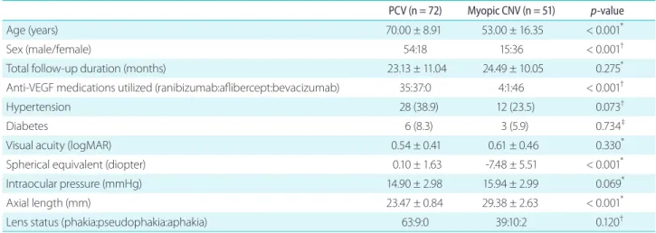
The mean age of the PCV group was 70.00 ± 8.91 years, which was significantly higher than that of the myopic CNV group (53.00 ± 16.35 years; p < 0.001). The proportion of women was higher in the myopic CNV group (25.0% vs. 70.6%; p <
0.001). The spherical equivalent (p < 0.001) and axial length (p < 0.001) in the myopic CNV group were significantly higher than those in the PCV group. The mean follow-up du- ration was 23.13 ± 11.04 months for PCV and 24.49 ± 10.05 months for myopic CNV. There were no significant differ- ences between the two groups (p = 0.275). In the PCV group, treatment included ranibizumab in 35 eyes and aflibercept in 37 eyes; in the myopic CNV group, treatment included ranibi- zumab in four eyes, aflibercept in one eye, and bevacizumab in 46 eyes (Table 1).
Subretinal hemorrhage (SRH), SRF, and intraretinal fluid (IRF) were found in 93.1%, 98.6%, and 22.2% of patients with PCV and 100%, 37.3%, and 15.7% of those with myopic CNV, respectively. There were no significant differences between the two groups for VA (p = 0.330) or CMT (p = 0.405; Table 2).
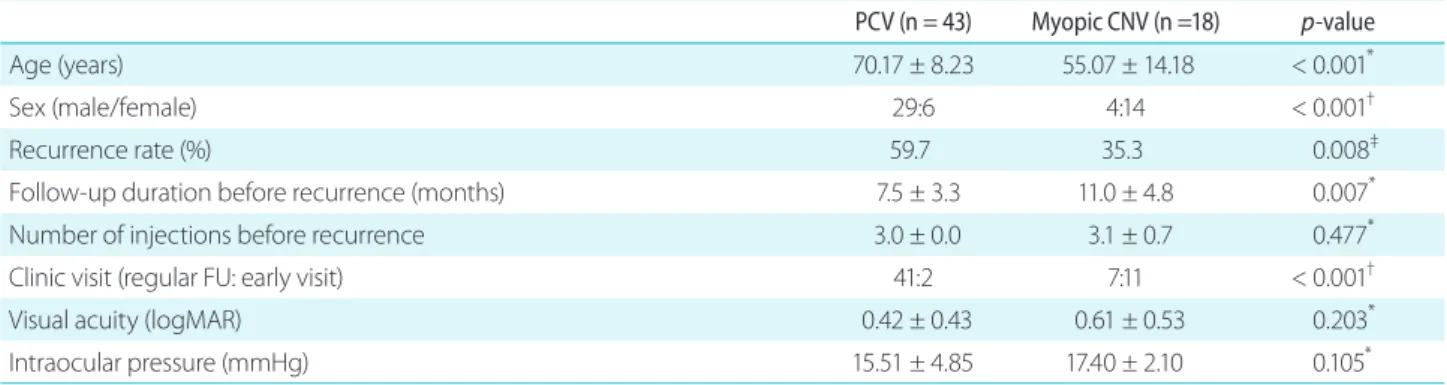
The rate of recurrence in the PCV group after achievement of a dry macula was 59.7% (43 eyes), which is significantly higher than that of the myopic CNV group (35.3%, 18 eyes;
p = 0.008). Recurrence was observed earlier in PCV (7.5 ± 3.3 months from diagnosis) than in myopic CNV (11.0 ± 4.8 months; p = 0.007). There were no significant differences in the number of injections between the two groups (3.0 ± 0.0 vs.
3.1 ± 0.7; p = 0.477). The recurrent lesion of PCV was found during routine follow-up examination in 95.3% of eyes. In contrast, recurrent myopic CNV was observed during routine
follow-up in 38.9% of eyes (p < 0.001), and the remaining patients visited the hospital earlier than in routine follow-up because of decreased visual acuity (11 eyes, 61.1%; Table 3).
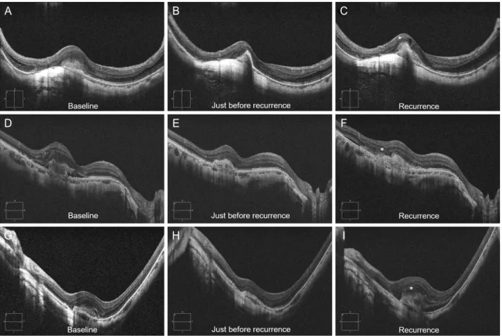
SRF was more common in PCV (93.0%) than in myopic CNV (22.2%; p < 0.001; Fig. 1), whereas subretinal hyperre- flective lesions with blurred margins on SD-OCT were more common in myopic CNV (88.9%) than in PCV (18.6%; p <
0.001; Fig. 2). Other factors, including SRH (p = 0.264) and IRF (p = 0.757), did not differ significantly between the two groups. CMT at the time of relapse was not significantly dif- ferent (Table 4).
CMT change from just before recurrence in patients with recurrent PCV and myopic CNV was 65.69 ± 48.51 μm and 40.93 ± 36.16 μm, respectively, which is a significant differ- ence (p = 0.024). In contrast to the change in CMT, the change
in visual acuity was significantly smaller in the PCV group (0.055 ± 0.158 logMAR) than in the myopic CNV group (0.361
± 0.399 logMAR; p = 0.001; Fig. 3).
Discussion
Numerous studies have reported the treatment outcomes of exudative AMD and myopic CNV. Kuroda et al. [23] re- ported that 65.7% of patients with neovascular AMD treated with three monthly ranibizumab injections experienced a recurrence during the first year. Another study that also used ranibizumab monotherapy reported that neovascularization recurred in 62.5% of patients [24]. Wakazono et al. [25]
reported that exudative AMD recurred in 43.7% of patients Table 1. Baseline characteristics of PCV and myopic CNV patients
PCV (n = 72) Myopic CNV (n = 51) p-value
Age (years) 70.00 ± 8.91 53.00 ± 16.35 < 0.001*
Sex (male/female) 54:18 15:36 < 0.001†
Total follow-up duration (months) 23.13 ± 11.04 24.49 ± 10.05 0.275*
Anti-VEGF medications utilized (ranibizumab:aflibercept:bevacizumab) 35:37:0 4:1:46 < 0.001†
Hypertension 28 (38.9) 12 (23.5) 0.073†
Diabetes 6 (8.3) 3 (5.9) 0.734‡
Visual acuity (logMAR) 0.54 ± 0.41 0.61 ± 0.46 0.330*
Spherical equivalent (diopter) 0.10 ± 1.63 -7.48 ± 5.51 < 0.001*
Intraocular pressure (mmHg) 14.90 ± 2.98 15.94 ± 2.99 0.069*
Axial length (mm) 23.47 ± 0.84 29.38 ± 2.63 < 0.001*
Lens status (phakia:pseudophakia:aphakia) 63:9:0 39:10:2 0.120†
Values are presented as mean ± standard deviation or number (%) unless otherwise indicated.
PCV = polypoidal choroidal vasculopathy; CNV = choroidal neovascularization; VEGF = vascular endothelial growth factor; logMAR = loga- rithm of the minimum angle of resolution.
*Independent t-test; †chi-square test; ‡Fisher's exact test.
Table 2. A comparison of baseline optical coherence tomography measurements and findings between PCV and myopic CNV patients PCV (n = 72) Myopic CNV (n = 51) p-value
Subretinal hemorrhage 67 (93.1) 51 (100.0) 0.076*
Subretinal fluid 71 (98.6) 19 (37.3) < 0.001*
Intraretinal fluid 16 (22.2) 8 (15.7) 0.368*
Central macular thickness (μm) 327.81 ± 105.05 343.60 ± 91.79 0.405†
Values are presented as mean ± standard deviation or number (%).
PCV = polypoidal choroidal vasculopathy; CNV = choroidal neovascularization.
*Chi-square test; †independent t-test.
after aflibercept treatment. Moon et al. [26] reported that 34.9% of patients with myopic CNV experienced recurrence, and two other studies reported that 23.3% and 22.7%, re- spectively, showed recurrence of CNV [27,28]. In the present study, the rate of recurrence of PCV was 59.7%, which is significantly higher than that of myopic CNV. The rates of recurrence for PCV and myopic CNV were not significantly different from previous results.
In this study, we focused on macular lesion characteristics and changes in visual acuity and CMT after recurrence in PCV and myopic CNV patients. At the time of recurrence, SRF was found in the majority of PCV patients but only in 22.2% of patients with myopic CNV, whereas subretinal hyperreflective lesion with blurred margin was more fre- quently observed with myopic CNV than with PCV (88.9%
vs. 18.6%). In addition, myopic CNV showed a significant Figure 1. Optical coherence tomography images showing recurrence after successful anti-vascular endothelial growth factor therapy in two polypoidal choroidal vasculopathy (PCV) patients (A, D). Eyes with PCV show a polypoidal lesion and double layer sign, which were treated by intravitreal aflibercept injection (B, E). After three injections, the subretinal fluid dried up (C, F). Recurrences were confirmed 5 and 3 months after initial treatment.
A
D
B
E
C
F
Table 3. Clinical characteristics of PCV and myopic CNV patients at recurrence
PCV (n = 43) Myopic CNV (n =18) p-value
Age (years) 70.17 ± 8.23 55.07 ± 14.18 < 0.001*
Sex (male/female) 29:6 4:14 < 0.001†
Recurrence rate (%) 59.7 35.3 0.008‡
Follow-up duration before recurrence (months) 7.5 ± 3.3 11.0 ± 4.8 0.007*
Number of injections before recurrence 3.0 ± 0.0 3.1 ± 0.7 0.477*
Clinic visit (regular FU: early visit) 41:2 7:11 < 0.001†
Visual acuity (logMAR) 0.42 ± 0.43 0.61 ± 0.53 0.203*
Intraocular pressure (mmHg) 15.51 ± 4.85 17.40 ± 2.10 0.105*
Values are presented as mean ± standard deviation unless otherwise indicated.
PCV = polypoidal choroidal vasculopathy; CNV = choroidal neovascularization; FU = follow up; logMAR = logarithm of the minimum an- gle of resolution.
*Mann–Whitney U test; †Fisher's exact test; ‡chi-square test.
decrease in visual acuity upon recurrence compared to PCV, and 61.1% of patients with recurrent myopic CNV com- plained of vision loss and visited the hospital earlier than their scheduled appointment.
Myopic CNV typically presents as type 2 CNV, is located between the neurosensory retina and retinal pigment epi- thelium layer, and has small dimensions. SD-OCT showed hyperreflective lesions located beneath the neurosensory Figure 2. Optical coherence tomography (OCT) images showing recurrence after successful anti-vascular endothelial growth factor therapy in three myopic choroidal neovascularization (CNV) patients (A, D, G). Eyes with myopic CNV show subretinal hemorrhage and subretinal hyperreflective mate- rials, which were treated with intravitreal bevacizumab injection (B, E, H). After three injections, the subretinal hemorrhage was absorbed, but subretinal hyperreflective materials remained with a clear margin (C, F, I). OCT images obtained 4, 5, and 6 months after the initial three injections show subretinal hyperreflective materials with a blurred margin (asterisk). Recurrences were confirmed, and additional injections were performed.
A
D
G
B
E
H
C
F
I
Table 4. A comparison of optical coherence tomography measurements and findings between PCV and myopic CNV patients at recurrence PCV (n = 43) Myopic CNV (n = 18) p-value
Subretinal hemorrhage 21 (48.8) 12 (66.7) 0.264*
Subretinal fluid 40 (93.0) 4 (22.2) < 0.001*
Intraretinal fluid 12 (27.9) 4 (22.2) 0.757*
Subretinal hyperreflective lesion with blurred margin 8 (18.6) 16 (88.9) < 0.001*
Central macular thickness (μm) 287.77 ± 71.75 308.87 ± 80.92 0.364†
Values are presented as mean ± standard deviation or number (%).
PCV = polypoidal choroidal vasculopathy; CNV = choroidal neovascularization.
*Fisher's exact test; †Mann–Whitney U test.
retina that could have been associated with subretinal fluid or intraretinal cysts. Subretinal hyperreflective lesions have been described in exudative AMD and are mostly associated with type 2 CNV [29-31]. Introini et al. [32] reported OCT findings in myopic CNV and described the main finding in myopic CNV as hyperreflective lesion with fuzzy borders.
They reported that hyperreflective lesions with fuzzy bor- ders are significantly associated with CNV activity.
Several studies have observed regression of the subretinal hyperreflective lesion with blurred margins after anti-VEGF treatment [32,33]. After regression of the lesion, well-defined hyperreflective lesions (possibly a fibrotic sequela) were ob-
served [33]. In this study, the majority of patients with recur- rent myopic CNV showed a subretinal hyperreflective lesion with clear margins just before recurrence (17 eyes, 94.4%), and the margin became hazy upon recurrence.
The increase in CMT was smaller at the time of recur- rence of myopic CNV than of PCV. However, visual acuity was significantly decreased compared to visual acuity in PCV patients. These results are similar to those in previous reports showing no significant changes in CMT after the initial anti-VEGF treatment despite increased visual acuity in myopic CNV [32]. Asai et al. [34] and associates reported that the intraretinal hyperreflective sign is a scarring reaction of the glial cells to hemorrhages and sheared synapses after invasion. The lack of integrity of the photoreceptor inner and outer segments (IS/OS) and external limiting membrane (ELM) line, which is indicative of damage to the photorecep- tors resulting from hemorrhage, is an important indicator of visual prognosis in various macular pathologies [35-37]. This subretinal hyperreflective lesion with blurred margins was observed in myopic CNV and may have led to damage of the IS/OS and ELM; this could have resulted in the decreased visual acuity.
In the present study, SRF was less common in myopic CNV than PCV at both baseline and recurrence. In addition, changes in CMT were significantly smaller in recurrent my- opic CNV than in PCV and could not be easily identified on OCT scan. In contrast to CNV secondary to AMD, myopic CNV showed minimal intraretinal and subretinal fluid [38].
This may be attributed to the attenuated nature of choroidal blood circulation in pathologic myopia [39,40] and preexist- ing thinning of the neurosensory retina secondary to myo- pic disease processes or hyperactivity of RPE, resulting in more effective pumping of fluid out of the subretinal space [38,41,42]. For these reasons, we presumed that small lesions or minimal changes, such as subretinal hyperreflective ma- terial with blurred margins, would be observed on the OCT scan in patients with recurrent myopic CNV.
When physicians follow up with patients with PCV or myopic CNV after the initial treatment, it is possible to ad- minister an additional injection of anti-VEGF for recurrence based on changes in CMT. Recurrence of PCV can be easily assessed based on increased thickness according to lesion characteristics, whereas the change in CMT in recurrent myopic CNV was not large. In patients with myopic CNV, a subretinal hyperreflective lesion with blurred margins was Figure 3. A comparison of changes in central macular thickness
(CMT) and best corrected visual acuity (BCVA; that at recurrence mi- nus that just before recurrence) in patients with polypoidal choroidal vasculopathy (PCV) or myopic choroidal neovascularization (CNV).
CMT showed a greater increase in the PCV group than in the myopic CNV group because of subretinal fluid (p = 0.024, A). However, BCVA was worse in the myopic CNV group than in the PCV group (p = 0.001, B). Error bars represent the standard error. LogMAR = logarithm of minimal angle of resolution.
80 60 40 20
0 PCV Myopic CNV
CMT changes (recurrence- just before recurrence,
μm)
Eyes p = 0.024
A
0.5 0.4 0.3 0.2 0.1
0 PCV Myopic CNV
BCVA changes (recurrence- just before recurrence, LogMAR)
Eyes p = 0.001
B
observed on SD-OCT at the time of recurrence. If a physi- cian diagnoses recurrence based on central macular thick- ness alone, recurrence of myopic CNV may be missed, and treatment time will be delayed.
This study had several limitations, including the relatively small number of patients and retrospective study design, which may have led to selection bias. This study popula- tion may not represent all PCV and myopic CNV patients.
However, the majority of PCV patients who experienced recurrence in this study showed SRF, and subretinal hyper- reflective lesions with blurred margins were observed during recurrence of myopic CNV.
In conclusion, new SRF was mainly observed in patients with recurrent PCV, whereas subretinal hyperreflective le- sions with a blurred margin were observed in patients with myopic CNV. Changes in central macular thickness after recurrence were greater in the PCV group than in the myo- pic CNV group, but visual acuity was worse in patients with myopic CNV than in those with PCV. Because patients with recurrent myopic CNV may not show clear changes in CMT, it is important to review minor changes in subretinal hyper- reflective lesions rather than changes in CMT.
Conflicts of Interest
The authors declare no conflicts of interest relevant to this article.
References
1. Yannuzzi LA, Sorenson J, Spaide RF, Lipson B. Idiopathic polyp- oidal choroidal vasculopathy (IPCV). Retina 1990;10:1-8.
2. Laude A, Cackett PD, Vithana EN, et al. Polypoidal choroidal vas- culopathy and neovascular age-related macular degeneration:
same or different disease? Prog Retin Eye Res 2010;29:19-29.
3. Chung SE, Kang SW, Lee JH, Kim YT. Choroidal thickness in polypoidal choroidal vasculopathy and exudative age-related macular degeneration. Ophthalmology 2011;118:840-5.
4. Jirarattanasopa P, Ooto S, Nakata I, et al. Choroidal thickness, vascular hyperpermeability, and complement factor H in age-related macular degeneration and polypoidal choroidal vasculopathy. Invest Ophthalmol Vis Sci 2012;53:3663-72.
5. Koizumi H, Yamagishi T, Yamazaki T, Kinoshita S. Relationship be- tween clinical characteristics of polypoidal choroidal vasculopa-
thy and choroidal vascular hyperpermeability. Am J Ophthalmol 2013;155:305-13.e1.
6. Sasahara M, Tsujikawa A, Musashi K, et al. Polypoidal choroidal vasculopathy with choroidal vascular hyperpermeability. Am J Ophthalmol 2006;142:601-7.
7. Kim SW, Oh J, Kwon SS, et al. Comparison of choroidal thickness among patients with healthy eyes, early age-related maculopa- thy, neovascular age-related macular degeneration, central se- rous chorioretinopathy, and polypoidal choroidal vasculopathy.
Retina 2011;31:1904-11.
8. Tano Y. Pathologic myopia: where are we now? Am J Ophthal- mol 2002;134:645-60.
9. Curtin BJ. The posterior staphyloma of pathologic myopia. Trans Am Ophthalmol Soc 1977;75:67-86.
10. Ikuno Y, Sayanagi K, Soga K, et al. Lacquer crack formation and choroidal neovascularization in pathologic myopia. Retina 2008;28:1124-31.
11. Avila MP, Weiter JJ, Jalkh AE, et al. Natural history of choroidal neovascularization in degenerative myopia. Ophthalmology 1984;91:1573-81.
12. Cohen SY, Laroche A, Leguen Y, et al. Etiology of choroidal neovas- cularization in young patients. Ophthalmology 1996;103:1241-4.
13. Grossniklaus HE, Green WR. Pathologic findings in pathologic myopia. Retina 1992;12:127-33.
14. Ruiz-Moreno JM, López-Gálvez MI, Donate J, et al. Myopic cho- roidal neovascularization. Ophthalmology 2011;118:2521-3.
15. Yoshida T, Ohno-Matsui K, Yasuzumi K, et al. Myopic choroi- dal neovascularization: a 10-year follow-up. Ophthalmology 2003;110:1297-305.
16. Hayashi K, Ohno-Matsui K, Shimada N, et al. Long-term pattern of progression of myopic maculopathy: a natural history study.
Ophthalmology 2010;117:1595-611, 611.e1-4.
17. Byeon SH, Lee SC, Oh HS, et al. Incidence and clinical patterns of polypoidal choroidal vasculopathy in Korean patients. Jpn J Ophthalmol 2008;52:57-62.
18. Neelam K, Cheung CM, Ohno-Matsui K, et al. Choroidal neo- vascularization in pathological myopia. Prog Retin Eye Res 2012;31:495-525.
19. Tong JP, Chan WM, Liu DT, et al. Aqueous humor levels of vascu- lar endothelial growth factor and pigment epithelium-derived factor in polypoidal choroidal vasculopathy and choroidal neo- vascularization. Am J Ophthalmol 2006;141:456-62.
20. Chan WM, Lai TY, Liu DT, Lam DS. Intravitreal bevacizumab (Avastin) for myopic choroidal neovascularization: six-month results of a prospective pilot study. Ophthalmology 2007;114:2190-6.
21. Koh AH, Chen LJ, Chen SJ, et al. Polypoidal choroidal vasculop- athy: evidence-based guidelines for clinical diagnosis and treat- ment. Retina 2013;33:686-716.
22. Tan CS, Ngo WK, Chen JP, et al. EVEREST study report 2: imaging and grading protocol, and baseline characteristics of a ran- domised controlled trial of polypoidal choroidal vasculopathy.
Br J Ophthalmol 2015;99:624-8.
23. Kuroda Y, Yamashiro K, Miyake M, et al. Factors associated with recurrence of age-related macular degeneration after anti-vas- cular endothelial growth factor treatment: a retrospective co- hort study. Ophthalmology 2015;122:2303-10.
24. Dadgostar H, Ventura AA, Chung JY, et al. Evaluation of injection frequency and visual acuity outcomes for ranibizumab mono- therapy in exudative age-related macular degeneration. Oph- thalmology 2009;116:1740-7.
25. Wakazono T, Yamashiro K, Oishi A, et al. Recurrence of choroidal neovascularization lesion activity after aflibercept treatment for age-related macular degeneration. Retina 2017;37:2062-8.
26. Moon BG, Cho AR, Lee J, et al. Improved visual outcome and low recurrence with early treatment with intravitreal anti-vascular endothelial growth factor in myopic choroidal neovasculariza- tion. Ophthalmologica 2017;237:128-38.
27. Lai TY, Luk FO, Lee GK, Lam DS. Long-term outcome of intravitreal anti-vascular endothelial growth factor therapy with bevacizumab or ranibizumab as primary treatment for subfoveal myopic choroi- dal neovascularization. Eye (Lond) 2012;26:1004-11.
28. Yang HS, Kim JG, Kim JT, Joe SG. Prognostic factors of eyes with naive subfoveal myopic choroidal neovascularization after intra- vitreal bevacizumab. Am J Ophthalmol 2013;156:1201-10.e2.
29. Coscas F, Querques G, Forte R, et al. Combined fluorescein angi- ography and spectral-domain optical coherence tomography imaging of classic choroidal neovascularization secondary to age-related macular degeneration before and after intravitreal ranibizumab injections. Retina 2012;32:1069-76.
30. Giani A, Esmaili DD, Luiselli C, et al. Displayed reflectivity of choroidal neovascular membranes by optical coherence to- mography correlates with presence of leakage by fluorescein angiography. Retina 2011;31:942-8.
31. Giani A, Luiselli C, Esmaili DD, et al. Spectral-domain optical co- herence tomography as an indicator of fluorescein angiography leakage from choroidal neovascularization. Invest Ophthalmol Vis Sci 2011;52:5579-86.
32. Introini U, Casalino G, Querques G, et al. Spectral-domain OCT in anti-VEGF treatment of myopic choroidal neovascularization.
Eye (Lond) 2012;26:976-82.
33. Bruyère E, Caillaux V, Cohen SY, et al. Spectral-domain optical coherence tomography of subretinal hyperreflective exuda- tion in myopic choroidal neovascularization. Am J Ophthalmol 2015;160:749-58.e1.
34. Asai T, Ikuno Y, Nishida K. Macular microstructures and prognos- tic factors in myopic subretinal hemorrhages. Invest Ophthal- mol Vis Sci 2014;55:226-32.
35. Shin HJ, Chung H, Kim HC. Association between foveal micro- structure and visual outcome in age-related macular degenera- tion. Retina 2011;31:1627-36.
36. Oishi A, Hata M, Shimozono M, et al. The significance of external limiting membrane status for visual acuity in age-related macu- lar degeneration. Am J Ophthalmol 2010;150:27-32.e1.
37. Hayashi H, Yamashiro K, Tsujikawa A, et al. Association between foveal photoreceptor integrity and visual outcome in neovas- cular age-related macular degeneration. Am J Ophthalmol 2009;148:83-9.e1.
38. Chan NS, Teo K, Cheung CM. Epidemiology and diagnosis of myopic choroidal neovascularization in Asia. Eye Contact Lens 2016;42:48-55.
39. Wakabayashi T, Ikuno Y. Choroidal filling delay in choroidal neo- vascularisation due to pathological myopia. Br J Ophthalmol 2010;94:611-5.
40. Avetisov ES, Savitskaya NF. Some features of ocular microcircula- tion in myopia. Ann Ophthalmol 1977;9:1261-4.
41. Miller H, Miller B, Ryan SJ. The role of retinal pigment epithelium in the involution of subretinal neovascularization. Invest Oph- thalmol Vis Sci 1986;27:1644-52.
42. Yoshida A, Ishiko S, Kojima M. Inward and outward permeability of the blood-retinal barrier in experimental myopia. Graefes Arch Clin Exp Ophthalmol 1996;234 Suppl 1:S239-42.