CONTINUED ROOT DEVELOPMENT AFTER AVULSION OF IMMATURE TEETH
Ju-Eun Lee, Young-Jin Kim, Hyun-Jung Kim, Soon-Hyeun Nam
Department of Pediatric Dentistry, School of Dentistry, Kyungpook National University
Traumatic injuries to immature permanent teeth are common and the results can be destructive. Although Hertwig’s epithelial root sheath is usually sensitive to trauma, it may resist damage from trauma thereby re- taining its vitality and continuing to calcify a root under favorable conditions.
This case report describes two cases of trauma to immature permanent incisors. The first case presents an avulsed maxillary central incisor which has been replanted. The other case shows completely avulsed mandibu- lar central incisors which have not been replanted. However, both cases subsequently show continued growing roots separated from the main roots.
This report highlights the ability of the immature pulp tissue to continue to form dentin and the robustness of Hertwig’s epithelial root sheath to initiate root development despite a traumatic injury.
Key words :Separated root, Immature teeth, Tooth avulsion, Hertwig’s epithelial root sheath Abstract
Ⅰ. Introduction
Traumatic injuries to immature permanent teeth are common, affecting 30% of children1). Of all dental in- juries, tooth avulsion can result in serious complications, such as loss of pulp vitality and root resorption2).
Stem cells from the apical papilla (SCAP) are general- ly considered crucial for proper development of tooth roots3). Many studies have reported continued root for- mation following trauma or infection and have highlight- ed the importance of Hertwig’s epithelial root sheath (HERS) in continued root growth4-9).
HERS is usually vulnerable to trauma; however, in some circumstances, it may survive and retain its ability
of root organization10,11).
Several reports have described that a separate root tip is developed apically to the existing root of immature tooth after trauma4-9). These reports have speculated that the detachment of SCAP and HERS by a traumatic episode accounts for the development of a separate root tip. Continued root growth in immature tooth with an infected pulp also has been reported even though the underlying mechanism is still unclear12).
This case report shows continued root development apart from the main roots after avulsion of immature permanent teeth and emphasizes the reparative power of immature pulp and HERS to complete their program of root formation.
Corresponding author : Soon-Hyeun Nam
Department of Pediatric Dentistry, School of Dentistry, Kyungpook National University, 2177 Dalgubeol-daero, Jung-gu, 700-412, Korea Tel: +82-53-600-7211 / Fax: +82-53-426-6608 / E-mail: [email protected]
Received January 10, 2013 / Revised May 14, 2013 / Accepted May 14, 2013
※This research was supported by Kyungpook National University Research Fund, 2013.
Ⅱ. Case Reports 1. Case Ⅰ
A 6-year-old girl visited the Department of Pediatric Dentistry at Kyungpook National University Dental Hospital for emergency care of maxillary central incisors following a fall-down. Clinical examination showed com- plete avulsion of the left central incisor from its socket and intrusion of the right central incisor. The teeth had immature roots with open apexes without apparent signs of tooth fracture or damage to the bone structure. The avulsed tooth was delivered by her parents being kept in milk for approximately 2 hours extra-orally. Under local anesthesia, the teeth were repositioned and splinted with semi-rigid resin-wire (Fig. 1a). Oral Antibiotics were pre- scribed to prevent possible infection. Four days after the accident, the patient visited our section to repair splint.
Three weeks later, splinting wire was removed. No pathologic swelling or sinus tract was found on intraoral examination. Both central incisors displayed moderate sensitivity on percussion and palpation tests. They did not respond to the cold test or electric pulp vitality test.
Two months after replantation, both teeth were within normal range on percussion and palpation tests.
However, the left central incisor did not respond to the cold test or electric pulp vitality test. Moreover, a drain- ing sinus tract was found in the mucolabial fold near the apex of the left central incisor. Endodontic treatment was started with a diagnosis of pulp necrosis of the left central incisor.
The access cavity was prepared after tooth isolation
with rubber dam. Then, a necrotic pulp was found. The canal length was determined by a parallel preoperative periapical radiograph. The canal was cleaned with #40 K-file and 5.25% sodium hypochlorite (Sultan, USA) 1mm short of the radiographic apex. The canal was dried with sterile paper points and a mixture of calcium hydroxide powder and saline was placed into the canal with a lentulo spiral. The access cavity was temporally sealed with Cavit (ESPE, Germany) and cotton pellet.
The patient returned one week later. She was asym- tomatic and the sinus tract was not present. The access cavity was opened and the canal again was irrigated with 5.25% sodium hypochlorite and saline. The canal was dried with sterile paper points and filled with Vitapex (Neo Dental, Japan). The tooth was restored with a dentin-bonded resin composite (Tetric ceram, Liechtenstein) (Fig. 1b).
At the 3-month recall after the accident, the patient was asymptomatic. However, interestingly enough, there was evidence of a developing radiopaque mass apical to the root end of the left central incisor on a periapical ra- diograph (Fig. 1c).
One year later, a periapical radiograph revealed con- tinued development of the radiopaque mass which ap- peared to be surrounded by a radiolucent band and radi- olucent central core, similar to the appearance of the pe- riodontal membrane and pulp canal of a root (Fig. 1d).
Two years later, a periapical radiograph showed pro- gressive enlargement of the radiopaque mass with calcif- ic change within the pulp canal. Also, we could find bony invasion within the pulp canal space of the right central incisor (Fig. 1e).
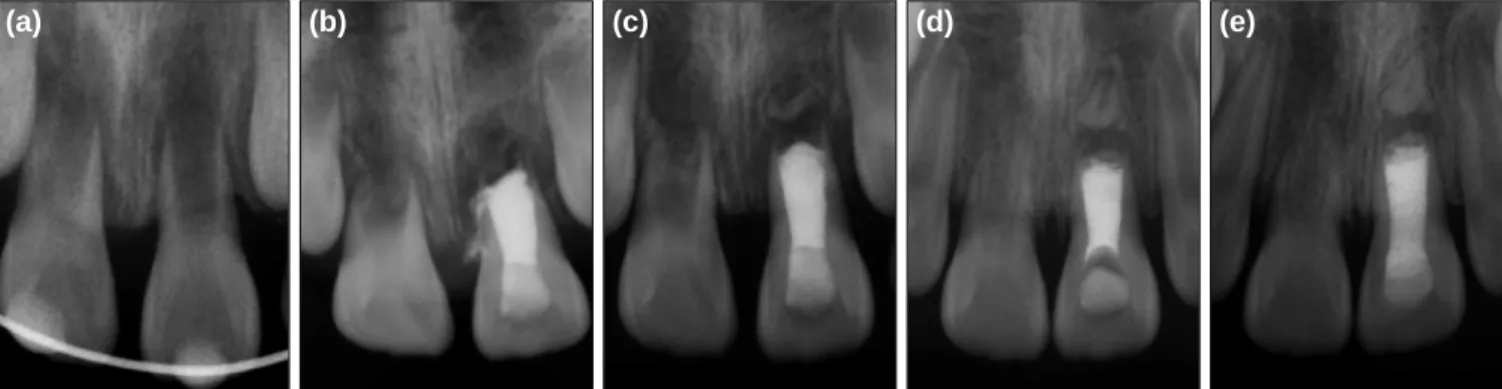
Fig. 1. Periapical views. (a) Initial visit; replanted maxillary central incisors with open apexes were observed. (b) Maxillary left central incisor at 2 months post-trauma; endodontic treatment was performed with a diagnosis of pulp necrosis. (c) Maxillary left central incisor at 3 months post-trauma; radiopaque mass apical to the maxillary left central incisor was evident. (d) Maxillary left central incisor at 1 year post-trauma; continuing development of the radiopaque mass at the apex was observed. (e) Maxillary left central incisor at 2 years post-trauma; the radiopaque mass had continued thickening with clo- sure of the apex.
(a) (b) (c) (d) (e)
2. Case Ⅱ
A 6-year-old girl tripped and knocked her face on the edge of a desk at home, resulting in completely avulsed mandibular central incisors.
She presented for treatment 4 days after the accident with a chief complaint of avlusion of the mandibular cen- tral incisors. Except for a small contusion of the gingiva around the mandibular central incisors, there were no other soft tissue injuries. The teeth could not be replaced into their sockets as she lost the teeth (Fig. 2a).
At the 3-month recall, the soft tissue had completely healed based on a clinical observation and the periapical radiograph indicated normal empty healing sockets with no evidence of residual dental hard tissue (Fig. 2b).
A review at 6 months after the initial accident showed the normal soft tissue architecture. However, a periapi- cal radiograph taken at the time revealed the formation of developing radiopaque masses on the areas corre- sponding to the sockets (Fig. 2c).
One year after the teeth had been lost, the develop- ment of the radiopaque masses continued without any sign of pathological changes. Well-defined periodontal ligament spaces could be traced within the alveolar bone (Fig. 2d). The soft tissue was clinically normal and the alveolar bone contour was in excellent condition.
Ⅲ. Discussion
Generally, HERS is considered responsible for the de- velopment of a tooth root. It may be involved in regulat- ing the differentiation of odontoblasts or cementoblasts that could give rise to further hard tissue formation13,14).
In addition, HERS may also play a role in preventing the invasion of periodontal ligament cells into the root canal, otherwise it would result in intracanal bone for- mation or partial/complete arrest of root development4).
Apical papilla, located apical to the developing pulp, is gelatinous soft tissue that is easily detached from the root apex15,16). Mesenchymal stem cells on apical papilla lead to root development, even in case of pulpal necrosis in immature teeth3).
Greer et al.17)reported that the pulp remnants left in the socket of an avulsed tooth led to continued develop- ment of root and calcification of dentin and cementum.
Histological or microscopic observation on the tooth sock- ets was not part of our case report but it is supposed that the pulp remnants from the avulsed teeth contin- ued to function to complete mineralization of roots with remaining HERS.
It is believed that normal root development and odon- toblastic differentiation would cease upon the destruction of HERS. However, some studies reported continued root formation after trauma and sometimes even after infec- tion4-9). In other reports, a separately growing root tip was observed in the apical area of the main root end af- ter trauma to an immature tooth, as SCAP and HERS can easily be detached by an external force due to their loose attachment to the apex5-9).
Yang et al.18) addressed that apexification treatment could result in such detachment of the tissue from the main root. Also, an excessive tooth mobility associated with pulpal infection is reported to be another contribut- ing factor for separation force12). In our case report, the external force directly related to a traumatic episode is regarded as accountable factor for the detachment be-
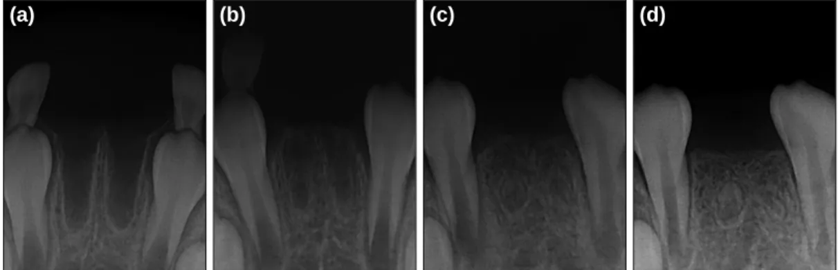
Fig. 2. Periapical views. (a) Initial visit; sockets of the completely avulsed mandibular central incisors were observed. (b) 3 months later, the sockets had completely healed without any sign of residual dental hard tissue. (c) 6 months later, radiopaque masses were found on the areas corresponding to the sockets. (d) 1 year later, continued development of the radiopaque masses was observed.
(a) (b) (c) (d)
tween root apex and HERS.
Although several reports have refered to isolated root formation, precise histology of the newly formed root has not been studied in detail. Gibson et al.5) examined a similar tooth fragment and revealed a confusing picture of disturbed dentinogenesis, resorption and repair that suggests the initial calcification of pulp stone formation.
In histologic evaluation, Yang et al.18) revealed that the separate root tip was composed of normal pulp, pre- dentin, dentin, and cementum, whereas the apical barri- er of the main root appeared to be composed of osteo- dentin, immature cementum, or immature bone. In this case report, histologic exanimation was not performed.
However, further histological evaluation could be carried out if the separated roots be removed.
The management of separate root fragments may be a awkward matter to the clinician. While Welbury and Walton9)removed the similar root tips due to associated infection, Greer et al.17) and Arrow19)did not remove the separated root fragments as they did not developed any complications. Also, in this case report, removal was not contemplated throughout the follow-up period as the separated root tips had contributed well to the mainte- nance of the alveolar ridge and soft tissue architecture without any sign of pathological changes.
Unless the separated root tip hinders orthodontic tooth movement or has any untoward pathosis, the option of removal should not be considered as removal could result in alveolar bone atrophy, subsequently causing esthetic problems on restoration treatment.
The replacement of pulp canal space with bony struc- ture was observed after the intrusive injury of the maxil- lary right central incisor in the first case. It is speculated that the partial or total damage to HERS may be re- sponsible4).
To conclude, HERS and SCAP, even when detached from the coronal root structure by an external force, could be capable of continuing the initiation of root de- velopment. Thus, in the case of avulsion of immature teeth, clinicians should be aware of the possibility that a new root tip can develop and have continued follow-ups of traumatized immature teeth for an accurate diagnosis.
Ⅳ. Summary
HERS is necessary for normal root development. After avulsion injury to an immature tooth, HERS can be de- tached from the coronal part and create a separated root
tip. This case report describes the separate root growth after trauma to immature teeth and highlights the im- portance of HERS in continued root formation.
References
1. Andreasen JO, Andreasen FM, Andersson L : Textbook and color atlas of traumatic injuries to the teeth, 4th edn. Blackwell Munksgaard, Copenhagen, Denmark 2007.
2. Andreasen JO, Ravn JJ : Epidemiology of traumatic dental injuries to primary and permanent teeth in a Danish population sample. Int J Oral Surg, 1:235-9, 1972.
3. Huang GT, Sonoyama W, Shi S, et al. : The hidden treasure in apical papilla : the potential role in pulp / dentin regeneration and bioroot engineering. J Endod, 34:645-51, 2008.
4. Andreasen JO, Borum MK, Andreasen FM : Replantation of 400 avulsed permanent incisors. 3.
Factors related to root growth. Endod Dent Traumatol, 11:69-75, 1995.
5. Gibson AC : Continued root development after trau- matic avulsion of partly-formed permanent incisor.
Br Dent J, 126:356-7, 1969.
6. Barker BC, Mayne JR : Some unusual cases of apexification subsequent to trauma. Oral Surg Oral Med Oral Pathol, 39:144-50, 1975.
7. Burley MA, Reece RD : Root formation following traumatic loss of an immature incisor: a case report.
Br Dent J, 141:315-6, 1976.
8. Smith BE, Thaler MN : Detached root apexogenesis.
Oral Surg Oral Med Oral Pathol, 73:129, 1992.
9. Welbury R, Walton AG : Continued apexogenesis of immature permanent incisors following trauma. Br Dent J, 187:643-4, 1999.
10. Rule DC : Some considerations in the clinical man- agement of traumatised permanent incisors with immature roots. Proc Br Paedod Soc, 3:33-5, 1973.
11. Cvek M : Prognosis of luxated non-vital maxillary incisors treated with calcium hydroxide and filled with gutta-percha. Endod Dent Traumtol, 8:45-55, 1992.
12. Jung IY, Kim ES, Lee CY, Lee SJ : Continued development of the root separated form the main root. J Endod, 37:711-4, 2011.
13. Ten Cate AR : A fine structural study of coronal and root dentinogenesis in the mouse : observation on
the so-called ‘von-Korff fibers’and their contribution to mantle dentin. J Anat, 125:183-97, 1978.
14. Sonoyama W, Seo BM, Yamaza T, Shi S : Human Hertwig’s epithelial root sheath cells play crucial roles in cementum formation. J Dent Res, 86:594-9, 2007.
15. Sonoyama W, Liu Y, Wang S, et al. : Mesenchymal stem cell-mediated functional tooth regeneration in swine, PloS One, 1:e79:1-8, 2006.
16. Sonoyama W, Liu Y, Huang GT, et al. : Characterization of the apical papilla and its resid-
ing stem cells from human immature permanent teeth: a pilot study. J Endod, 34:166-71, 2008.
17. Greer JM, Moule AJ, Greer PJ : Resumed tooth development following avulsion of a permanent cen- tral incisor. Int Endod J, 29:266-70, 1996.
18. Yang SF, Yang ZP, Chang KW : Continuing root formation following apexification treatment. Endod Dent Traumatol, 6:232-5, 1990.
19. Arrow P : An unusual healing of a replanted perma- nent lateral incisor. Aust Dent J, 54:57-60, 2009.
주요어: 분리된 치근, 미성숙 영구치, 완전 탈구, Hertwig 상피 근초
미성숙 영구치의 탈구성 외상 이후 계속된 치근 성장
이주은∙김영진∙김현정∙남순현 경북대학교 치의학전문대학원 소아치과학교실
외상에 의한 미성숙 영구치의 손상은 유치열에서 영구치열로 이환되는 8~10세경에 가장 빈발하며 전체 외상 환자의 높은 빈도를 차지한다. 외상에 대한 결과는 치아의 파절, 전위, 함입, 정출, 탈구 등의 경조직 손상 뿐 아니라 치수, 치주인대, Hertwig 상피 근초, 치조골, 치은 및 구강점막 등의 치아 인접조직의 손상도 포함한다.
일반적으로 Hertwig 상피 근초는 외상성 손상에 취약하지만, 때때로 감염이나 외상에 의한 손상을 견디고 생활력을 유지 하여 치근성장에 대한 정상적인 기능을 수행하는 것이 보고된 바 있다.
본 증례에서는 외상에 의해 완전 탈구된 미성숙 영구치를 가진 두 명의 환자에 대해 보고하고자 한다. 첫 번째 환자의 경우 탈구된 상악 중절치를 재식하였고 두 번째 환자의 경우 탈구된 하악 중절치를 재식하지 않았다. 하지만 두 환자 모두에서 탈 구된 치아의 치조와 부위에 분리된 치근의 계속적인 성장을 보이는 바 이를 보고하고자 하며, 나아가 계속적인 치근형성에 있어서 미성숙 치수 조직과 Hertwig 상피 근초의 생활력 보존이 결정적임을 알리고자 한다.
국문초록