고지방식이에 의해 비만이 유도된 Zucker Rats에서 상백피분말의 비만 개선효과
충북대학교 수의과대학1, 박정휘 한의원2, 충북대학교병원 진단검사의학과3
홍성희1․김동규1․이남진1․조정희1․강종구1․김윤배1․박정휘2․황석연3
Anti-Obesitic Effect of Mulberry Root-Bark (Mori radicis Cortex) in Zucker Rats with High Lipid Diet Induced-Obesity
Seong-Hee Hong1, Dong-Kyu Kim1, Nam-Jin Lee1, Jung-Hee Cho1, Jong-Koo Kang1, Yun-Bae Kim1, Jung-Hui Park2, and Seock-Yeon Hwang3
College of Veterinary Medicine and Research Institute of Veterinary Medicine, Chungbuk National University, Cheongju 361-763, Korea1
Park, Jung-Hui Oriental Medical Clinic, Daejeon 302-122, Korea2
Department of Clinical Laboratory Medicine, Chungbuk National University Hospital, Cheongju 360-240, Korea3
This study was performed to investigate the anti-obesitic effect of mulberry root-bark on male and female Zucker FA/FA or FA/fa rats. Obesity in the rats was induced by feeding high-lipid diet contained 3% corn oil and 1% cholesterol. Experimental groups in male and female rats were assigned to normal diet group (normal control), high-lipid diet group(positive control) and 3% mulberry root-bark powder in high-lipid diet group (MRC). The mulberry treated-group showed decreases of body weight, FER (food efficiency ratio) value and lipid peroxidation in the liver and increase of high density lipoprotein cholesterol (HDL-C) value, compared with positive control groups. Our findings suggest that mulberry root-bark has a potential role in preventing or improving obesity from the following points of view, body weight, serum lipids and antioxidant enzyme activities.
Key Words : Zucker rats, Mulberry root, Anti-obesity 대한임상검사학회지 : 37권 제2호, 129-137, 2005
1)
I. 서 론
비만은 현대사회에서 가장 일반적인 대사성 질병이다.
동물 실험결과 비만의 원인은 1) 특발(spontaneous
교신저자 : 황석연, (우)361-713 충북 청주시 개신동 62, 충북대학교병원 진단검사의학과
Tel : 043-269-6257 E-mail : [email protected]
naturally occurring), 2) 유전적 요인(한 가지 이상 특이 유전자의 돌연변이), 3) 식이성 원인 4) 신경내분비적 원 인 등으로 나눌 수 있지만 그 중에서 과잉에너지 축적이 가장 직접적으로 관계되며 비만의 95%는 단순 비만에 속 한다(Hill, 2002).
비만 치료에 있어서 항비만약물의 이용이 중요하지만 시중에 유통되고 있는 항비만약물은 그 부작용이 문제가 되고 있는데, sibutramine의 심혈관계에 부작용, orlistat의
위장관계열 부작용 등이 그것이다(Fujioka 등, 2000;
McMahon 등, 2000; Leung 등, 2003). 이에 따라 부작용 없는 대체 약물을 개발하기 위한 많은 실험이 시행되고 있는데, 상백피를 주재료로 한 본 연구 또한 그 중 하나이 다.
상백피(mulberry root-bark, Mori radicis Cortex)는 뽕나 무(Morus alba L.) 또는 동속 식(Moraceae)의 뿌리껍질로 서 한방에서는 사폐화(폐의 열을 내림), 리이변(대소변을 잘 보게 함), 산어혈(어혈을 풀어 줌), 하기행수(기운[열]
을 내리게 하고 수분을 조절함), 지수청담(기침을 멎게 하 고 담을 삭임), 소염 작용 등이 탁월하여 해열진해거담제 로 널리 사용되고 있으며, 민간에서는 사람이 뱀에 물렸 을 때나 특발성 괴저에 바르고 목욕하였다고 기록되어 있다(Nomura 등, 1976; 문, 1984).
본 실험은 유전적으로 비만 유전자를 갖고 있는 Zucker rat 중 lean type인 Zucker FA/FA와 Zucker FA/fa rat를 사 용하였다. 비만유전자에 의해 지방세포에서 분비되는 단 백질 leptin은 시상하부의 포만중추에 작용하여 식욕과 에 너지 소모 조절에 관여하는데, obese Zucker rat의 경우 leptin receptor의 결손에 의한 leptin signaling 결핍과 관계 되어 비만과 과식증이 나타나게 된(Chua, 1996; Beck 등, 2002). 또한 Zucker rat의 leptin signaling 결핍은 식이성 비만을 유발시키는 혈중, 위, 시상하부의 펩타이드성 호 르몬인 ghrelin과 음의 상관관계가 있다고 보고되었다 (Kojima 등, 1999; Tschop 등, 2000; Tschop 등, 2001;
Beck 등, 2002). Lean Zucker rat(FA/FA and FA/fa)의 혈 장 ghrelin농도는 obese Zucker rat보다 낮지만, 사람 비만 의 경우 ghrelin의 혈중 농도가 낮기 때문에 이 실험에서 는 사람의 비만에 접근하고자 lean Zucker rat를 사용하였 (Tschop 등, 2001; Beck 등, 2004). 이 실험에서는 고지방 식이로 비만을 유도한 lean Zucker rat에 상백피 분말 (mulberry leaves powder)을 8주간 식이로 공급하여 체중 변화, 혈중 내 glucose, 혈청 생화학적 검사 및 지질과산화 를 통하여 항비만 효과를 확인하고자 하였다.
Ⅱ. 재료 및 방법
1. 시험물질
상백피 분말은 뽕나무 뿌리를 건조시켜 분말화하여 3%
의 농도로 사용하였으며, 콜레스테롤(Junsei Chemical
Co., Ltd. Japan), corn-oil(Junsei Chemical Co., Ltd. Japan) 과 그 외 실험에 사용한 시약은 특급 시약을 사용하였다.
2. 실험동물 및 실험설계
실험동물은 5주령의 male Zucker fa/fa rat(33마리, 이하 male rat)와 female Zucker fa/fa rat(33마리, 이하 female rat)를 구입하여 1주간 순화 후 3개의 시험군인 정상대조 군, 양성대조군, 시험물질 급여군에 대해 각 실험군당 암 수 11마리씩 22마리를 배정하였다.
시험물질급여군은 3% 상백피 분말(MRC), 1% 콜레스 테롤 그리고 3% corn oil을 가루사료에 혼합하여 8주간 급여하였으며, 대조군(normal group)과 양성대조군 (positive group)도 각각 일반가루사료와 고지방가루식이 (1% 콜레스테롤, 3% corn oil)를 공급하였다. 실험동물의 사육조건은 온도 23±2℃, 상대습도 50±10%, 환기횟수 10-12/hr, 명암주기 12시간(07:00 점등-19:00 소등), 조도 200 Lux로 유지하였고, 실험동물용 가루사료((주)샘타코 바이오코리아, 경기도 오산시)와 여과된 정제수를 자유로 이 섭취할 수 있도록 공급하였다.
동물의 체중과 사료섭취량은 매주 측정하였고, 임상증 상은 매일 관찰하였다. 8주 후에 동물을 부검하고 혈액을 채취하였고, 간 조직은 ice-cold 0.9% 생리식염수로 liver perfusion을 한 후에 간을 적출하고 생리식염수로 간의 혈 액을 제거한 후, 액체 질소에 동결하여 지질과산화를 측 정하였다.
3. 혈액학적 검사
부검 전일 18시간 동안 절식시킨 동물을 에테르 마취 하에 배대동맥에서 전혈을 채혈하여 응고되지 않도록 ethylenediaminetetraacetic acid(EDTA)와 혼합한 후 임상 병리학적 방법에 준하여 검사를 시행하였다.
4. 혈청 생화학적 검사
부검 전일 18시간 동안 절식시킨 동물을 에테르 마취 하에 배대동맥에서 전혈을 채혈하여 얻은 혈액을 실온에 30분간 방치하여 응고시킨 후, 원심분리(3,000 rpm × 10 min)해서 얻은 혈청을 자동분석기(Hitachi-747, Hitachi medical, Japan)를 이용하여 glucose를 포함한 생화학적 parameter를 임상병리학적 시험법에 준하여 측정하였다.
200 250 300 350 400 450 500
0 1 2 3 4 5 6 7 8
week
body weight(g) .
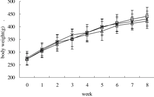
Fig. 1. Changes in body weights of male Zucker rats during treatment with MRC for 8weeks. × , normal control group (normal diets); □, positive control group (high-lipid diets); △, MRC 3%
group (3% mulberry root-bark powder in high-lipid diets). Body weight of MRC group was decreased as compared with those of negative control and positive control group.
5. 조직 중 지질과산화 측정
조직 내 지질과산화(lipid peroxidation)의 측정은 malondialdehyde(MDA) 유사물질인 1,1,3,3-tetaethoxy- propane(TEP)을 standard로 하는 thiobarbituric acid(TBA) 법으로 측정하였다. 간 적출 후 질소 냉동시킨 간 조직을 세절한 후 얼음으로 냉각시킨 9배의 PBS 완충액을 가하 고 동시에 얼음으로 냉각시키면서 homogenizer로 균질화 하였다. Homogenate 1 mL에 1 mL의 8.1% sodium dodecyl sulfate solution, 2 mL의 20% acetic acid solution 을 넣고 진탕한 다음 1 mL의 0.8% TBA solution을 넣고 95℃에서 60분간 반응시킨 후 냉각시켜 상층액을 취하여 분광광도계를 이용하여 532 nm에서 흡광도를 측정하였 다.
6. 통계학적 분석
모든 실험결과는 평균치±표준편차로 표시하였으며 Levene's test를 통하여 분산이 동질성을 갖는 경우 one-way analysis of variance(ANOVA)를 실시하여 유의 성이 관찰되면 대조군과의 유의차가 있는 실험군을 알아 내기 위하여 Dunnett's t-test 실시하여 p<0.05일 때 통계 학적으로 유의성이 있다고 판정하였다.
Ⅲ. 결 과
1. 체중, 사료효율 및 장기무게 변화
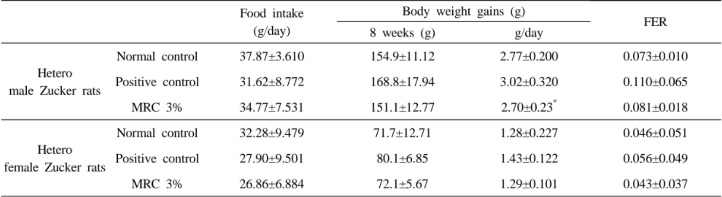
8주간 수컷 rat에서는 양성대조군의 체중이 161% 증가 하였고, 상백피 분말 급여군(이하 MRC군)에서는 153%가 증가하였다(Fig. 1). 한편 암컷 rat에서는 각각 정상대조군 과 양성대조군에서 139%, 143%의 체중 증가율을 보였고, MRC군에서는 139%의 체중증가를 보였다(Fig. 2). 또한 1 일 체중 증가량은 양성 수컷 rat에서 3.02±0.320 g/day인 데 반해 MRC 수컷 rat는 2.70± 0.23g/day 에 그쳐, 유의한 감소를 보였고, 암컷 rat에서도 1일 체중 증가량이 감소하 는 경향을 보였다(Table 1). MRC군의 사료효율도 수컷 0.081±0.018 g/day, 암컷 0.043±0.037 g/day로써 정상대조 군과 비슷한 수치를 보였으며 이는 양성대조군군에 비해 낮은 수치였다(Table 1).
수컷 rat에서 정상대조군의 상대적 간 무게는 3.14±0.145 g이고, 양성대조군과 MRC군의 간 무게는 각 각 3.54±0.183 g과 3.62±0.275 g으로 정상대조군에 비해 유의하게 증가하였다(p<0.05). 암컷 rat에서 양성대조군의 간 무게는 3.28±0.301 g, MRC군은 3.39±0.151 g으로 관 찰되었다(Table 2). 신장의 무게는 모든 군에서 유의한 차 이를 보이지 않았다.
170 200 230 260 290
0 1 2 3 4 5 6 7 8
week
body weight(g *
+
+,*
++
Fig. 2. Changes in body weights of female Zucker rats during treatment with MRC for 8weeks. × : normal control group (normal diets), □ : positive control group (high-lipid diets), △ : MRC 3% group (3%
mulberry root-bark powder in high-lipid diets). Body weight of MRC group was decreased significantly as compared with that of positive control group.
* significantly different from normal control (p<0.05).
+, ++ significantly different from positive control (p<0.05, p<0.01).
Table 1. Effects of MRC on food intake, body weight gains and food efficiency ratio (FER) (mean±SD) Food intake
(g/day)
Body weight gains (g) 8 weeks (g) g/day FER
Hetero male Zucker rats
Normal control 37.87±3.610 154.9±11.12 2.77±0.200 0.073±0.010 Positive control 31.62±8.772 168.8±17.94 3.02±0.320 0.110±0.065
MRC 3% 34.77±7.531 151.1±12.77 2.70±0.23* 0.081±0.018
Hetero female Zucker rats
Normal control 32.28±9.479 71.7±12.71 1.28±0.227 0.046±0.051 Positive control 27.90±9.501 80.1±6.85 1.43±0.122 0.056±0.049
MRC 3% 26.86±6.884 72.1±5.67 1.29±0.101 0.043±0.037
* Significantly different from positive control (p<0.05).
FER : food efficiency ratio, MRC : Mori radicis Cortex.
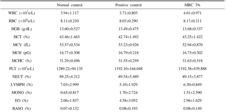
2. 혈액 및 혈청 생화학적 결과
백혈구 및 혈액의 세포성분을 측정한 결과 수컷 rat에 서 정상대조군에 비해 양성대조군은 MCV, MCH에서 감 소하였고(p<0.05), 시험물질투여군(MRC group)군은 양성 대조군에 비해 MCH가 증가하였다(p<0.05)(Table 3, 4).
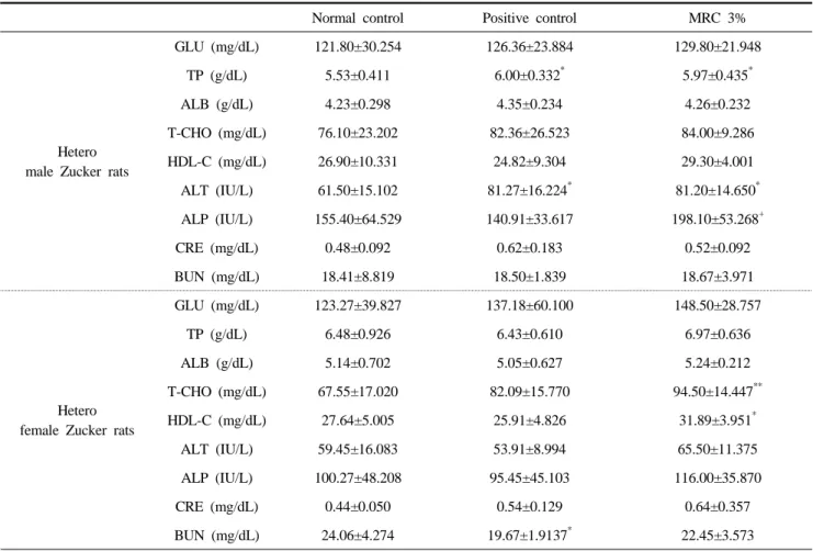
한편 총 콜레스테롤을 포함한 혈액생화학적 항목을 측정 한 결과 수컷 rat에서 총 단백과 ALT 효소의 수치는 양성 대조군과 MRC군이 음성대조군에 비하여 증가하였고, ALP 효소는 MRC군이 음성대조군과 비교하여 증가하였
다(p<0.05). 암컷 rat의 MRC군은 총 콜레스테롤의 수치가 음성대조군에 비해 증가하였고(p<0.01), HDL-C수치는 양 성대조군에 비하여 증가하였다(p<0.05). HDL-C의 증가경 향은 수컷 rat의 MRC군에서도 각각 관찰되었다(Table 5).
3. 조직 중 지질과산화 측정
수컷 rat의 간 내 지질분석결과에서 정상대조군은 1.74±0.083 mmol로 나타났으며 이에 비해 양성대조군은 2.43±0.174 mmol로써 유의한 차이를 볼 수 있었다
Table 2. The organ weight of male Zucker rats treated with MRC for 8 weeks (mean±SD)
Absolute organ weight Relative organ weight
Liver (g) Kidney (L)(g) Kidney (R)(g) Liver (g) Kidney (L)(g) Kidney (R)(g)
Hetero male Zucker rats
Normal
Control 13.68±1.032 1.51±0.111 1.46±0.118 3.14±0.145 0.35±0.028 0.34±0.028 Positive
control 15.63±1.915 1.49±0.123 1.47±0.122 3.54±0.183* 0.34±0.029 0.34±0.023 MRC 3% 15.48 ±1.919 1.45±0.099 1.45±0.071 3.62±0.275* 0.34±0.029 0.34±0.025
Hetero female Zucker rats
Normal
Control 7.32±0.428 0.89±0.102 0.90±0.130 2.87±0.090 0.34±0.039 0.36±0.054 Positive
control 8.67±0.941 0.95±0.126 0.90±0.132 3.28±0.301* 0.36±0.047 0.34±0.048 MRC 3% 8.74±0.518 0.87±0.081 0.91±0.067 3.39±0.151* 0.34±0.031 0.35±0.022
* Significantly different from normal control (p<0.05).
0.00 0.50 1.00 1.50 2.00 2.50 3.00
m a le fe m a le
lipidperoxidation
++
**
++
++
CON CHOL MRC CON CHOL MRC
Fig. 3. Content of lipid peroxidation of hetero male Zucker rats after treatment with MRC for 8 weeks. CON is normal control group (normal diets), CHOL is positive control group(high-lipid diets), MRC is 3% mulberry roots-bark powder in high-lipid diets group. Lipid peroxidation value of MRC group was decreased as compared with that of positive control. Specially, in female, lipid poroxidation level of MRC group was decreased significiantly (p<0.01).
** significantly different from positive control (p<0.01).
++ significantly different from normal control (p<0.01).
(p<0.01). 또한 MRC군은 2.27±0.069 mmol로 양성대조군 에 비해 감소하는 경향을 보였다(Fig. 3).
암컷 rat의 경우, 정상대조군의 간 내 지질은 1.72±
0.160 mmol인데 양성대조군은 2.41±0.131 mmol로 유의
한 차이를 보였으며(p<0.01), 또한 MRC군은 1.96± 0.206 mmol로써 양성대조군에 비해 유의하게 감소(p<0.01)하였 다(Fig. 3).
Table 3. The defference of the complete blood cell count (CBC) of male Zucker rats treated with MRC for 8 weeks (mean±SD)
Normal control Positive control MRC 3%
WBC (×103u/L) 6.41±2.460 5.81±1.618 6.14±0.792
RBC (×106u/L) 8.39±0.397 8.71±0.446 8.65±0.177
HGB (g/dl) 13.88±0.609 13.88±0.763 14.10±0.254
HCT (%) 45.01±2.543 45.48±2.751 45.35±0.840
MCV (fL) 53.63±0.950 52.20±1.463* 52.46±0.608
MCH (pG) 16.55±0.237 15.92±0.316* 16.30±0.205+
MCHC (%) 30.85±0.657 30.53±0.617 31.09±0.166
PLT (×103u/L) 1171.30±126.573 1205.80±206.757 1325.00±103.142
NEUT (%) 86.36±8.916 90.21±6.954 92.03±1.059
LYMPH (%) 7.33±2.173 5.90±1.937 7.02±1.074
MONO (%) 1.03±1.649 2.86±5.772 0.08±0.149
EO (%) 3.83±5.999 1.42±1.131 1.08±0.688
BASO (%) 0.48±1.177 0.17±0.260 0.05±0.127
* Significantly different from normal control (p<0.05).
+ Significantly different from positive control (p<0.05).
WBC : white blood cell, RBC : Red blood cell, HGB : hemoglobin, HCT : hematocrit, MCV : mean corpuscular volume, MCH : mean corpuscular hemoglobin, MCHC : mean corpuscular hemoglobin content, PLT : platelet, NEUT : neutrophil, LYMPH : lymphocyte, MONO : monocyte, EO : eosinophil, BASO : basophil.
Table 4. The defference of the CBC in female Zucker rats treated with MRC for 8 weeks (mean±SD)
Normal control Positive control MRC 3%
WBC (×103u/L) 3.94±1.117 3.71±0.803 4.61±0.971
RBC (×106u/L) 8.11±0.210 8.03±0.290 8.17±0.211
HGB (g/dL) 13.60±0.527 13.49±0.475 13.68±0.337
HCT (%) 43.46±1.463 42.74±1.492 43.25±1.422
MCV (fL) 53.57±0.534 53.23±0.926 52.94±0.870
MCH (pG) 16.77±0.308 16.79±0.218 16.75±0.302
MCHC (%) 31.29±0.496 31.55±0.259 31.63±0.518
PLT (×103u/L) 1289.22±94.135 1192.10±164.048 1192.38±439.888
NEUT (%) 89.25±4.312 89.54±5.489 89.15±3.877
LYMPH (%) 7.03±2.999 5.10±1.929 6.30±0.849
MONO (%) 0.65±0.817 1.70±2.724 1.51±2.590
EO (%) 2.06±1.837 4.58±3.052 2.96±1.629
BASO (%) 0.07±0.132 0.08±0.193 0.08±0.149
WBC : white blood cell, RBC : Red blood cell, HGB : hemoglobin, HCT : hematocrit, MCV : mean corpuscular volume, MCH : mean corpuscular hemoglobin, MCHC : mean corpuscular hemoglobin content, PLT : platelet, NEUT : neutrophil, LYMPH : lymphocyte, MONO : monocyte, EO : eosinophil, BASO : basophil.
Table 5. The defference of the blood chemistry in Zucker rats treated with MRC for 8 weeks (mean±SD)
Normal control Positive control MRC 3%
Hetero male Zucker rats
GLU (mg/dL) 121.80±30.254 126.36±23.884 129.80±21.948
TP (g/dL) 5.53±0.411 6.00±0.332* 5.97±0.435*
ALB (g/dL) 4.23±0.298 4.35±0.234 4.26±0.232
T-CHO (mg/dL) 76.10±23.202 82.36±26.523 84.00±9.286
HDL-C (mg/dL) 26.90±10.331 24.82±9.304 29.30±4.001
ALT (IU/L) 61.50±15.102 81.27±16.224* 81.20±14.650* ALP (IU/L) 155.40±64.529 140.91±33.617 198.10±53.268+
CRE (mg/dL) 0.48±0.092 0.62±0.183 0.52±0.092
BUN (mg/dL) 18.41±8.819 18.50±1.839 18.67±3.971
Hetero female Zucker rats
GLU (mg/dL) 123.27±39.827 137.18±60.100 148.50±28.757
TP (g/dL) 6.48±0.926 6.43±0.610 6.97±0.636
ALB (g/dL) 5.14±0.702 5.05±0.627 5.24±0.212
T-CHO (mg/dL) 67.55±17.020 82.09±15.770 94.50±14.447**
HDL-C (mg/dL) 27.64±5.005 25.91±4.826 31.89±3.951*
ALT (IU/L) 59.45±16.083 53.91±8.994 65.50±11.375
ALP (IU/L) 100.27±48.208 95.45±45.103 116.00±35.870
CRE (mg/dL) 0.44±0.050 0.54±0.129 0.64±0.357
BUN (mg/dL) 24.06±4.274 19.67±1.9137* 22.45±3.573
*, ** Significantly different from normal control (p<0.05, p<0.01).
+ Significantly different from positive control (p<0.05, p<0.01).
GLU : glucose, TP : total protein, ALB : albumin, T-CHO : total-cholesterol, HDL-C : high density lipoprotein cholesterol, ALT : alanine aminotransferase, ALP : alkaline phosphatase, CRE : creatinine, BUN : Blood Urea Nitrogen.
Ⅳ. 고 찰
비만 Zucker rat는 지방세포에서 유래하는 항비만 인자 로써 강력한 섭식억제 작용과 에너지소비 촉진작용을 가 진 펩티드 호르몬인 렙틴이 수용체 수준에서 결핍되어 비만이 유도되는 유전적 실험동물 모델이다(Beck 등, 2002). 이러한 비만 유전자를 가지고 있는 Zucker rat의 유전자형 중 lean type의 Zucker FA/FA rat와 Zucker FA/fa에 고지방식이로 비만을 유도하여 상백피 분말의 항비만 효과와 간 보호 효과를 알아보았다.
8주간의 시험물질 급여 후 각 실험동물 간 체중증가에 대해서 수컷과 암컷, 모두 MRC군의 체중증가억제가 보 였고, 특히 암컷의 3주, 5주, 6주의 체중은 고지방사료군 인 양성대조군에 비교했을 때 유의한 차이가 관찰되었다.
사료효율 또한 MRC군은 정상식이군과 비슷한 수치를 보 였다. 이는 처음 체중이 유의한 차이가 없을 경우 사료 효율이 떨어진다는 것이 체지방 축적이 적게 된다고 말 할 수 있다는 연구(Lee 등, 1992)로부터 MRC가 체지방 축적 억제기능을 가진다고 해석할 수 있다.
실험 종료 후 채취한 간과 신장의 중량을 비교해 본 결 과, 간을 제외한 다른 조직에서는 큰 차이가 없었지만 간 의 경우 정상대조군에 비해 고지방식이를 먹인 군에서 모 두 무게가 증가하였다. 이것은 고지방 섭취시 간 무게가 증가한다는 다른 연구 보고와 일치되는 결과였다 (Wursch, 1979; Park, 1994).
상백엽 분말의 간에 대한 보호 작용을 알아보기 위해 간의 지질과산화 함량을 측정하였다. 지질과산화 반응은 여러 경로에서 생성된 반응성 산소종(reactive oxygen
species [ROS])을 포함하는 유리기(free radical)가 효소와 핵산을 비롯한 세포의 구성성분을 공격함에 따라 (Freeman과 James, 1982), 지단백 과산화(Daugherty와 Roselaar, 1995; Steinberg, 1995; Berliner과 Heinecke, 1996), 평활근 세포 비대(Ushio-Fukai 등, 1996), 혈관내피 세포 기능장애 등 세포의 구조적 및 기능적 손상을 초래 하는 반응이다(Marui 등, 1993; Harrison, 1997). 따라서 지질과산화물의 양이 적을수록 세포보호 작용을 갖는다 는 것을 알 수 있었다. 간의 과산화지질을 측정한 결과 암․수컷 모두 MRC군이 양성대조군군에 비해 감소했으 며, 특히 암컷에서의 유의한 감소가 관찰되었다. 이로부 터 상백피 분말이 항산화작용을 하여 간 보호효과를 나 타낸다는 사실을 알 수 있었다. 이 결과는 Lim(2001)의 연구에서 상백피의 과산화지질 생성을 억제한다는 결과 와 유사하게 나타났다.
혈액의 세포성분 측정에서는 별다른 변화가 관찰되지 않았지만 혈청 생화학 측정에서 MRC군의 암․수컷 HDL-C의 농도가 증가하였다. 또한 총콜레스테롤에 대한 HDL-C의 비율(HDL-C/T-Cho)도 수컷에서 정상대조군에 서 35.35%, 양성대조군에서 30.13%, MRC 군에서 34.88%가 나왔고, 암컷에서도 각각 40.92%, 31.56%, 33.74%가 관찰되었다. 혈중 HDL-C의 농도가 사람에서 심혈관질환에 반비례한다는 기존의 연구를 고려해 볼 때 (Troms, 1977; Corti 등, 1995; Uri 등, 1997), 상백피 분말 이 혈중 HDL-C의 농도를 높이고 총 콜레스테롤 중 HDL-C이 차지하는 비율을 높임으로써 관상동맥경화증 을 비롯한 각종 동맥경화질환을 예방할 수 있을 것이라 생각된다.
위 결과를 종합하여 보면, 상백피는 사료효율을 감소시 켜 체중감소를 유도하고, 지질과산화를 억제하는 항산화 기능을 하며, 특히 HDL-C의 혈중농도를 증가시켜 관상 동맥질환을 예방하는 기능을 한다고 사료된다.
V. 결 론
상백피 분말의 비만개선 효과를 비만이 유도된 Zucker rat에서 검토해본 결과 MRC group의 혈중 HDL-C 농도 가 정상대조군과 양성대조군에 비하여 증가함을 볼 수 있 었고, 또한 총콜레스테롤에 대한 HDL-C의 비율도 증가 하였다. HDL-C 농도와 비율의 증가로부터 상백피 분말 이 동맥경화질환을 감소시킬 수 있다고 사료되며, 추후
상백피의 체내 작용 기전 규명실험과 그에 따른 응용 실 험이 수행되어야 되어야 한다고 생각된다.
참 고 문 헌
1. Beck B, Musse N, Stricker-Krongrad A. Ghrelin, macronutrient intake and dietary preferences in long- evans rats. Biochemical and Biophysical Research Communications 292(4):1031-1035, 2002.
2. Beck B, Richy S, Stricker-Krongrad A. Feeding response to ghrelin agonist and antagonist in lean and obese Zucker rats. Life Sciences 76: 473-478, 2004.
3. Berliner JA, Heinecke JW. The role of oxidized lipoproteins in atherogenesis. Free Radic Biol Med 20:707-727, 1996.
4. Chua Jr, Chung WK, Wu-Peng XS, Zhang Y, Liu SM, Tartaglia L, Leibel RL. Phenotypes of mouse diabetes and rat fatty mutations in the OB (leptin) receptor. Science 271(5251):994-996, 1996.
5. Corti MC, Guralink JM, Salive ME. HDL cholesterol predicts coronary heart disease mortality in older persons. JAMA 274:539-544, 1995.
6. Daugherty A, Roselaar SE. Lipoprotein oxidation is a mediator of atherogenesis: insights from pharma- cological studies. Cardiovasc Res 29:297-311, 1995.
7. Freeman BA, James DC. Biology of disease. Free radicals and tissue injury. Lab Invest 47:412-426, 1982.
8. Fujioka K, Seaton TB, Rowe E, Jelinek CA, Raskin P, Levovitz HE, Weinstein SP. Weight loss with sibutramine improves glycaemic control and other metabolic parameters in obese patients with type 2 diabetes mellitus. Diabetes, Obesity and Metabolism 2(3):175-187, 2000.
9. Harrison DG. Endothelial function and oxidant stress.
Clin Cardiol 20:11-17, 1997.
10. Hill JO. An overview of the etiology of obesity. In Fairbrun CG, Broenell KD(eds). Eating disorders and obesity. p460-466, The Guilford Press, New York, 2002.
11. Kojima M, Hosoda H, Date Y, Nakazato M, Matsuo
H, Kangawa K. Ghrelin is a growth-hormone- realeasing acylated peptide from stomach. Nature 402:656-660, 1999.
12. Lee H, Choi BK, Lee WC, Park CI, Furugawa Y, Kimura S. Effect of dietary protein levels, caffeine and green tea on body fat deposition in wistar rats. J Korean Soc Food Nutr 21:595-600, 1992.
13. Leung WYS, Thomas GN, Chan JCN, Tomlinson B.
Weight management and current options inpharmaco- therapy: orlistat and subutramine, Clinical Thera- peutics 25(1):58-80, 2003.
14. Marui N, Offermann MK, Swerlick R, Kunsch C, Rosen CA, Ahmad M, Alexander RW, Medgord RM.
Vascular cell adhesion molecule-1(VCAM-1) gene transcription and expression are regulated through an antioxidant sensitive mechanism in human vascular endothelial cells. J Clin Invest 92:1866-1874, 1993.
15. McMahon FG, Fujioka K, Singh BN, Mendel CM, Rowe E, Rolston K, Johnson F, Mooradian AD.
Efficacy and safety of sibutramine in obese white and African American patients with hypertension: a 1-year, double-blind, placebo-controlled, multicenter trial. Archives of Internet Medicine 160(14):2185- 2191, 2000.
16. Nomura T, Fukai T, Yamada S, Katayanagi M.
Phenolic constituents of the cultivated mulberry tree (Morus alba. L.). Chem Pharm Bull 24(11):2898, 1976.
17. Park OJ. Plasma lipids and fecal excretion of lipids in rats fed a high fat diet, a high cholesterol diet or a low fat/high sucrose diet. Kor J Nutr 27(8):785-794, 1994.
18. Steinberg D. Role of exidized LDL and antioxidants in atherosclerosis. Adv Exp Med Biol 369:39-48, 1995.
19. Troms HS. High density lipoprotein and coronary heart disease: a prospective casecontrol study. Lancet 1:965-967, 1977.
20. Tschop M, Wawarta R, Riepl RL, Friedrich S, Bidilngmaier M, Landgraf R, Folwaczny C. Post- prandial decrease of circulating human ghrelin levels.
Journal of Endocrinological Investigation 24 (6):
RC19-RC21, 2001.
21. Tschop M, Weyer C, Tataranni PA, Devanarayan V, Ravussin E, Heiman ML. Circulating ghrelin levels are decreased in human obesity. Diabetes 50 (4):707- 709, 2001.
22. Tschop M, Smiley DL, Heiman ML. Ghrelin induces adiposity in rodents. Nature 407:908-913, 2000.
23. Uri G, Shlomit Y, Jack HM. Isolated low HDL cholesterol as a risk factor for coronary heart, disease mortality. A 21-year follow-up of 8000 men.
Arterioscler Thromb Vasc Biol 17:107-113, 1997.
24. Ushio-Fukai M, Zafari AM, Fukui T, Ishizuka N, Griendlling KK. P22phox is a critical component of the superoxide generating NADH/NADPH oxidase system and regulates angiotensin Ⅱ-induced hyper- trophy in vascular smooth muscle cells. J Biol Chem 271:23317-23321, 1996.
25. Wursch P. Influence of tannin-rich Caro Pod fiber on the cholesterol metabolism in the rat. J Nutr 109(4), 685-692, 1979.
26. 문관심. 약초의 성분과 이용. p146, 백과사전출판사, 서울, 1984.
27. Lim S. Antilipidperoxidative effects of Morus alba in diabetic mMice. 대전대학교 한의학연구소 논문집 10(1):483-487, 2001.