Ⅰ. 서 론
악관절의 연골은 연골세포와 세포외 기질로 구성되어 있 다. 연골세포는 조골세포와는 다른 특징을 가지고 있는 세 포로 연골과 연관된 세포외 기질 단백질을 합성하며 이들 단백질은 인장강도에 저항하면서 기계적인 부하에 생체가 적응하게끔 하여 준다. 이러한 단백질의 발현은 유전자와 주변 환경인자와의 상호작용에 의하여 조절되는 데, 단백질 의 발현에 관여하는 것으로 알려진 환경인자로는 세포외 기 질 성분과 같은 용해성 중간자도 있고 기계적인 요소도 있 다
1). 악관절에 대한 해로운 자극은 연골세포에도 좋지 않는
영향을 주게됨으로 이는 악관절의 병적인 상태를 야기할 수 있다.
2)그러므로 이러한 단백질의 발현 양상을 조사하는 것 은 악관절의 병리를 이해하는 데 중요하다고 하겠다.
최근에 2형 교원섬유를 발현하는 사람에서 얻어진 몇 개 의 연골세포주가 개발된 바 있다
3). 이들은 연골세포의 다양 한 기능을 연구하는 데 이용되어 왔다
4-6). 연골세포의 기계 적인 자극에 대한 반응은 관절연골의 세포외 기질 항상성을 유지하는 데 필수적인 것으로 인식되어왔다. 전단력이나 수 압과 같은 기계적인 힘은 정상 관절의 부하운동 중에 일어 나는 것으로 세포 반응에 영향을 미치고 결과적으로 연골 대사에 영향을 미치게 된다.
김신엽∙김성곤*∙최제용**∙남동석
서울대학교 치과대학 치과교정학교실, 한림대학교 성심병원 구강악안면외과*, 경북대학교 의과대학 생화학교실**
전단력이 연골세포에 미치는 영향에 관한 연구
THE SHEAR STRESS PROTEOME OF CHONDROCYTES
Shin-Yeop Kim, Seong-Gon Kim * , Je-Yong Choi ** , Dong-Seok Nahm
Department of Orthodontics, School of Dentistry, Seoul National University, Department of Oral and Maxillofacial Surgery, Sacred Heart Hospital, Hallym University*,
Department of Biochemistry, School of Medicine, Kyoungpook National University**
The objective of this study is screening the shear stress related proteins in chondrocytes using two- dimensional electrophresis and MALDI-TOF. C-28/I2 cell line were grown The fluid-induced shear stress (FISS) was applied using a cone viscometer at a rotational velocity of 80rpm for periods of 12 hours.
Control cultures were tested under identical conditions without mechanical load application. Collected sam- ples were used for the two-dimensional electrophoresis and MALDI-TOF. The identified proteins were cal- cyclin, RPE-spondin, interleukin-2, extracellular signal regulated kinase (ERK), lamin B2, porA protein, and RET-ELE1 protein. All of them showed a decreased expression. In conclusion, seven proteins were identified as a shear stress related proteins in chondrocytes. As the destruction of articular cartilage is one of main pathogenesis of TMJ internal derangement, this study will give useful information for the under- standing of the molecular aspect of TMJ disease.
Key words : Chondrocyte, MALDI-TOF, FISS
Abstract
연골세포의 전단력에 대한 반응은 자극의 형태, 지속 시 간, 강도에 영향을 받게 된다. 비록 전단력이 연골세포의 대 사활동을 어떻게 바꾸는 지에 대한 명확한 결론은 아직 없 는 상태이지만 동물세포에서 수행한 실험에서는 유체역학 으로 유도된 전단력이 연골 형성에 영향을 미치는 것으로 보고되어 있다.
7)높은 전단력이 가해지는 부위에서는 연골 의 변성과 관절의 기능 상실을 가져오게 된다. 전단력은 기 계적인 부하의 양상이나 강도가 변한 경우 악관절과 같은 diarthroidal joint의 파괴를 가져오는 주된 원인으로 보고 된 바 있다
8).
배양된 세포에서 수행되는 실험에서는 기계적인 자극이 세포 내 신호전달 체계나 유전자 발현, 성장, 분화 및 세포 사멸에 어떠한 영향을 미치는 지를 알아낼 수 있다. 따라서 본 연구의 목적은 연골세포에서 전단력과 연관된 단백질 발 현을 2차원 전기영동법과 matrix assisted laser desorp- tion ionization-time of flight (MALDI-TOF)를 이용하 여 밝히는 데 있다.
Ⅱ. 재료 및 방법
세포 배양 및 유체역학 유도 전단력의 적용
C-28/I2 cell line (Immortalized human juvenile costal chondrocyte, Beth Israel Deaconess Medical Center, Harvard Institute of Medicine, USA)을 이용하 여 실험을 수행하였다 (Fig. 1). 상기 세포주를 Ham’s F12/Dulbecco’s modified Eagle’s medium (Gibco, BRL, Gaithersburg, MD)에서 키워졌고 배양액에는 1%
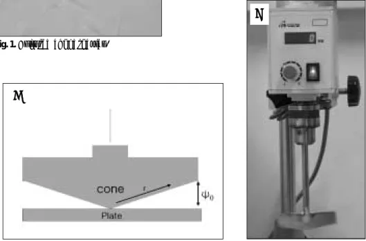
penicillin/streptomycin, bFGF (100 μg/ml), 그리고 10% fetal bovine serum (FBS)를 함유하고 있었다. 배양 된 세포는 전단력을 가하기 24시간 전에 PBS로 세 차례 세 척을 시행하였고 10ml의 혈장이 포함되어 있지않는 배양액 을 넣었다. 전단력은 cone viscometer를 이용하여 가하였 으며 회전 속도는 80rpm이었으며 힘을 가한 시간은 12시 간이었다 (Fig. 2). 대조군은 같은 조건에서 전단력을 가하 지 않은 상태에서 배양하였다. 시편은 24시간 후에 얻었으 며 영하 70도에서 다음 실험 전까지 보관하였다.
2차원 전기영동법
2차원 전기 영동법은 이전에 발표된 논문의 방법대로 수 행하였다.
8)배양된 세포 시편을 500 μl의 buffer I (0.3%
SDS, 200 mM DTT, 28 mM Tris-HCl,)에 녹여서 부유
Fig. 1. Cultured chondrocytes.
Fig. 2. A A.. Diagram of the apparatus (r: radius, ψ
0: angle). B B.. The apparatus of shear stress generator.
A A
B
B
액 형태로 만들었다. 이는 유리구슬을 이용하여 혼합기에서 4분간 흔들어 줌으로서 균일화하였다. 이어서 원심분리를 13,000rpm에서 15분 4도에서 실시하였을 때 수용성 분획 과 불수용성 분획으로 나누어진다. 이중에서 수용성 분획을 금번 실험에 사용하였다. 이어서 100도에서 5분간 노출시 킨 후 얼음에서 식혔다. 그리고 24 μl의 buffer II (24 mM Tris, 476 mM Tris-HCl, 50 mM MgCl
2, 1mg/mL Dnase I (Gibco BRL, Grand Island, NY), 0.25 mg/ml RNase A (Sigma Chemical, St. Louis, MO))를 첨가하 였다. 반응은 4도에서 15분간 진행시켰으며 전체 부피의 4 배의 차가운 acetone을 가하여 반응을 중지시켰다. 얼음에 서 20분간 두게 되면 단백질 침적이 생기게 된다. 단백질은 12000 rpm에서 10분간 원심분리하여 모았으며 15 μl의 buffer pH 4-8 (540 mg/ml urea, 10 mg/ml DTT, 2%
v/v Ampholyte 4-8 (Millipore, Bedford, MA), 0.52%
v/v Triton X-100)에 용해시켰다. 고해상도 2차원 전기영 동법은 Lopez 등
10)에 의하여 기술된 변형된 방법을 이용하 여 수행하였다. Bio-Rad Protein IEF Cell (Bio-Rad Laboratories, Hercules, CA)을 이용하여 1차 분리하였 고, Millipore Investigator system (Milipore)을 이용하 여 2차분리하였다. 건조된 gels을 storage phosphor screen (Packard Instrument Company, Downers Grove, IL)에 48시간 노출시켰으며 각 spot의 정량적 분석 은 OptiQuant Image Analysis Software (Packard Instrument Company)를 이용하여 수행되었다.
MALDI-TOF
MALDI-TOF는 이전에 보고된 논문을 참조하여 수행하 였다.
11)Trypsin을 시편에 첨가한 후 혼합물은 37도에서 5
시간 배양하였다. 이어서 5 μl의 10% trifluoroacetic acid (TFA)를 첨가하여 단백질 분해를 중단시켰다. 부가적으로 chymotrypsin 처리한 TLCK (1-chloro-3-tosylamido-7- amino-2-heptanone-HCl) treated (2μl of 2mg/ml)도 사용하였다. MALDI-TOF는 Bruker Ultraflex TOF (Bruker Daltonics Inc., Germany)를 이용하여 수행되었 으며 이는 직선형 질량분석기로 N2 laser를 이용한 양이온 작동모드형 질량분석기이다. 레이저의 강도는 신호를 얻어 지기에 충분한 정도로 가하였으며 가속전압은 28kV로 정 하였다. 장비는 본 실험에 들어가기에 앞서서 알려진 단백 질/펩타이드 시편으로 시험하였다. 분석에 사용된 시편은 0.1% TFA에 50pmol/μl의 농도로 용해시킨 것을 사용하 였다.
Ⅲ. 실험 결과
본 실험에서는 전단력을 연골세포에 가한 경우 그 발현이 현저히 변하는 단백질을 모두 7개 찾아내었다 (Table 1).
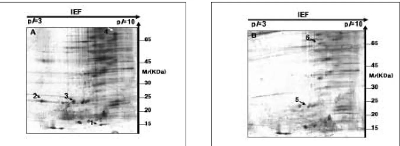
전체적인 단백질의 발현 양상은 전단력을 가한 경우가 대조 군과 비교하여 상당히 다르게 나타남을 알 수 있었다 (Fig.
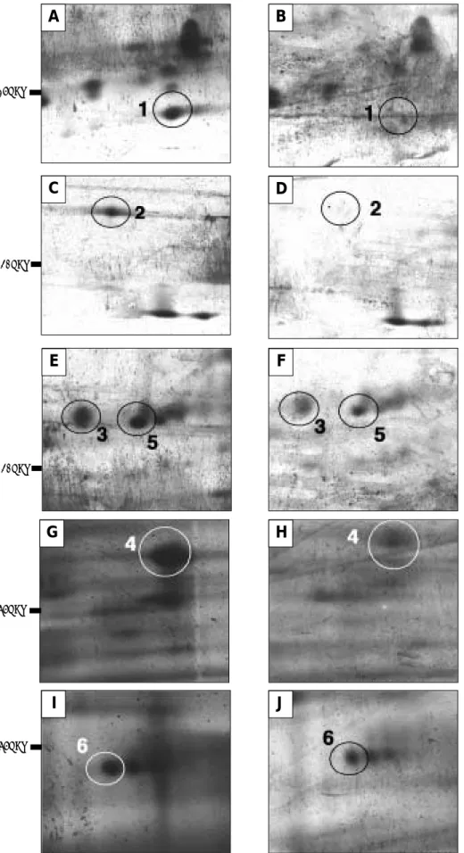
3). Calcyclin은 전단력을 가한 경우 연골세포에서 그 발현 이 현저히 저하되어 거의 관찰되지 않았다 (Fig. 4A, B).
RPE-spondin과 interleukin 2는 분자량과 pI 값이 유사 하여 같은 spot에서 나왔는데, 이 역시 발현이 현저히 감소 하여 실험군에서는 spot이 거의 관찰되지 않았다 (Fig.
4C, D). 이 이외에도 extracellular regulated kinase (ERK), PorA protein (Fig. 4E, F), lamin B2 (Fig.
4G, H), 및 RET-ELE1 protein (Fig. 4I, J)은 실험군에 서 발현은 관찰되나 현저히 감소되는 단백질로 분석되었다.
Fig. 3. Silver-stained 2-DE analytical gels of proteins of derived from compared control and experiment of A A:: CONTROL B
B:: EXPERIMENT (SHEAR STRESS). The numbered spots are listed in Table 1.
Fig. 4. Sections of 2-D separation of labeled proteins from control (A, C, E, G, and I) and stressed cells (B, D, F, H, and J). The identification numbers are those used in Table 1 and correspond as follows: 1 (calcyclin), 2(RPE-spondin, interleukin-2), 3(extracellular signal regulated kinase), 4(lamin B2), 5(porA protein), and 6(RET-ELE1 protein).
A B
C D
E F
G
J H
I
15KDa
65KDa
65KDa 20KDa
20KDa
Ⅳ. 총괄 및 고찰
생체 내에는 수용성 물질로 다양한 효과를 내는 인자들이 많은 데 이들은 염증성 cytokines, 성장인자나 호르몬들로 연골 내 대사 과정과 연관되어 기계적인 힘에 따라 그 발현 이 변화될 가능성이 있으며 결과적으로 세포외 기질의 변화 를 초래하게 된다. 각각의 분자들은 관절이 처해진 환경에 따라 그 발현이 증감되게 된다. 이론적인 모형에서는 전단 력이 관절염에 영향을 미치는 것으로 밝혀져 있다
12,13). 수학 적인 모형과 동물실험에서의 결과는 악관절에서 변형된 부 하와 관절염 상태에 밀접한 연관이 있음이 밝혀진 바 있다.
관절 연골의 손상과 연관이 있는 기계적인 부하는 역시 골 관절염 발생의 위험 요소일 것이다. 실험실 연구에서 연골 에 대한 위해성 기계적 압박이 강도에 비례하여 세포사멸을 증가시킨다는 보고가 있다
13). 비록 MALDI-TOF를 통하여 전체 아미노산 서열을 분석할 수는 없으나 MALDI-TOF는 미지의 단백질을 확인하는 데 있어서 상당히 신뢰할 수 있 는 방법이다. 이번 실험에서 모두 7개의 단백질이 전단력과 관련하여 발현이 감소되는 단백질로 확인되었다 (Table 1). 이들 중에서 calcyclin, interleukin-2 및 ERK는 기계 적인 스트레스와 연관이 있는 단백질로 보고된 바 있다.
Calcyclin는 칼슘과 결합하는 단백질로 분자량은 10-15 kDa이다
14). 이 단백질의 기능은 세포질 내 칼슘 이온 농도 의 조절과 연관이 있고
15), exocytosis
16), 단백질 분해
17)및 세포 주기 조절
18)을 담당하고 있다. 이 단백질은 다양한 종 류의 세포에서 발현된다
19). Calcyclin의 발현이 기계적인 힘에 반응하여 변한다는 것이 폐에서는 보고된 바 있다
20). 그리고 calcyclin의 발현 증가는 가벼운 기계적 자극에서 상
피세포와 기질세포의 상호작용으로 세포증식이 일어나는 경우에 관찰할 수 있다
21). 연골세포에서 calcyclin의 발현이 어떠한 의미를 가지는 지에 대하여는 아직 보고된 바 없다.
이 단백질이 칼슘과 결합하는 단백질이고 세포 증식에 관여 하는 것을 감안하면 이의 발현 감소는 과도한 기계적 자극 에서 연골세포의 증식이 억제되는 것과 연관이 있을 수 있다.
임파구에 의하여 연골세포가 파괴되는 것은 류마티스성 관절염에서 면역체계가 관절의 파괴에 어떻게 기여하는 지 를 명확하게 보여준다. Interleukin 1 (IL-1)은 세포외 기 질의 변환주기를 증가시키고 metalloprotease의 합성을 촉 진함으로서 연골에 영향을 미친다. 이러한 효과는 세포 표 면에 위치한 수용체 (IL-1R)를 통하여 일어난다. 특정 농 도의 IL-2는 IL-1R의 발현을 감소시킨다
22). 그러므로 IL-2 는 수용체의 발현을 조절함으로서 연골에서 세포외 기질 대 사를 조절할 수 있다. IL-2의 발현도 이번 실험에서는 감소 됨을 보였다 (Fig. 4C, D).
격리된 연골세포에 cytokines
23), 성장인자
24), matrix adhesion
25), 혹은 기계적인 스트레스
26,27)를 가하는 경우 각 각의 자극은 ERK를 활성화시켜 이들 자극에 대하여 세포가 반응하고 조절할 수 있게 한다. ERK는 mitogen-activated protein kinases (MAPKs)에 속하는 단백질로서 세포 밖 에서 오는 자극을 핵으로 전달하는 역할을 함으로서 다양한 유전자의 발현 및 조절에 영향을 미치게 된다
28). 연골세포에 서는 ERK에 의하여 조절되는 기능으로는 증식
24), 탈분화
29), 세포사멸
24)그리고 세포외 기질 물질의 합성
26)이 있다. 그리 고 단백질 분해효소의 억제자로 작용한다
30). 이번 실험에서 는 ERK의 발현이 감소된 것으로 나타났다 (Fig. 4E, F).
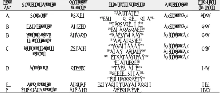
Table 1. Proteins identified in each spot by peptide mass fingerprinting using MALDI-TOF.
Spot Candidate Protein Accession
Sequenced peptides Modification Sequence
No. number coverage
1 Calcyclin BCHUY
56
LMEDLDR
62Oxidaton(M) 14.0%
105
SPEGGGGAGARGGAWP
1152 RPE-Spondin Q96J64
67
DPACFARGWR
76Oxidaton(M) 7.0%
37
TLWMALCALSR
472 Interleukin-2 Q8NFA4
18MLTFKFYMPK
27Oxidation(M) 7.0%
Extracellular
291MLVLDACIR
2993 signal regulated JC6138
130
IQFLVYQMMK
139Oxidation(M)
3.2%
kinase
260
NYMKGLPELEK
270Oxidation(M)
228
GSDHLDQLKEIMK
240Oxidation(M)
1
RVLDETAR
284 Lamin B2 A45023
390DQSLGNWR
3979.1%
50
7TTSRGCYVM
51555
SEVELAAALSDK
665 PorA protein Q9ZEQ6 SAYKPAYVDENKMVHAAVVG 95%
6 RET-ELE1 protein Q9UM84 PISSAEMTFR 92.3%
스트레스에 의하여 발현이 증가되는 샤페론으로는 Hsp70과 Hsp25가 있으며 이들은 모두 핵으로 전달되는 신호를 조절하며 핵 내에 위치하게 된다
31). 이들 샤페론의 발현은 세포 내 기포 형성이나 lamin을 함유하고 있는 구조 물의 파괴시에 나타난다
30). Lamin B2의 발현이 본 연구에 서는 강한 전단력을 가하는 경우 감소되는 것으로 나타났다 (Fig. 4G, H). 이는 아마도 스트레스와 연관되어 나타나는 현상으로 사료된다. 이번 연구에서 확인된 단백질로는 RPE-spondin, porA protein, RET-ELE1 protein이 있 지만 (Fig. 3), 이들 단백질이 기계적인 자극과 연관되어 연 구된 논문은 아직 없다. 따라서 이는 추후 연구를 통하여 밝 혀져야 할 것으로 사료된다.
결론적으로 이번 연구에서는 전단력과 관련하여 연골세포 에서 발현이 감소되는 단백질로 일곱개를 확인하였으며, 관 절 연골의 파괴가 악관절 내장증의 주된 발병과정이기에 이 들 단백질의 확인은 추후 악관절 질환의 분자생물학적 연구 에 기초 자료로 사용될 수 있으리라 사료된다.
Acknowledgement
The authors thank Mary B. Goldring (Massach- usetts General Hospital, Charlestown, MA) for her giving cell line kindly.
참고문헌