Original Article
원고 접수일 2011년 12월 2일, 원고 수정일 2012년 1월 25일, 게재 확정일 2012년 3월 20일
책임저자 김수관
(501-759) 광주시 동구 서석동 375번지, 조선대학교 치의학전문대학원 구강악안면 외과학교실
Tel: 062-220-3815, Fax: 062-228-7316, E-mail: [email protected]
RECEIVED December 2, 2011, REVISED January 25, 2012, ACCEPTED March 20, 2012
Correspondence to Su-Gwan Kim
Department of Oral and Maxillofacial Surgery, School of Dentistry, Chosun University
421, Seoseok-dong, Dong-gu, Gwangju 501-759, Korea
Tel: 82-62-220-3815, Fax: 82-62-228-7316, E-mail: [email protected]
CC This is an open access article distributed under the terms of the Creative Commons Attribution Non-Commercial License (http://creativecommons.org/licenses/
by-nc/3.0) which permits unrestricted non-commercial use, distribution, and reproduction in any medium, provided the original work is properly cited.
수산화인회석 코팅 임플란트와 Sandblasted, Large-grit and Acid-etched Implant의 임상적 초기 안정성 비교
임형섭ㆍ김수관ㆍ오지수
조선대학교 치의학전문대학원 구강악안면외과학교실
Abstract
Comparison of Clinical Initial Stability of Hydroxy-apatite Coated Implant and Sandblasted, Large-grit and Acid-etched Implant
Hyoung-Sup Lim, Su-Gwan Kim, Ji-Su Oh
Department of Oral and Maxillofacial Surgery, School of Dentistry, Chosun University
Purpose: This study attempts to compare the stability of hydroxy-apatite coating implant with that of sandblasted, large-grit and acid-etched surface implant at an early state of installation.
Methods: 35 implants were installed in 18 patients, who had visited hospital for implant installation. The early stability at operation, 6 weeks and 12 weeks after operation using OsstellTM mentor (Integration Diagnostics, Savedalen, Sweden) and PeriotestⓇ (Siemens AG, Benssheim, Germany) were measured, and subsequently analyzed statistically.
Results: OsstellTM mentor value of hydroxy-apatite coated implant (HAPTITE) was measured as 70.14±9.07 at the stage of installation, 76.98±5.25 at 6 weeks and 80.28±4.23 at 12 weeks after installation. A statistically significant increase in measurement value was observed after 6 weeks and 12 weeks than when implants were placed. In case of IMPLANTIUM (DENTIUM Co. Ltd., Seoul, Korea), the measurement value was 74.68±7.42 at installation, 79.03±4.39 at 6 weeks and 80.59±3.59 at 12 weeks after installation. In addition, a statistically significant increase in the value was observed when comparative analysis of the value at after installation and 12 weeks after installation was carried out. However, no significant difference between HAPTITE and IMPLANTIUM was observed. The average measurement value of periotestⓇ was 1.94±3.90 at installation of HAPTITE, 4.03±1.48 at 6 weeks and 5.00±1.71 at 12 weeks after installation. Moreover, whilst comparing the value at after installation and 12 weeks after installation, statistically significant decrease in the value was observed. In case of IMPLANTIUM, the average measurement value was measured as 4.25±1.76 at installation, 4.76±0.97 at 6 weeks and
5.18±0.91 at 12 weeks after installation and no statistically significant difference was observed. Furthermore, no statistically significant difference was observed between HAPTITE and IMPLANTIUM.
Conclusion: In this study, both the implants demonstrated favorable early stability at the time of measurement using OsstellTM mentor and PeriotestⓇ. Moreover, based on the observed results, both HAPTITE and IMPLANTIUM are considered as potent to exhibit clinically stable and prognostic results.
Key words: Coating, Dental implant, Stability
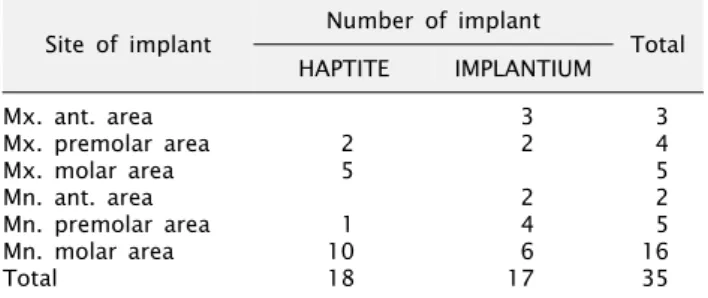
Table 1. Location of implant
Site of implant Number of implant
Total HAPTITE IMPLANTIUM
Mx. ant. area 3 3
Mx. premolar area 2 2 4
Mx. molar area 5 5
Mn. ant. area 2 2
Mn. premolar area 1 4 5
Mn. molar area 10 6 16
Total 18 17 35
HAPTITE, hydroxy-apatite coated implant; Mx., maxilla; ant., anterior; Mn., mandible.
서 론
Brånemark 등[1]에 의해 처음 임플란트가 소개된 이후, 임플 란트는 치아 결손을 회복할 수 있는 일반적인 치과 치료로서 인식되고 있으며, 최근에는 임플란트의 성공률을 90∼95%로 높 은 성공률을 보이고 있다[2-7].
티타늄은 표면에 안정적인 산화 피막을 형성하며, 생체 적합성 이 높은 특징을 가지고 있고, 다른 금속 재료에 비해 골과 결합이 빠르고 낮은 탄성 계수를 가지는 특성으로 치과 임플란트뿐만 아니라 많은 다른 임상 분야에서 다양하게 사용되고 있다[8,9].
하지만 최근 골질이 좋지 않은 상악 구치부의 초기 안정성 증대와 발치 후 즉시 식립 임플란트 등의 연구 등으로 임플란트의 초기 골형성 촉진을 위해 다양한 표면처리 기술들이 개발되고 있다 [10-12]. 다양한 임플란트 표면처리 방식 중 수산화인회석 코팅은 임플란트 표면에 수산화인회석을 plasma spray시켜 depres- sion, undercut, porosity를 만들어서 표면을 불규칙한 형태로 만드는 방법으로 골형성을 증가시키고, 임플란트의 골유착을 촉 진시키는 방법으로 최근 많은 연구에서 좋은 임상적 결과를 나타 내고 있다[13-23]. 또 하나의 임플란트 표면처리 방식으로는 sandblasted, large-grit and acid-etched (SLA) surface가 있 다. 이는 blasting을 시행한 후 acid-etching을 시행함으로써 blasting 시 남아 있는 입자들을 acid로 세척하여 표면의 오염을 줄이고, 산 자체의 효과인 산부식으로 작은 함몰을 형성하는 방식 으로, 조기 골유착에 효과적임을 보고하는 논문들이 발표되고 있다[24,25].
본 연구에서 사용된 수산화인회석 코팅 임플란트인 hydroxy-apa- tite coated implant (HAPTITE, DENTIS Co. Ltd., Daegu, Korea)는 기존의 일반적인 plasma sprayed hydroxy-apatite (HA) coating의 문제점인 불균일한 코팅과 박리현상을 보완하기 위해 진공상태의 상온에서 '상온초박막코팅(super high speed resorbable blast media coating)' 기법을 사용하여 코팅 두께를 2 μm로 줄인 임플란트이며, microthread 상방 1 mm 구간을 resorbable blasting media (RBM) 표면의 safe zone으로 처리 하여 연조직에 의한 수산화인회석 코팅층의 노출을 방지하는 장점 이 있다. 또한 SLA surface implant인 IMPLANTIUM (DENTIUM Co. Ltd., Seoul, Korea)은 대표적인 SLA surface implant로서 현재 높은 성공률을 보인다. 이에 본 연구에서는 HAPTITE와 IMPLANTIUM의 식립 초기의 안정성에 대해 비교해보고자 하였 다.
연구방법
본 연구는 2009년 10월부터 2011년 6월 사이에 임플란트 식립 을 주소로 조선대학교 치과병원 임플란트센터를 내원하여 방문한
환자 중 본 연구에 동의하에 임상 시험에 참가한 환자 18명을 대상으로 하였다. 환자들은 HAPTITE와 SLA surface implant인 IMPLANTIUM를 식립 받았으며, 18명에서 35개의 임플란트가 식립되었다. 실험의 오차를 줄이기 위해 한 명의 술자로부터 시술 을 받았다. 남자가 11명, 여자가 7명이었으며, 조절되지 않는 전신질환을 가진 환자는 실험에서 제외하였으며, 전신질환을 가 진 환자들은 고혈압 4명, 당뇨 2명이었으나 내과적으로 잘 조절되 었다.
환자들의 연령, 성별, 식립 부위, 직경 및 길이와 임플란트 식립 시와 식립 6주 후, 12주 후의 Osstell
TMmentor (Integration Diagnostics, Savedalen, Sweden)와 Periotest
Ⓡ(Siemens AG, Benssheim, Germany)를 이용하여 초기 안정도를 측정하 였으며, 이를 통계 분석하였다. 동일 임플란트의 주수별 수치 검정에는 ANOVA (SPSS for windows ver. 12.00, SPSS Inc., Chicago, IL, USA)가 사용되었으며, 같은 주수별 다른 임플란트 의 수치 검정에는 independent t-test (SPSS for windows ver.
12.00)가 사용되었고 유의 수준 0.05에서 검정되었다.
결 과
HAPTITE의 경우 11명에서 18개의 임플란트가 식립되었고, IMPLANTIUM의 경우 7명에서 17개의 임플란트가 식립되었다.
임플란트의 식립 분포를 보면 HAPTITE의 경우 하악 대구치부가
10개, 상악 대구치부 5개, 상악 소구치부 2개, 하악 소구치부
1개였으며, IMPLANTIUM의 경우 하악 대구치부 6개, 하악 소구
치부 4개, 상악 전치부 3개, 상악 소구치와 하악 전치부에 각각
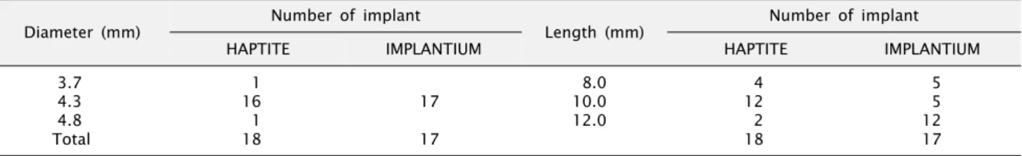
2개의 임플란트가 식립되었다(Table 1). 식립된 임플란트의 폭경
은 HAPTITE의 경우 4.3 mm가 16개, 4.8 mm와 3.7 mm가
각각 1개씩 식립되었으며, IMPLANTIUM은 17개 모두 4.3 mm
가 식립되었다. 임플란트의 길이는 8 mm에서 12 mm까지 식립
되었으며, HAPTITE의 경우 10 mm가 12개로 가장 많았으며,
12 mm 4개, 8 mm 2개가 식립되었다. IMPLANTIUM의 경우 12
mm가 7개, 10 mm와 8 mm가 각각 5개씩 식립되었다(Table 2).
Table 4. Value of Periotest
ⓇPeriod HAPTITE IMPLANTIUM
Fixation 1.94±3.90 (7, 3)* 4.25±1.76 (7, 1)
After 6 weeks 4.03±1.48 (6, 0) 4.76±0.97 (6, 3)
After 12 weeks 5.18±0.91 (7, 1)* 5.18±0.91 (7, 3)
Periostest values (PTVs).
HAPTITE, hydroxy-apatite coated implant.
*Statistically signigicant difference between fixation and 12 weeks (P=0.007).
Table 3. Value of Osstell
TM mentorPeriod HAPTITE IMPLANTIUM
Fixation 70.14±9.07 (57, 83)*† 74.68±7.42 (54, 83)††
After 6 weeks 76.98±5.25 (67, 85)* 79.03±4.39 (70, 85)
After 12 weeks 80.28±4.23 (70, 85)† 80.59±3.59 (73, 86)††
Implant stability quotient (ISQ).
HAPTITE, hydroxy-apatite coated implant.
*Statistically signigicant difference between fixation and 6 weeks (P=0.004).
†Statistically signigicant difference between fixation and 12 weeks (P< 0.001).
††Statistically signigicant difference between fixation and 12 weeks (P=0.007).
Table 2. Diameter and length of implant
Diameter (mm) Number of implant
Length (mm) Number of implant
HAPTITE IMPLANTIUM HAPTITE IMPLANTIUM
3.7 1 8.0 4 5
4.3 16 17 10.0 12 5
4.8 1 12.0 2 12
Total 18 17 18 17
HAPTITE, hydroxy-apatite coated implant.
Osstell
TMmentor 측정값은 HAPTITE의 경우 식립 시 70.14± 9.07 (최저값: 57, 최고값: 83)로 조사되었고, 6주 후 측정된 평균값은 76.98±5.25 (최저값: 67, 최고값: 85)였으며, 12주 후 측정된 평균값은 80.28±4.23 (최저값: 70, 최고값: 85) 으로 측정되었다. 식립 직후와 6주 후, 식립 직후와 12주 후 통계적으로 유의할만한 수치 증가가 있었다( P =0.004, <0.001).
IMPLANTIUM의 경우 식립 시 측정값은 평균 74.68±7.42 (최저 값: 54, 최고값: 83)였으며, 6주 후에는 79.03±4.39 (최저값:
70, 최고값: 85)로 조사되었다. 12주 후에는 80.59±3.59 (최저 값: 73, 최고값: 86)로 측정되어, 식립 직후와 12주 후 비교 시 유의할만한 수치 증가가 관찰되었다( P =0.007). HAPTITE와 IMPLANTIUM 간의 유의적 차이는 없었다(Table 3). Periotest
Ⓡ측정값은 HAPTITE의 경우 식립 시 평균값은 1.94±3.90 (최저 값: 7, 최고값: 3)이었으며, 6주 후에는 4.03±1.48 (최저값:
6, 최고값: 0), 12주 후에는 5.00±1.71 (최저값: 7, 최고값:
1)로 측정되었고, 식립 직후와 12주 후를 비교할 때 유의할만한 수치 감소가 있었다( P =0.007). IMPLANTIUM의 경우 식립 시 평균값은 4.25±1.76 (최저값: 7, 최고값: 1)이었으며, 6주 후에는 4.76±0.97 (최저값: 6, 최고값: 3), 12주 후에는
5.18± 0.91 (최저값: 7, 최고값: 3)로 측정되었고 통계적으로
유의할만한 차이는 없었다. 또한 HAPTITE와 IMPLANTIUM 간 에 통계적으로 유의한 차이는 없었다(Table 4).
고 찰
최근 임플란트 표면처리에 대한 많은 연구가 진행되고 있다.
이는 임플란트 표면이 임플란트와 골과의 골유착에 있어 중요한 역할을 하기 때문이다[11,12]. 1991년 Buser 등[25]은 여러 가지 표면 특성을 임플란트의 동물 실험에서 불규칙하고 거친 표면을 가진 임플란트가 골유착이 더 좋음을 보고하였다. 초기 임플란트 표면은 machined surface를 가졌으며, 이는 기계적인 가공을 통해 매끈한 평활 표면을 만드는 방법이다. 이후 임플란트 표면처 리에 대해 다양한 방법들이 소개되어 왔고 그 종류로는 수산화인 회석 코팅 표면(hydroxyapatite coating surface), 티타늄 플라 즈마 분사 표면(titanium plasma sprayed surface, TPS), 입자 분사 표면(blasted surface), 산부식 처리 표면(acid etching sur- face), 입자 분사 후 산부식 처리 표면(blasted and etched sur- face) 등이 대표적이다.
수산화인회석은 골조직의 구성 성분 중 하나로서 골과 뛰어난
생체 친화성을 보이며, 골 조직과의 결합성이 매우 우수한 것으로
보고되고 있다[26-30]. 이러한 이유로 수산화인회석을 이용한 임 플란트의 표면처리가 지속적으로 연구되고 있다. 1990년 Kohri 등[31]은 수산화인회석 코팅 임플란트가 초기 골반응이 우수하다 고 보고하였고, 1992년 Golec와 Krauser[32]는 수산화인회석 코팅 임플란트가 골과 임플란트 접촉량이 순수 티타늄재료보다 증가하였고, 계면 강도를 비교하였을 때, 약 5∼8배 정도 증가한 다고 보고하였다. Lee 등[21]은 dog을 이용한 동물실험에서 HA coated implant가 RBM surface implant에 비해 6주와 12주의 new bone formation (NBF) rate를 비교한 실험에서 6주 후 NBF rate가 통계적으로 유의하게 높았다고 보고하였다. Kim 등[22]은 가토의 대퇴골에 3종류의 수산화인회석 코팅 임플란트에 대한 제거회전력 비교 연구에서 Haptite가 Tapered Screw-Vent (Zimmer Inc., Warsaw, IN, USA)와 BioTite-H (DIO Co.
Ltd., Busan, Korea)에 비해 식립 초기 만족스러운 제거회전력값 을 보였다고 보고하였다. 본 연구에서도 HAPTITE의 경우 식립 직후와 6주 후, 식립 직후와 12주 후의 Osstell
TMmentor 수치와 식립 직후와 12주 후의 Periotest
Ⓡ수치에서 유의할만한 초기 안정성을 보였다.
임플란트의 거친 표면을 만들기 위해 Bowers 등[33]은 sand- blasting을 시행하였다. 하지만 sandblasting 시 사용되는 Al2O3 의 위해성에 대한 논란이 발생하면서 sandblasting 후 산부식을 시키는 방법인 SLA가 개발되었다. SLA 임플란트는 일차적으로 sandblasting 과정에서 macrorough한 "valleys"를 형성하고, 이차적으로 산부식 과정에서 microrough한 "micropits"을 형성 하여 표면 거칠기를 증가시킨다[34]. 또한 표면 오염물질들을 제 거하여 세포 부착을 방해하지 않는 alumina free한 임플란트 표면을 만든다[35,36]. SLA 표면처리 임플란트는 다른 표면처리 임플란트에 비해 빠르게 골-임플란트 접촉이 발생한다고 보고되 고 있다[37]. 또한 1995년 Martin 등[38]은 SLA 처리 임플란트가 TPS 코팅 임플란트에 비해 조골세포의 알카리성 인산효소 활성이 더 높았고, 임플란트에 접하고 있는 골세포 역시 분화가 잘되어 골 형성세포에 가깝다고 보고한 바 있다. 본 연구에서 사용된 IMPLANTIUM은 SLA 표면처리의 특징을 잘 가지고 있는 임플란 트로, 2011년 Jo 등[39]은 후향적 임상 연구를 통해 양호한 임상적 결과 및 100%의 생존율을 보고하였다. 본 연구에서 IMPLANTIUM 의 경우 Osstell
TMmentor의 수치 중 식립 직후와 12주 후에서만 유의적 차이를 보였으나, HAPTITE의 수치와 비교하여 유의할만 한 차이를 보이지 않았다.
2004년 Kwak 등[40]은 성견에서 장골에서 임플란트 표면처리 방법에 따른 골유착에 대한 비교 평가에서 machined surface와 RBM 임플란트에 비해 HA 코팅 임플란트와 SLA 임플란트에서 통계적으로 유의할만큼 양호한 골 유착을 얻었음을 보고한 바 있다. 본 연구에서는 HA 코팅 임플란트인 HAPTITE와 SLA 임플 란트인 IMPLANTIUM을 비교하였을 때 유의할만한 수치 차이를
보이지는 않았으나, 모두 양호한 초기 안정성을 보이고 있다.
하지만 본 연구에서는 증례의 수가 적고 식립부위를 한정하지 못하였다. 향후 추가적인 증례를 통한 연구 및 식립부위의 제한 및 골질에 따른 비교를 통한 추가적인 연구가 필요할 것으로 생각한다.
결 론
본 연구에서는 HA coating 임플란트인 HAPTITE와 SLA 임플 란트인 IMPLANTIUM의 초기 안정성에 대해 비교하고자 하였다.
두 임플란트 모두 Osstell
TMmentor와 Periotest
Ⓡ에서 양호한 초기 안정성을 보이며, 이를 통해 HAPTITE와 IMPLANTIUM 모두 임상적으로 안정적이며 예지성 있는 결과를 나타낼 수 있을 것으로 생각한다. 또한 HAPTITE는 일반적인 plasma sprayed HA coating의 문제점인 불균일한 코팅과 박리현상을 충분히 보 완할 수 있는 임플란트 재료라 생각한다.
References