원고 접수일 2012년 5월 17일, 원고 수정일 2012년 12월 12일, 게재 확정일 2013년 3월 20일
책임저자 국민석
(500-757) 광주시 북구 용봉로 77, 전남대학교 치의학전문대학원 구강악안면외과학 교실
Tel: 062-220-5436, Fax: 062-220-5437, E-mail: [email protected]
RECEIVED May 17, 2012, REVISED December 12, 2012, ACCEPTED March 20, 2013
Correspondence to Min-Suk Kook
Department of Oral and Maxillofacial Surgery, School of Dentistry, Chonnam National University
77, Yongbong-ro, Buk-gu, Gwangju 500-757, Korea
Tel: 82-62-220-5436, Fax: 82-62-220-5437, E-mail: [email protected]
CC This is an open access article distributed under the terms of the Creative Commons Attribution Non-Commercial License (http://creativecommons.org/licenses/
by-nc/3.0) which permits unrestricted non-commercial use, distribution, and reproduction in any medium, provided the original work is properly cited.
Le Fort I 골절단술을 이용한 상악골 전진 후 안정성에 관한 3차원적 연구
오철중ㆍ허정우ㆍ정 광ㆍ조민성ㆍ정승곤ㆍ박홍주ㆍ오희균ㆍ유선열ㆍ국민석
전남대학교 치의학전문대학원 구강악안면외과학교실
Abstract
Three Dimensional Study on the Postoperative Stability after Advancement of Maxilla Using Le Fort I Osteotomy
Chul-Jung Oh, Jung-Woo Hur, Kwang Chung, Min-Sung Cho, Seunggon Jung, Hong-Ju Park, Hee-Kyun Oh, Sun-Youl Ryu, Min-Suk Kook
Department of Oral and Maxillofacial Surgery, School of Dentistry, Chonnam National University
Purpose: This study evaluated postoperative maxillary stabilities in patients with skeletal Class III malocclusion who were taken both maxillary advancement surgery and mandibular retrusive surgery, using Le Fort I osteotomy, through three-dimensional computed tomography.
Methods: We selected 14 patients who were taken postoperative three-dimensional computerized tomography at the time before surgery, immediately after surgery, six months after surgery among the patients undergone both maxillary advancement surgery using Le Fort I osteotomy and mandibular retrusive surgery using bilateral sagittal split ramus osteotomy. We measured and compared the vertical distance of A-point and posterior nasal spine (PNS), the horizontal distance of A-point and PNS in transverse plane and coronal plane of the three-dimensional reconstructed images, respectively.
Results: In transverse plane, the distance difference between immediately after surgery (S1) and immediately before surgery (S0) of A-point was 0.04±1.80 mm, S2 and S0 was 0.15±1.69 mm, and between S1 and S2 was 0.11±0.58 mm. There were no significant differences between these data (P>0.05). In transverse plane, the distance between S1-S0 of PNS was
3.87±2.37 mm, S2-S0 of PNS was 3.79±2.39 mm, and S1-S2 of PNS was 0.08±0.18 mm. There were significant differences between these data (P<0.05). In coronal plane, the distance between S1-S0 of A-point was 3.99±0.86 mm, S2-S0 was 3.57±1.09 mm, and S1-S2 was 0.42±0.42 mm. There were significant differences between these data (P<0.05). In coronal plane, the distance between S1-S0 of PNS was 3.82±0.96 mm, S2-S0 was 3.43±0.91 mm, and S1-S2 was 0.39±0.49 mm. There were significant differences between these data (P<0.05). In transverse plane, it was estimated that PNS has no statistical postoperative stability in the same direction. In coronal plane, it was estimated that both A-point and PNS had no statistical postoperative stability (P<0.05).
Conclusion: Clinically, the operation plan needs to take into account of the maxillary relapse.
Key words: Le Fort I osteotomy, Three dimensional computed tomography, Maxillary relapse, Postoperative stability
서 론
구강악안면 기형증은 선천성, 발육성 원인으로 인해 성장, 발육 기간 동안 악골격계가 비정상적으로 변화를 일으키며 치열 및 구강, 악골계 기능에 보상적인 변화를 초래하는 질환이다. 따라서 저작, 발음 등 기능적 저하는 물론 잘못 발육된 악골격계로 인한 비심미적 안모로 환자에게 심리적으로 나쁜 영향을 준다[1].
악교정 수술은 치아 교정 치료 방법으로 만족할 만한 치열의 교합, 안모의 개선이 힘들 경우 악골 자체의 공간적 변화를 통해 악골 간의 조화 및 심미적 안모 개선을 위해서 악교정 수술을 시행하게 된다[2,3]. 악안면기형 환자에서 악교정 수술을 통해 기능적, 심미적 회복을 얻기 위해서는 술 전 정확한 진단과 치료 계획이 수립되어야 하며 술 후 안정성 확립이 필수적이다[4-7].
상악골 열성장에 대한 전진수술은 1939년 Axhausen이 부정 유합된 상악골에 대해 Le Fort I 골절단술을 최초로 사용하였으며, 1967년 Hoge-man과 Wilmar가 구개열 환자의 상악골을 Le Fort I 골절단술을 사용하여 보고하였다.
Le Fort I 상악골 전진술을 받은 환자에 있어 가장 큰 문제는 상악골의 후방 회귀일 것이다. Le Fort I 골절단술은 안정된 술식 으로 보고되었으나[8-12], Proffit 등[5,13]은 골격성 제 III급 부정 교합 환자에서 상악골 전방이동 및 하악골 후방이동이 동시에 시행된 경우 상악 수술만 한 경우보다 안정적이라 하였으며, 술 후 고정방법에 상관없이 1년 후 약 20% 정도에서 2∼4 mm의 상악골 후방 회귀가 발생한다고 보고하였다. 또한 Bell과 Scheideman[2]은 상악골의 전방이동 후 2∼3개월 내에 약 2 mm의 후방 회귀가 일어난다고 하였다.
그러나 양악 수술 후 상악골 회귀에 대한 연구들은 측면 두부 계측 방사선 사진을 이용하여 투사도를 제작하고 계측점을 이용하 여 평가한 방법이 대부분이며, 상의 왜곡, 확대, 구조물 간의 중첩 등으로 정확한 정보를 제공하지 못하는 경우가 많아 Le Fort I 골절단술을 이용한 상악골 전진술 후 안정성에 대해 논란이 있는 것이 사실이다.
최근 3차원 전산화단층촬영 기술의 발전과 보편화로 수술 전과 후, 3차원 전산화단층촬영을 이용한 악교정 수술 환자의 진단 및 평가가 가능하게 되었고, 이렇게 얻어진 3차원 전산화단층촬영 영상은 측면 두부 계측 방사선 사진에 비해 표면, 부피, 선형거리 의 정량화를 용이하게 하였다. 그러나 Le Fort I 골절단술을 이용 한 상악골 전진술 후 상악골 회귀에 대한 3차원 전산화단층촬영을 이용한 연구는 흔치 않은 실정이다.
이에 본 연구는 3차원 전산화단층촬영을 이용하여 Le Fort I 골절단술을 이용한 상악골 전진술 및 하악지 시상분할 골절단술 을 이용한 하악골 후방이동술을 동시에 받은 골격성 제III급 부정 교합 환자에 있어 술 후 상악골 안정성에 대하여 평가, 보고하는 바이다.
연구방법
1. 연구 대상
전남대학교병원 구강악안면외과에서 골격성 제III급 부정교합 으로 진단받고 2010년 4월부터 2011년 1월까지 Le Fort I 골절단 술을 이용한 상악골 전진술 및 하악지 시상분할 골절단술을 이용 한 하악골 후방이동술을 동시에 받은 환자 중 술 전, 수술 직후 및 술 후 6개월 경과 후 3차원 전산화단층촬영을 시행한 환자 총 14예를 대상으로 하였다. 남성이 5예, 여성이 9예였으며, 평균 나이는 21.9세로 18세에서 36세의 연령분포를 보였다.
Le Fort I 골절단술은 최소 2 mm에서 최대 6 mm까지 상악골 전진술을 시행하였으며 1예를 제외한 모든 경우에서 상악골 상하 방이동술 및 안면고경경사 수정술이 같이 시행되었다. Le Fort I 골절단술과 하악지 시상분할 골절단술 후 상하악 모두 mini- plate와 screw를 이용하여 고정하였다.
2. 연구 방법
Le Fort I 골절단술을 통한 상악골 전진술 후 상악골의 후방 회귀를 평가하기 위해 술 전, 수술 직후 및 술 후 6개월 후에 전산화단층촬영을 시행하였다. V worksTM 4.0 (Cybermed Inc., Seoul, Korea)을 이용하여 촬영된 전산화단층촬영 이미지 들을 3차원으로 재구성하였다. 3차원으로 재구성된 이미지에서 양악 수술 후 변화하지 않는 Frankfort horizontal plane (FH plane)과 양측 porion을 지나고 FH plane에 수직인 평면을 기준으로 양악 수술 후 변화하는 계측점을 측정하였다.
1) 전산화단층촬영 검사
남성 5명, 여성 9명에 대해 전산화단층촬영기(CTI pro, GE co, USA)를 이용하여 수술 전과 수술 직후 및 술 후 6개월 경과 후 안면골 전산화단층촬영을 시행하였다. 촬영된 이미지는 국제 의료 영상 표준(Digital Imaging Communication in Medicine) 파일로 저장하였다.
2) 기준평면 설정
기준평면(reference plane)으로서 3차원 전산화단층촬영 영 상에 대해 가로평면(transverse plane)과 관상평면(coronal plane)을 설정하였다. 가로평면(transverse plane)은 양측 po- rion과 우측 orbitale을 연결하여 형성하는 FH plane으로 하였으 며, 관상평면(coronal plane)은 양측 porion을 지나며 FH plane 과 수직으로 만나는 평면으로 설정하였다.
3) 계측 항목 및 통계학적 분석
술 전, 수술 직후와 술 후 6개월 경과 후 전산화단층촬영을
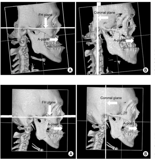
Fig. 1. Reference plane and point using before Le Fort I osteotomy and bilateral sagittal split ramus osteotomy in this study. (A) Tran- sverse plane and A-point. (B) Cor- onal plane and A-point. FH plane, Frankfort horizontal plane.
Fig. 2. Reference plane and point using after Le Fort I osteotomy and bilateral sagittal split ramus oste- otomy in this study. (A) Transverse plane and A-point. (B) Coronal plane and A-point. FH plane, Frankfort horizontal plane.
시행하고 3차원으로 재구성한 이미지의 가로평면에 대해 A-point 및 후비극의 수직거리를, 관상평면에서 A-point 및 후비극의 수평 거리를 측정하였다(Fig. 1, 2).
그리고 각 환자마다 측정된 수술 직후(S1)-술 전(S0) 간의 거리 차, 술 후 6개월 경과 후(S2)-술 전(S0) 간의 거리 차 및 수술 직후(S1)-술 후 6개월 경과 후(S2) 간의 거리차에 대해 평균 및 표준편차를 구하고, 수술에 의한 이동량을 의미하는 수술 직후 (S1)-술 전(S0) 간의 거리 차에 대해 술 후 6개월간의 회귀량을 의미하는 수술 직후(S1)-술 후 6개월 경과 후(S2) 간의 거리 차를 two tailed-paired T test를 시행하여 평가하였다.
결 과
전산화단층촬영 후 재구성된 3차원 영상에서 술 전, 수술 직후 및 술 후 6개월 경과 후 경조직의 기준점에 대한 각 계측항목을 측정한 결과는 다음과 같다(Table 1, 2).
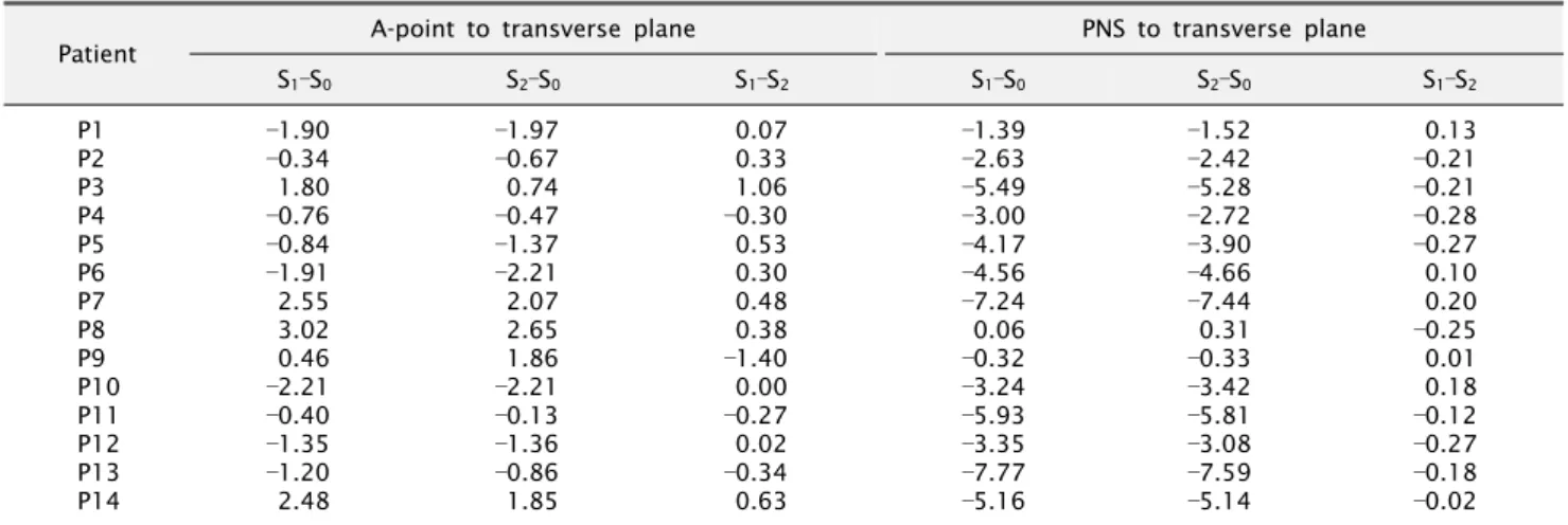
가로평면에 대해 A-point의 수술 직후(S1)와 술 전(S0) 차는
0.04±1.80 mm, 술 후 6개월 경과 후(S2)와 술 전(S0) 차는
0.15±1.69 mm, 수술 직후(S1)와 술 후 6개월 경과 후(S2) 차는 0.11±0.58 mm로 나타났으며, S1-S0 값에 대해 S1-S2 값을 two tailed-paired T test를 시행한 결과 유의한 차이는 없는 것으로 평가되었다(P>0.05) (Table 3).
가로평면에 대해 posterior nasal spine (PNS)의 S1-S0는
3.87±2.37 mm, S2-S0는 3.79±2.39 mm, S1-S2는 0.08±
0.18 mm로 나타났다. S1-S0 값에 대해 S1-S2 값은 유의한 차이 가 있는 것으로 평가되었다(P<0.05) (Table 3).
관상평면에 대해 A-point의 S1-S0는 3.99±0.86 mm, S2-S0는 3.57±1.09 mm, S1-S2는 0.42±0.42 mm로 나타났다. 14명 환자 모두 평균 4 mm 전방이동한 것으로 나타났으며 후방 회귀량 은 평균 0.42 mm였다. S1-S0 값에 S1-S2 값을 two tailed-paired T test를 시행한 결과 유의한 차이가 있는 것으로 평가되었다(P
<0.05) (Table 4).
관상평면에 대해 PNS의 S1-S0는 3.82±0.96 mm, S2-S0는 3.43±0.91 mm, S1-S2는 0.39±0.49 mm로 나타났다. 14명 환자 모두 평균 3.8 mm 전방이동한 것으로 나타났으며 후방 회귀량은 평균 0.4 mm였다. S1-S0 값에 S1-S2 값을 two
Table 1. The variation of A-point and PNS to transverse plane (unit: mm)
Patient A-point to transverse plane PNS to transverse plane
S1–S0 S2–S0 S1–S2 S1–S0 S2–S0 S1–S2
P1 P2 P3 P4 P5 P6 P7 P8 P9 P10 P11 P12 P13 P14
1.90
0.34 1.80
0.76
0.84
1.91 2.55 3.02 0.46
2.21
0.40
1.35
1.20 2.48
1.97
0.67 0.74
0.47
1.37
2.21 2.07 2.65 1.86
2.21
0.13
1.36
0.86 1.85
0.07 0.33 1.06
0.30 0.53 0.30 0.48 0.38
1.40 0.00
0.27 0.02
0.34 0.63
1.39
2.63
5.49
3.00
4.17
4.56
7.24 0.06
0.32
3.24
5.93
3.35
7.77
5.16
1.52
2.42
5.28
2.72
3.90
4.66
7.44 0.31
0.33
3.42
5.81
3.08
7.59
5.14
0.13
0.21
0.21
0.28
0.27 0.10 0.20
0.25 0.01 0.18
0.12
0.27
0.18
0.02 PNS, posterior nasal spine.
Table 2. The variation of A-point and PNS to coronal plane (unit: mm)
Patient A-point to coronal plane PNS to coronal plane
S1–S0 S2–S0 S1–S2 S1–S0 S2–S0 S1–S2
P1 P2 P3 P4 P5 P6 P7 P8 P9 P10 P11 P12 P13 P14
4.57 3.28 3.49 3.15 3.72 4.76 3.86 3.67 4.51 5.27 5.78 3.72 2.85 3.18
4.21 2.05 3.08 2.41 3.23 4.48 3.79 3.16 4.43 4.29 6.18 3.50 2.72 2.43
0.36 1.23 0.41 0.74 0.50 0.28 0.08 0.51 0.08 0.98
0.40 0.22 0.13 0.75
4.24 3.32 3.28 3.59 3.97 4.21 3.48 3.99 4.65 4.44 6.24 2.81 2.19 3.12
4.21 3.15 2.42 3.54 3.22 3.52 3.38 3.52 4.13 4.31 5.44 2.15 3.04 2.05
0.03 0.17 0.86 0.05 0.75 0.69 0.10 0.47 0.52 0.13 0.80 0.66
0.85 1.07 PNS, posterior nasal spine.
Table 3. The average immediate surgical (S1-S0), total (S2-S0), and follow-up (S1-S2) changes of A-point and PNS to transverse plane (unit: mm)
A-point PNS
S1-S0
S2-S0
S1-S2
0.04±1.80
0.15±1.69 0.11±0.58
3.87±2.37
3.79±2.39
0.08±0.18*
*P <0.05.
Values are presented as mean±standard deviation.
PNS, posterior nasal spine.
Table 4. The average immediate surgical (S1-S0), total (S2-S0), and follow-up (S1-S2) changes of A-point and PNS to coronal plane (unit: mm)
A-point PNS
S1-S0
S2-S0
S1-S2
3.99±0.86 3.57±1.09 0.42±0.42*
3.82±0.96 3.43±0.91 0.39±0.49*
*P <0.05.
Values are presented as mean±standard deviation.
PNS, posterior nasal spine.
tailed-paired T test를 시행한 결과 유의한 차이가 있는 것으로 평가되었다(P<0.05) (Table 4).
고 찰
양악 교정 수술은 기능적 측면뿐만 아니라 심미적으로 안모를
개선할 수 있으며[14], 최선의 교합 관계와 심미적 향상을 위해서 는 적절한 환자 분석 및 수술-교정 복합치료가 시행되어야 한다.
Hall 등[15]은 하악골 상행지 골절단 시 근막의 박리와 외상으로 인한 혈액 공급 불량으로 근심측 절단골 첨단부의 괴사가 발생할 수 있다고 하였다. Le Fort I 골절단술 시행 시 감염, 출혈, 비중격 변형, 비공의 변형 및 술 후 회귀 등이 발생할 수 있다.
악교정 수술 후 회귀 경향은 악간 고정 기간 중과 후에 많이 발생한다고 하며[16-20], 회귀에 관여하는 요소로는 하악 하연 평면각의 크기, 술 전 및 술 후 교정 치료 시행 여부 및 저작근과 하악골 주위의 골막이나 피부 등 결체조직의 장력이 있다[21,22].
그러나 술 후 회귀는 발생 빈도 및 원인에 대한 논란이 많다.
Le Fort I 골절단술의 술 후 회귀에 관한 연구에서 측모 두부 규격 방사선사진을 이용한 계측점 오차가 회귀값 오차의 원인으로 추정되어 왔다[23]. 이를 방지하기 위해서 Houston[24]과 Houston 등[25]은 측정 및 관찰자 제한성의 특이적 신뢰 한계를 확립하기 위한 주기적 재측정이 필요하며 연구자의 잠재적 기대 또한 오차의 원인이 될 수 있으므로 측정값은 무작위 추출하거나 이중 은폐 실험 설계(double blind experimental design)하도 록 권하였다.
최근 경조직 분석을 위해 3차원적 분석 방법들이 고안되어 사용되고 있다. 안면 경조직 분석은 안면 연조직처럼 직접 측정이 어렵고 측면 두부 계측 방사선 사진 등과 같은 2차원 방사선 사진들은 다른 조직과의 중첩으로 인해 오차들이 발생할 수 있다 는 단점이 있다. 이에 반해 안면부 전산화단층촬영으로 얻은 이미 지는 다각도로 회전이 가능하며 원하는 부위에 대해 분할할 수 있으며 일반 방사선사진에서 발생할 수 있는 중첩으로 인한 오차 를 줄일 수 있다.
본 연구에서는 제III급 부정교합 환자에 있어 3차원 전산화단층 촬영을 이용하여 Le Fort I 골절단술을 이용한 상악골 전진술 및 하악골 후방이동을 동시에 시행 후 상악골 안정성에 대하여 평가하였다.
상악골의 회귀에 있어 술 후 이동에 대한 많은 연구가 진행되었 다. 수직적 관계에서 Bishara 등[26]은 상악골의 상방이동술 후 상악골은 상방으로 계속 이동하고, 전방부가 후방부보다 2배 이상 상방이동한다고 하였다. Rotter와 Zeitler[27]는 상악골의 상방이 동술에서 술 후 변화의 절대치는 상방이동하고, 후방에서 약간 크지만 통계적 유의성이 없다고 하였다. Im 등[28]은 계측점들의 술 후 수직적 변화는 상, 하 양방향으로 이동하는 경향을 보이나 통계적으로 유의한 차이가 없었다고 하였다.
본 연구에서는 상악골의 수직적 관계에서 가로평면에 대한 A-point의 S1-S2 값이 0.11±0.58 mm로 S1-S0 값에 대해 통계적 유의성을 보이지 않아 상악골의 술 후 안정성이 있다고 평가되었 으나(P>0.05), PNS의 S1-S2 값이 0.08±0.18 mm로 S1-S0
값에 대해 유의적 차이를 보여 술 후 안정성이 없다는 결과를 보였다(P<0.05). 하지만 그 양에서 수술에 의한 PNS의 평균 상방이동량의 2% 정도로 그 양이 미비하였다. 이러한 차이는 전방부보다 후방부의 상방이동량이 큰 상방이동술을 대부분의 수술에서 시행하였기 때문으로 판단된다.
수평적 관계에서 Le Fort I 골절단술 후 회귀에 대해 Carlotti와 Schendel[12]은 상악골의 전방이동 및 하악골 이동 여부에 상관
없이 안정된 결과를 나타낸다고 하였으며, Law 등[29]은 상악골의 후방이동술은 술 후 안정된 상태로 유지한다고 하였다. 그러나 Bell과 Scheideman[2]은 상악골의 이동량과 회귀는 상관이 없지 만 술 후 초기 2∼3개월 내에 약 2 mm의 회귀가 일어난다고 하였으며, Proffit 등[13]도 골격성 III급 부정교합 환자에서 상악 골의 전방이동 후 약 20%에서 1년 후에 2∼4 mm의 후방 회귀가 발생한다고 하였다. 또한 Araujo 등[10]은 Le Fort I 골절단술은 안정적 술식이지만, 전진술은 안정성에 문제가 있고 특히 골이식 을 시행하지 않은 군에서 후방이동이 유의성 있게 크다고 하였다.
본 연구에서는 관상평면에 대한 A-point 및 후비극의 S1-S2
값은 각각 0.42±0.42 mm와 0.39±0.49 mm로 수술에 의한 평균 전방이동량의 10% 정도의 회귀량을 보여 수평적으로 통계적 술 후 안정성이 없는 것으로 평가되었다(P<0.05).
상악의 안정성에 영향을 미치는 인자로 반흔조직의 수축, 비중 격의 부적합, 부적절한 고정, 상악 고정 시 저항 등이 있을 수 있다[30]. 또한 고정방법에 따른 회귀성의 비교에서 Bishara 등 [26]은 비견고 고정(nonrigid fixation) 시 술 후 이동량은 20∼
65%로 보고하였고, Weiss 등[31]은 상악골 전진술 후 수평면에서 견고 고정군과 비견고 고정군 모두 6주에서 1년 사이에 상악의 전방기준점이 후방으로 재위치 되는 경향이 있다고 하였다. 그리 고 Seo 등[32]은 전진량에 따른 회귀성향을 알아보기 위해 환자를 두 그룹으로 나누어 분석한 결과 6 mm 이상으로 전진량이 많은 그룹에서 전진량이 적은 그룹보다 약간 높은 회귀성향을 보였으나 통계적 유의성은 없었다고 하였다. 상악골의 회귀를 줄이기 위해 수술 계획 수립 시 고정 방법과 전진량 등에 대해 고려하여야 한다. 또한 수술 전, 후의 교정 치료는 술 후 재발률을 줄이고 술 후 안정성 유지에 좋은 결과를 보여줄 수 있다[21].
본 연구는 Le Fort I 골절단술을 이용한 상악골 전진술 및 하악지 시상분할 골절단술을 이용한 하악골 후방이동술을 동시에 받은 골격성 제III급 부정교합 환자에 있어 술 후 상악골 안정성에 대하여 평가하였다. 가로평면에 대해 A-point는 수직적 방향으로 통계적 술 후 안정성이 있는 것으로 평가되었지만(P>0.05), PNS은 수직적 방향으로 통계적 술 후 안정성이 없는 것으로 평가되었다. 또한 관상평면에 대해 A-point 및 PNS은 수평적으 로 통계적 술 후 안정성이 없는 것으로 평가되었다(P<0.05).
따라서 임상적으로 Le Fort I 골절단술을 이용한 상악골 전진술 시 상악골의 회귀를 고려하여 수술 계획을 수립하여야 할 것으로 생각된다. 이에 대한 추가적인 추적 관찰 및 연구가 요구되며, 규격화되지 않은 적은 수의 표본을 대상으로 연구한 점을 보완하 기 위하여 표본의 규격화 및 표본 수의 확대를 통한 연구가 시행되 어야 할 것으로 생각된다.
결 론
본 연구는 3차원 전산화단층촬영을 이용하여 Le Fort I 골절단 술을 이용한 상악골 전진술 및 하악골 후방이동술을 동시에 받은 골격성 제III급 부정교합 환자에 있어 술 후 상악골 안정성에 대하여 평가하였다.
그 결과 수직적으로 이동량이 큰 후비극과 수평적으로 전방이 동된 A-point의 경우 술 후 통계적 안정성이 없는 것으로 평가되 었다. 따라서 임상적으로 Le Fort I 골절단술을 이용한 상악골 전진술 시 상악골의 회귀를 고려하여 수술 계획을 수립하여야 할 것으로 생각된다. 또한 술 후 상악골의 회귀를 줄이기 위한 노력으로 수술 계획 수립 시 이동량과 고정 방법 등에 대한 고려가 필요하며, 수술 전, 후의 교정 치료를 통해 술 후 재발률 감소와 술 후 안정성 유지에 좋은 결과를 얻을 수 있을 것으로 생각된다.
References
1. McNeill RW, Proffit WR, White RP. Cephalometric prediction for orthodontic surgery. Angle Orthod 1972;42:154-64.
2. Bell WH, Scheideman GB. Correction of vertical maxillary deficiency: stability and soft tissue changes. J Oral Surg 1981;39:666-70.
3. Bell WH, Proffit WR, White RP. Surgical correction of dentofa- cial deformities. Philadelphia: W.B, Saunders Co.; 1980. p.1329-67.
4. Wolford LM, Chemello PD, Hilliard FW. Occlusal plane al- teration in orthognathic surgery. J Oral Maxillofac Surg 1993;
51:730-40.
5. Proffit WR, Phillips C, Turvey TA. Stability after surgi- cal-orthodontic corrective of skeletal Class III malocclusion.
3. Combined maxillary and mandibular procedures. Int J Adult Orthodon Orthognath Surg 1991;6:211-25.
6. Satrom KD, Sinclair PM, Wolford LM. The stability of double jaw surgery: a comparison of rigid versus wire fixation. Am J Orthod Dentofacial Orthop 1991;99:550-63.
7. Choi SW, Park HS, Cha IH. A study on accuracy of the maxillary repositioning in orthognathic surgery by the ex- ternal measuring technique. J Korean Assoc Oral Maxillofac Surg 1996;22:537-43.
8. Bell WH, McBride KL. Correction of the long face syndrome by Le Fort I osteotomy. A report on some new technical modifications and treatment results. Oral Surg Oral Med Oral Pathol 1977;44:493-520.
9. Schendel SA, Eisenfeld J, Bell WH, Epker BN, Mishelevich DJ. The long face syndrome: vertical maxillary excess. Am J Orthod 1976;70:398-408.
10. Araujo A, Schendel SA, Wolford LM, Epker BN. Total maxil- lary advancement with and without bone grafting. J Oral Surg 1978;36:849-58.
11. Bell WH, Jacobs JD. Tridimensional planning for surgical/
orthodontic treatment of mandibular excess. Am J Orthod 1981;80:263-88.
12. Carlotti AE Jr, Schendel SA. An analysis of factors influenc- ing stability of surgical advancement of the maxilla by the Le Fort I osteotomy. J Oral Maxillofac Surg 1987;45:924-8.
13. Proffit WR, Phillips C, Prewitt JW, Turvey TA. Stability after surgical-orthodontic correction of skeletal Class III malocclusion.
2. Maxillary advancement. Int J Adult Orthodon Orthognath Surg 1991;6:71-80.
14. Riedel RA. Esthetics and its relation to orthodontic therapy.
Angle Orthod 1950;20:168-78.
15. Hall HD, Chase DC, Payor LG. Evaluation and refinement of the intraoral vertical subcondylar osteotomy. J Oral Surg 1975;
33:333-41.
16. Ive J, McNeill RW, West RA. Mandibular advancement: skel- etal and dental changes during fixation. J Oral Surg 1977;35:
881-6.
17. Kohn MW. Analysis of relapse after mandibular advancement surgery. J Oral Surg 1978;36:676-84.
18. McNeill RW, Hooley JR, Sundberg RJ. Skeletal relapse during intermaxillary fixation. J Oral Surg 1973;31:212-27.
19. Poulton DR, Ware WH. Surgical-orthodontic treatment of se- vere mandibular retrusion. Am J Orthod 1971;59:244-65.
20. Poulton DR, Ware WH. Surgical-orthodontic treatment of se- vere mandibular retrusion. II. Am J Orthod 1973;63:237-55.
21. Um IW, Min BI. Analysis on the relapse after orthognathic surgery for mandibular progathism in cephalography. J Korean Assoc Oral Maxillofac Surg 1986;12:145-58.
22. Rotskoff KS, Herbosa EG, Nickels B. Correction of condylar displacement following intraoral vertical ramus osteotomy. J Oral Maxillofac Surg 1991;49:366-72.
23. Bjork A, Solow B. Measurement on radiographs. J Dent Res 1962;41:672-83.
24. Houston WJ. The analysis of errors in orthodontic measure- ments. Am J Orthod 1983;83:382-90.
25. Houston WJ, Maher RE, McElroy D, Sherriff M. Sources of error in measurements from cephalometric radiographs. Eur J Orthod 1986;8:149-51.
26. Bishara SE, Chu GW, Jakobsen JR. Stability of the LeFort I one-piece maxillary osteotomy. Am J Orthod Dentofacial Orthop 1988;94:184-200.
27. Rotter BE, Zeitler DL. Stability of the Le Fort I maxillary os- teotomy after rigid internal fixation. J Oral Maxillofac Surg 1999;57:1080-8.
28. Im YH, Ko SO, Shin HK. A study on the postoperative sta- bility of lefort I osteotomy in the two-jaw surgery of the skeletal class III malocclusion patients. J Korean Assoc Oral Maxillofac Surg 2001;27:397-403.
29. Law JH, Rotskoff KS, Smith RJ. Stability following combined maxillary and mandibular osteotomies treated with rigid in- ternal fixation. J Oral Maxillofac Surg 1989;47:128-36.
30. Welch TB. Stability in the correction of dentofacial deform- ities: a comprehensive review. J Oral Maxillofac Surg 1989;
47:1142-9.
31. Weiss MJ, Patty S, Phillips C. Dental and skeletal stability following maxillary advancement. J Dent Res 1989;68:259-65.
32. Seo MG, Koo SY, Kim EJ, Leem DH, Shin HK, Ko SO. The skeletal stability of Le Fort I maxillary advancement. J Korean Assoc Maxillofac Plast Reconstr Surg 2010;32:149-53.