Korean J. Environ. Biol. 36(3) : 291~298(2018) https://doi.org/10.11626/KJEB.2018.36.3.291
INTRODUCTION
Benthic cyanobacteria are classified according to the sub- strates; epiphytic (submerged plants or other algae), epilith- ic (rocks or stones), epipelic (mud) and epizoic (crustacean or arthropod). Some of cyanobacteria that have symbiotic relationship with lichen or bryophytes exposed to the atmo- sphere (Goyal 1997). Rocks exposed to the atmosphere are inhabited by extremophilic cyanobacteria that play a basic role as primary producers in a variety of subaerial habitats such as cliffs and pinnacles of dry and humid regions (Pen- tecost and Whitton 2000), hot and cold deserts (Wynn-Wil- liams 2000; Vincent 2007) and caves (Hoffmann 2002), and might contribute to the future colonization of other planets (Grilli Caiola and Billi 2007; Billi 2012).
Cyanobacteria have both positive and negative influences on human and environment. They are an important compo-
nent of microbial food webs and grazed biomass in water environments (Gonzalez et al. 1998; Charpy 2005). Spiruli- na had been used to food for livestock and human, and they are being developed as substitutional foods for their high content of protein, vitamin, mineral and so on (Graham et al. 2009).
But nowadays, freshwater habitats are threatened world- wide because of cyanobacteria, such as Oscillatoria, and Aphanizomenon. They produces odorous materials like Geosmin and 2-MIB (Robarts and Zohary 1987; Codd 1995; Graham et al. 2009; Whitton 2012). Therefore, con- tinuous researches of cyanobacteria, especially Microcystis, Anabaena, Oscillatoria, and Aphanizomenon are being con- ducted worldwide (Park and Kim 1995; Park 2005; Lee et al. 2017).
The 4,617 taxa of cyanobacteria have been reported to AlgaeBase (Guiry and Guiry 2018), and 377 taxa have been reported in Korea (Kim 2015; Song and Lee 2017; Yim et al. 2017). Cephalothrix and Cyanophanon have been reported each 2 taxa and Toxifilum reported 1 taxa in world-
* Corresponding author: Ok Min Lee, Tel. 031-249-9643, Fax. 031-241-0860, E-mail. [email protected]
ⓒ2018. Korean Society of Environmental Biology.
A Study of Seven Unrecorded Species of Benthic and Subaerophytic Cyanobacteria (Cyanophyceae, Cyanophyta) in Korea
Byoung Cheol Yim, Jee Hwan Kim
1, Sung Ro Yoon, Nam Ju Lee and Ok Min Lee*
Department of Life Science, College of Natural Science, Kyonggi University, Suwon 16227, Republic of Korea
1
Bioresources Culture Collection Division, Nakdonggang National Institute of Biological Resources, Sangju 37242, Republic of Korea
Abstract - Samples were collected from benthic and subaerophytic habitats of fresh and brackish water in Korea from April 2017 to April 2018. Accordingly, three genera and seven species were newly recorded in Korea. Three unrecorded genera were Cephalothrix, Cyanophanon and Toxifilum, and seven species were Anagnostidinema pseudacutissimum, Calothrix elenkinii, Cephalothrix komarekiana, Cyanophanon mirabile, Cyanosarcina chroococcoides, Leptolyngbya ectocarpi and Toxifilum mysidocida. Anagnostidinema pseudacutissimum and Cephalothrix komarekiana had been previously reported to inhabit freshwater, but these were found to be present in brackish water during the period of the present study. Also, Cyanosarcina chroococcoides previously reported to inhabit moorland waters and swamps, were found to be present in subaerophytic habitat.
Keywords : benthic, brackish water, cyanobacteria, subaerophytic
<Original article>
wide (Guiry and Guiry 2018).
For this study, we collected unrecorded cyanobacteria from fresh and brackish waters to add the cyanophycean flora of Korea.
MATERIALS AND METHODS
We collected benthic and subaerophuytic cyanobacteria from six sites in Korea from April 2017 to April 2018 (Fig.
1; Tables 1, 2). Benthic cyanobacteria were collected scrub- bing off submerged plants or rocks using soft brush. Sub- aerophytes were collected from lichen or bryophytes using sharp spatula. Each sample was sealed in sterile bottles and transported them to the laboratory (Crispim et al. 2004).
Measurement of salinity was performed by marine tester (DMT-10, DYS, Korea).
The samples were examined under BX53 light micro- scope at ×400-1,000 (Olympus, Tokyo, Japan) and photo- graphed using AxioCam HRC camera (Carl Zeiss, Oberko- chen, Germany). The taxonomic classification system was based on Komárek et al. (2014) and Algaebase (Guiry and Guiry 2018). In this study, identification of cyanobacteia re- ferred to Hirose et al. (1977), Prescott (1982), Chung (1993), Komárek and Anagnostidis (1999, 2005), John et al. (2011) and Komárek (2013).
RESULTS AND DISCUSSION
Three genera and seven species of benthic and subaero- phytic cyanobacteria were newly recorded in Korea. The newly recorded genera were Cephalothrix, Cyanophanon and Toxifilum, and the newly recorded species were Anag-
Table 1. Sampling sites in Korea from April 2017 to April 2018
Site Local name Latitude (N) Longitude(E)
st.1 33, Dobong-ro, 157-gil, Dobong-gu, Seoul 37.673434 127.042158
st.2 58, Madong-gil, Samhyang-eup, Muan-gun, Jeollanam-do 34.845626 126.412473
st.3 598-63, Gyeongam-dong, Gunsan-si, Jeollabuk-do 35.981633 126.727461
st.4 129-1, Sincheon-ri, Seongsan-eup, Seogwipo-si, Jeju-do 33.351350 126.857913
st.5 833-1, Ojo-ri, Seongsan-eup, Seogwipo-si, Jeju-do 33.460851 126.914538
st.6 1956-49, Oedo 2-dong, Jeju-si, Jeju-do 33.493495 126.427391
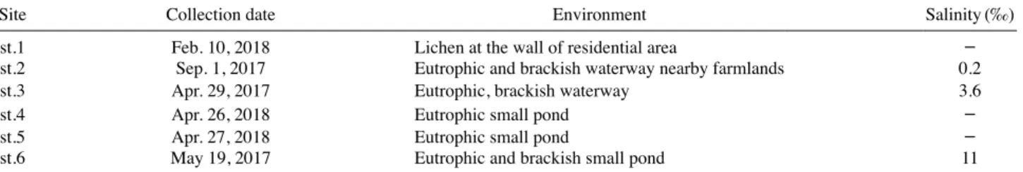
Table 2. The environments of sampling sites from April 2017 to April 2018
Site Collection date Environment Salinity(‰)
st.1 Feb. 10, 2018 Lichen at the wall of residential area -
st.2 Sep. 1, 2017 Eutrophic and brackish waterway nearby farmlands 0.2
st.3 Apr. 29, 2017 Eutrophic, brackish waterway 3.6
st.4 Apr. 26, 2018 Eutrophic small pond -
st.5 Apr. 27, 2018 Eutrophic small pond -
st.6 May 19, 2017 Eutrophic and brackish small pond 11
Fig. 1. The map showing the six sampling sites in Korea from April 2017 to April 2018.
nostidinema pseudacutissimum, Calothrix elenkinii, Cepha- lothrix komarekiana, Cyanophanon mirabile, Cyanosarcina chroococcoides, Leptolyngbya ectocarpi and Toxifilum mysidocida.
Order Chroococcales Schaffner 1922 Family Chroococcaceae Rabenhorst 1863 Genus Cyanosarcina Kovácik 1988
Cyanosarcina chroococcoides (Geitler) Kovácik 1988 (Fig. 2)
Synonym: Myxosarcina chroococcoides Geitler 1927
The colonies are microscopic and shaped subspherical or irregularly rounded with different sizes. The colony is composed of densely and irregularly aggregated packets of cells in different sizes. The mucilaginous envelope that sur- rounds the colony is thin (about 1 μm thick), firm and co- lourless. The mucilaginous envelope tightly surrounds the cell packets, sometimes diffluent in old colonies. The cell is irregularly rounded and olive-green or blue-green colored.
The cell diameter is 8-12 μm.
Ecology: This species lives in moorland waters and swamps. Maybe they have wide distribution over the whole temperate zone (Komárek and Anagnostidis 1999). In this
study, we collected this species from subaerophytic sub- strate (the wall of residential area).
Distribution: Europe: Spain (Alvarez-Cobelas and Gal- lardo 1988); South America: Argentina (Tell 1985); Asia:
Japan (Hirose et al. 1977).
Site of collection: The wall of residential area, Dobong-gu, Seoul (February 10, 2018).
Specimen Locality: ACKU2018MD01
Order Nostocales Borzì 1914
Family Rivulariaceae Bornet and Flahault 1886 Genus Calothrix Agardh ex Bornet and Flahault 1886 Calothrix elenkinii Kossinskaja 1924 (Fig. 3)
The thallus is composed of elongated clusters, also producing solitary filaments. Filaments exist solitarily, in groups or clusters. Filaments are fasciculated, arcuated, and entangled together at the bases. Sheaths are thin, not lamel- lated, colorless; they are open at the apex and usually lon- ger than the trichomes. The width excluding the sheath is 5.5-7 μm at the basements and 5-6.5 μm wide at the middle parts. There are solitary heterocysts at the basement’s apex, with 4.5-7 μm width. At the basements, constricted at the cross-wall strongly appears, but slightly constricted cross- walls appear at the opposite end (apical regions). Trichomes
Fig. 2. Microscopic images of Cyanosarcina chroococcoides(Geitler) Kovácik. The researchers focused on sub-colonies comprising of pellets of cells (A–C) and on fine mucilagi- nous envelope (D). Scale bar: 10 μm.
A B
C D
Fig. 3. Microscopic images of Calothrix elenkinii Kossinskaja 2016. The researchers focused on heterocysts (red arrows), basement (A) and apex (B) and showed overall appearance of trichomes (C, D). Scale bar: 10 μm.
A B
C D
are blue-green or olive-green colored and apical cells are narrowed and rounded. Cells are quadratic or a little shorter than long, but elongated and longer than wide in the upper part.
Ecology: This species appears in freshwater and benthic habitat. They lives in streams and soils (Komárek 2013).
We collected it from eutrophic small pond.
Distribution: Europe: Spain (Mateo et al. 2006); South America: Argentina (Tell 1985); Asia: Russia (Medvedeva and Nikulina 2014).
Site of collection: Gotari-mot, Seongsan-eup, Seogwipo-si, Jeju-do (April 26, 2018).
Specimen Locality: ACKU2018IR03
Order Oscillatoriales Schaffner 1922
Family Coleofasciculaceae Komárek, Kastovsky, Mares and Johansen 2014
Genus Anagnostidinema Strunecký et al. 2017 Anagnostidinema pseudacutissimum (Geitler)
Strunecký, Bohunická, J.R. Johansen and J. Komárek 2017 (Fig. 4)
Synonym: Oscillatoria pseudacutissima Geitler
Phormidium pseudacutissimum (Geitler) Anagnostidis and Komárek 1988
Geitlerinema pseudacutissimum (Geitler) Anagnostidis 1989
The thallus is blue-green colored. Trichomes are straight or slightly coiled and motile with anticlockwise rotation.
Trichomes are unconstricted or slightly constricted at cross- walls and slightly attenuated at the ends and bent. Apical cells are hooked and roundly-pointed. Cell contents have prominent carotenoid granules, usually near the cross-walls.
Cells are colored bright blue-green and 1.2-3 times longer than wide, with 1.2-1.4 μm width.
Ecology: This species appears in freshwater and benthic habitat (Komárek and Anagnostidis 2005). In this study, we collected it from eutrophic and brackish waterway (salinity 0.2‰).
Distribution: Europe: Netherlands (Veen et al. 2015).
Site of collection: Madong-waterway, Samhyang-eup, Muan-gun, Jeollanam-do (September 1, 2017).
Specimen Locality: ACKU2017IR11
Family Phormidiaceae Anagnostidis and Komárek 1988 Genus Cephalothrix C.F.S. Malone et al. 2015
The thallus is fasciculated and blue-green colored. Tri- chomes are cylindrical and straight, slightly attenuated, sometimes bent at the end. Trichomes are unconstricted or slightly constricted at the cross-walls. Sheaths are faculta- tive hyaline, firm and attached to trichome or wider. Apical cells are strongly or slightly capitate and sometimes with conical calyptra. Hormogonia formation occurred by bicon- cave necridic cells. Cells are wider than long, length is 2.0- 3.4 μm and width is 4.2-6.5 μm.
Cephalothrix komarekiana C.F.S. Malone et al. 2015 (Fig. 5)
Filaments are cylindrical and straight, but at the end, bent and slightly attenuated. Filaments are constricted or uncon- stricted at the cross-walls. Sheaths are facultative hyaline, firm and narrow or wide. Hormogonia formation occurred by biconcave necridic cells. At the end of the cells, capitate slightly or strongly appears. Cell contents have facultative
Fig. 4. Microscopic images of Anagnostidinema pseudacutissimum (Geitler) Strunecký, Bohunická, JR Johansen & J Komárek.The researchers showed overall appearance of trichomes (A) and focused on apex (B, C) and middle parts of trichomes (D). Scale bar: 10 μm.
A B
C D
aerotopes. Cells are wider than long, length is 1.2-2.5 μm and width is 5.5-7 μm.
Ecology: This species appears in freshwater and benthic habitat. They lives in an alkaline lake in the Brazilian Pan- tanal wetlands, Mato Grosso do Sul State, Brazil (Malone et al. 2015). In this study, we collected it from brackish small stream (salinity 3.6‰).
Distribution: South America: Brazil (Malone et al. 2015).
Site of collection: Gyeongpo-gyo, Gyeongam-dong, Gun- san-si, Jeollabuk-do (April 29, 2017).
Specimen Locality: ACKU2018IR04
Order Synechococcales Hoffmann, Komárek and Kastovsky 2005
Family Chamaesiphonaceae Borzì 1878 Genus Cyanophanon Geitler 1955
Cells are heteropolar, and solitary or in groups. They are elongated and attached to the substrate by end. The cells are long cylindrical when old. Cell content is homogeneous or with solitary granules, usually with visible, separate chromatoplasma (lateral position of thylakoids). Cells are pale blue-green, olive-green or light reddish colored. Re- production is occurred by simultaneous or successive from the apex of the cylindrical cells, rarely along the whole cell;
division by transverse fission. Exocytes are separated one by one, or a great part cell is transformed into a row of exo- cytes.
Cyanophanon mirabile Geitler 1955 (Fig. 6)
Cells appear in solitary or in groups. The cells are elon- gate, cylindrical, straight or arcuated. The apex of the cell is rounded and they divided into a row of cylindrical to barrel-shaped exocytes in the upper parts. Cell contents are usually homogeneous and are pale olive-green, grey-blue or pinkish in color. Cell length is 2.2-4.5 μm and width is 0.7- 1.2 μm.
Ecology: This species occurs in freshwater species and ep- iphytes on another filamentous algae. They lives on rocks with flowing water, streams and waterfalls with clear, kath- arobic water, usually in mountains (Komárek and Anagnos- tidis 2005). We collected it from eutrophic pond.
Distribution: Europe: Czech Republic (Gadea et al. 2013).
Site of collection: Ojo-ri pond, Seongsan-eup, Seogwi- po-si, Jeju-do (April 27, 2018).
Specimen Locality: ACKU2018MD04
Family Leptolyngbyaceae Komárek, Kastovsky, Mares and Johansen 2014
Genus Leptolyngbya Anagnostidis and Komárek 1988
Fig. 5. Microscopic images of Cephalothrix komarekiana CFSMalone et al. The researchers showed overall bundle of trichomes. The red arrows indicate calyptra. Scale bar: 10 μm.
A B
C D
Fig. 6. Microscopic images of Cyanophanon mirabile Geitler.
They appear to be attached at surface of other cyanobacte- ria (A) and chlorophytes (B–D). Scale bar: 10 μm.
A B
C D
Leptolyngbya ectocarpi (Gomont) Anagnostidis and Komárek 1988 (Fig. 7)
Synonym: Phormidium ectocarpi Gomont 1899
Thallus is thin, membranaceous and carmine-red or brownish-red in color. Filaments are densely and irregularly entangled or parallel-arranged. Sheaths are mostly color- less, diffluent, mucilaginous, rarely distinct. Trichomes are constricted at the diaphanous, ungranulated cross-walls and are either slightly attenuated. Apical cells are rounded or conical-rounded, lengthened and without calyptra. Cell con- tents are homogeneous with distinct chromatoplasma. Cells are isodiametric longer than wide. The length is 1.2-1.5 μm and the width is 1.4-1.7 μm.
Ecology: This species appears in marine and epiphytic, epizoic habitat. They lives epiphytic on various seaweeds, epizoic on various animals, mud and walls of glass vessels with marine water widely distributed in littoral and sublit- toral zones of coasts (Komárek and Anagnostidis 2005). We collected it from eutrophic brackish pond (salinity 11‰).
Distribution: Europe: Britain (Whitton et al. 2003), Spain (Gallardo et al. 2016); Indian Ocean Islands: Mauritius (Sil- va and Pienaar 2000).
Site of collection: Mai-mot, Oedo 2-dong, Jeju-si, Jeju-do (May 19, 2017).
Specimen Locality: ACKU2017IR04
Family Pseudanabaenaceae Anagnostidis and Komárek 1988
Genus Toxifilum Zimba, Huang, Foley and Linton 2017 Filaments are composed of a uniseriate trichome. Sheath is usually present, thin, colorless. Filaments are gliding and attenuated at the end. Cells are longer than wide and they have peripheral thylakoids. Filaments and single cells are bright green colored. Both external and cross cell walls are undulated.
Toxifilum mysidocida Zimba, Huang, Foley and Linton 2017 (Fig. 8)
Filaments are rarely solitary and free floating, usually forming clusters or mats on substrate. Filaments are undu- lated, with thin sheaths, and are unbranched. Filaments are narrowing at the ends and exhibit gliding motility. Cells are cylindrical, with peripherally arranged thylakoids. Poly- phosphate bodies and cyanophycin granules occur in cell corners with micro plasmodesmata present between cross cell walls. Cell’s length is 3-10 μm and width is 2.2-2.5 μm.
Ecology: This species appears in marine and benthic hab- itat. They lives in Rincon Bayou salt marsh, Nueces Bay, TX, USA (Zimba et al. 2017). We collected it from brackish small stream (salinity 3.6‰).
Distribution: North America: Texas (Zimba et al. 2017).
Site of collection: Gyeongpo-gyo, Gyeongam-dong, Gun- san-si, Jeollabuk-do (April 29, 2017).
Specimen Locality: ACKU2018IR05
Fig. 8. Microscopic images of Toxifilum mysidocida Zimba et al.
The researchers showed overall appearance of trichomes.
The red arrows indicate aerotopes. Scale bar: 10 μm.
A B C
Fig. 7. Microscopic images of Leptolyngbya ectocarpi (Gomont) Anagnostidis & Komárek. The researchers showed overall appearance eof trichomes (A, B) and focused on pointed apex of trichomes (C, D). Scale bar: 10 μm.
A B
C D
ACKNOWLEDGEMENTS
This study was supported by a grant from the National Institute of Biological Resources (NIBR201801205) and the Nakdonggang National Institute of Biological Resources [(The project on collection of freshwater algal strains (Year- 2), 2018)], funded by the Ministry of Environment (MOE) of the Republic of Korea.
REFERENCES
Billi D. 2012. Anhydrobiotic Rock- Inhabiting Cyanobacteria:
Potential for Astrobiology and Biotechnology. pp. 119–132.
In Adaptation of Microbial Life Organisms in Extreme En- vironments: Research and Application (Stan-Lotter H eds.).
Springer, Wien New York.
Charpy L. 2005. Importance of photosynthetic picoplankton in coral reef ecosystems. Vie Milieu. 55:217–224.
Chung J. 1993. The Microscopic Illustrations of the Freshwater Algae of Korea. Academy Publishing Co., Seoul.
Codd GA. 1995. Cyanobacterial toxins: occurrence, properties and biological significance. Water Sci. Technol. 32:149–
156.
Crispim CA, CC Gaylarde and PM Gaylarde. 2004. Biofilmson church walls in Porto Alegre, RS, Brazil, with special at- tention to Cyanobacteria. Int. Biodeterior. Biodegradation 54:121–124.
Gadea I, M Rodilla, J Sospedra, S Falco and T Morata. 2013.
Seasonal dynamics of the phytoplankton community in the Gandía coastal area, Southern Gulf of Valencia. Thalassas.
29:35–58.
Gallardo T, I Bárbara, J Afonso-Carrillo, R Bermejo, M Al- tamirano, A Gómez Garreta, MC Barceló Martí, J Rull Lluch, E Ballesteros and J De la Rosa. 2016. Nueva lista crítica de las algas bentónicas marinas de España. Boletín Informativo de la Sociedad Española de Ficología 51:7–52.
Gonzalez JM, J-P Torreton, P Dufour and L Charpy. 1998.
Temporal and spatial dynamics of the pelagic microbial food web in an atoll lagoon. Aquat. Microb. Ecol. 16:53–
Goyal SK. 1997. Algae and soil environment. Phykos. 36:1–13.64.
Graham LE, JM Graham and LW Wilcox. 2009. Algae. Second edition. Pearson Benjamin Cummings. San Francisco.
Grilli Caiola M and D Billi. 2007. Chroococcidiopsis from desert to Mars. pp. 555–568. In Algae and Cyanobacteria in Extreme Environments (Seckbach J ed.). Springer, Dor- drecht.
Guiry MD and GM Guiry. 2018. AlgaeBase. World-wide elec- tronic publication, National University of Ireland, Galway.
http://www.algaebase.org. accessed on 20 Aug. 2018.
Hirose HM, T Akiyama, H Imahori, H Kasaki, S Kumano, H Kobayasi, E Takahashi, T Tsumura, M Hirano and T Yamagishi. 1977. Illustrations of the Japanese Freshwater Algae. Uchidarokakugo Publishing Co. Ltd., Tokyo.
Hoffmann L. 2002. Caves and Other Low-light Environ- ments: Aerophytic Photoautotrophic Microorganisms. pp.
835–843. In Encyclopedia of Environmental Microbiology (Bitton G ed.). Wiley, New York.
John DM, BA Whitton and AJ Brook. 2011. The Freshwater Algal Flora of the British Isles. An Identification Guide to Freshwater and Terrestrial Algae. Second edition. Cam- bridge: Cambridge University Press.
Kim HS. 2015. National List of Species of Korea: Blue-green Algae. The National Institute of Biological Resources Pub, Incheon.
Komárek J. 2013. Cyanoprokaryota. 3. Heteracystous Genera.
Süsswasserflora von Mitteleuropa 19/3. Spektrum Akade- mischer Verlag, Heidelberg.
Komárek J and K Anagnostidis. 1999. Cyanoprokaryota. 1.
Chroococcales. Süsswasserflora von Mitteleuropa 19/1.
Spektrum Akademischer Verlag, Heidelberg.
Komárek J and K Anagnostidis. 2005. Cyanoprokaryota. 2.
Oscillatoriales. Süsswasserflora von Mitteleuropa 19/2.
Spektrum Akademischer Verlag, Heidelberg.
Komarek J, J Kastovsky, J Mares and JR Johansen. 2014.
Taxonomic classification of cyanoprokaryotes (cyanobac- terial genera) 2014, using a polyphasic approach. Preslia 86:295–335.
Lee EH, KS Cho and AJ Son. 2017. Detection and quantifi- cation of toxin-producing Microcystis aeruginosa strain in water by NanoGene assay. J. Microbiol. Biotechnol.
27:808–815 (in Korean).
Malone CFS, J Rigonato, HD Laughinghouse, ÉC Schmidt, ZL Bouzon, A Wilmotte, MF Fiore and CL Sant’Anna. 2015.
Cephalothrix gen. nov. (Cyanobacteria): towards an intra- specific phylogenetic evaluation by multilocus analyses.
Int. J. Syst. Evol. Microbiol. 69:2993–3007.
Mateo P, I Douterelo, E Berrendero and E Perona. 2006. Phys- iological differences between two species of cyanobacteria in relation to phosphorus limitation. J. Phycol. 42:61–66.
Medvedeva LA and TV Nikulina. 2014. Catalogue of Fresh- water Algae of the Southern Part of the Russian Far East.
Dalnauka, Vladivostok.
Park JG. 2005. Developmental characteristic of cyanobacte- rial bloom in lake Daecheong. Korean J. Environ. Biol.
23:304–314 (in Korean).
Park SK and JH Kim. 1995. Cross correlation analysis of en- vironmental factors affecting water-bloom of Microcystis aeruginosa (Cyanophyta). Kor. J. Limnol. 28:381–391.
Pentecost A and BA Whitton. 2000. Limestones. pp. 257–279.
In The Ecology of Cyanobacteria: Their Diversity in Time and Space (Whitton BA eds.). Kluwer, Dordrecht.
Prescott GW. 1982. Algae of the Western Great Lakes area.
WC Brown Company Publishers, USA.
Robarts RD and T Zohary. 1987. Temperature effects on photosynthetic capacity, respiration and growth–rates of bloom–forming cyanobacteria. N. Z. J. Mar. Freshwat. Res.
21:391–399.
Silva SMF and RN Pienaar. 2000. Some benthic marine cyano- phyceae of Mauritius. Bot. Mar. 43:11–27.
Song MA and OM Lee. 2017. A study of six newly recorded species of cyanobacteria (Cyanophyceae, Cyanophyta) in Korea. J. Species Res. 6:154–162.
Tell G. 1985. Catálogo de las algas de agua dulce de la República Argentina. Bibl. Phycol. 70:1–283.
Veen A, CHJ Hof, FAC Kouwets and T Berkhout. 2015. Rijk- swaterstaat Waterdienst, Informatiehuis Water Taxa Wa- termanagement the Netherlands (TWN). The Netherlands:
Laboratory for Hydrobiological Analysis, Rijkswaterstaat.
http://ipt.nlbif.nl/ipt/resource?r=checklist-twn. accessed on 20 Aug. 2018.
Vincent WF. 2007. Cold Tolerance in Cyanobacteria and Life
in the Cryosphere. pp. 289–301. In Algae and Cyanobac- teria in Extreme Environments (Seckbach J ed.). Springer, Heidelberg.
Whitton BA. 2012. Ecology of Cyanobacteria II: Their Diver- sity in Space and Time. Springer, Dordrecht.
Whitton BA, DM John, MG Kelly and EY Haworth. 2003. A Coded List of Freshwater Algae of the British Isles. Second Edition. World-wide Web electronic publication. accessed on 20 Aug. 2018.
Wynn-Williams DD. 2000. Cyanobacteria in Desert-life at the Limit? pp. 341–366. In The Ecology of Cyanobacteria.
Their Diversity in Time and Space (Whitton BA eds.). Klu- wer Academic Publishers, Dordrecht.
Yim BC, MA Song, SD Bang, SR Yoon and OM Lee. 2017.
Notes on six unrecorded indigenous species of filamentous cyanobacteria (Cyanophyceae, Cyanophyta) in Korea. Ko- rean J. Environ. Biol. 35:296–304.
Zimba PV, IS Huang, JE Foley and EW Linton. 2017. Iden- tification of a new-to-science cyanobacterium, Toxifilum mysidocida gen. nov. and sp. nov. (Cyanobacteria, Cyano- phyceae). J. Phycol. 53:188–197.
Received: 20 August 2018 Revised: 7 September 2018 Revision accepted: 10 September 2018