∙Received: March 28, 2014. Accepted: April 21,2014
∙Corresponding author : Jin-Wook Choi
Department of Nuclear Medicine, Seoul National University Hospital, 101 Daehak-ro, Jongno-gu, Seoul 110-744, Korea
Tel: +82-2-2072-2532, Fax: +82-2-766-9083 E-mail: [email protected]
Original Article Adaptive Statistical Iterative Reconstruction 기법을
이용한 Bone SPECT/CT 검사에서 피폭량 감소 방안
서울대학교병원 핵의학과1, 서울대학교 의과대학 핵의학교실2
최진욱1⋅최현준1⋅박찬록1⋅조성욱1⋅김진의1⋅이재성1,2⋅이동수1,2
Reducing Dose in SPECT/CT Using Adaptive Statistical Iterative Reconstruction Technique
Jin-Wook Choi1, Hyeon-Jun Choi1, Chan-Rok Park1, Sung-Wook Cho1, Jin-Eui Kim1, Jae-Sung Lee1,2 and Dong-Soo Lee1,2
1Department of Nuclear Medicine, Seoul National University Hospital, Seoul, Korea
2Department of Nuclear Medicine, Seoul National University College of Medicine, Seoul, Korea
Purpose: Adaptive statistical iterative reconstruction (ASIR) technique is a reconstruction method of CT image using statistical noise modeling which is known to reduce image noise and to preserve image quality despite reducing radiation dose. The aim of this study is to evaluate images using ASIR on bone SPECT/CT which is primarily performed in our hospital. Materials and Methods: We compared the images of applied ASIR (ASIR level: 20-80%) and none ASIR by changing the mA based on 120 kVp, 100 mA using Discovery NM/CT 670 (GE, U.S.A). First, we evaluated attenuation correction in SPECT image by changing the ASIR level using Anthropomorphic phantom. Second, we compared the contrast to noise ratio (CNR), image noise and spatial resolution in CT image using ACR phantom. Third, after selecting the ASIR level applicable patient using lower torso phantom, we examined 2 patients who followed up bone SPECT/CT and we performed blind test. Results:
The degree of attenuation correction in SPECT image showed no significant difference between applied ASIR and none ASIR (P>0.05). When applied ASIR, the noise of CT image were reduced at least 17 up to 52% by changing the mA. The CNR of image with ASIR was maintained more than 0.8 at 40 mA (ASIR 60%) while those without ASIR showed 0.42 at standard 40 mA. In comparison of the high contrast object, we distinguished 12 line pairs/cm at 40 mA regardless of appling ASIR. Comparison of the patients image applied ASIR level 60% (40 mA) which found out by spine image of lower torso phantom showed no signigicant difference between applied ASIR and none ASIR in blind test. The CTDIvol and DLP for applied ASIR 60% showed decreased by 60%, 60% on average than using standard mA. Conclusion: The study show that the radiation dose in SPECT/CT using ASIR can be reduced despite degradation of SPECT and CT images. In addition, higher ASIR level could be possibly applied characteristics of SPECT/CT that region of interest is limited to bone. (Korean J Nucl Med Technol 2014;18(1):134-139)
Key Words : SPECT/CT, ASIR
서 론
SPECT/CT는 SPECT image와 CT image를 동시에 획득
하여 진단의 질을 높일 수 있어 최근 그 빈도가 늘어나는 추 세이다. 하지만 SPECT image 획득을 위한 방사성동위원소 와 CT image 획득을 위한 X-ray에 의한 2중 피폭으로 환자 의 피폭량이 증가할 수밖에 없는 것 또한 사실이다. 때문에 여러 병원들과 장비회사들이 환자의 방사선 피폭 감소를 위 해 많은 연구와 새로운 소프트웨어를 개발하고 있다.1) 본 논 문은 환자의 방사선 피폭 감소를 위해 개발되어진 여러 소 프트웨어 중 GE사에서 개발되어진 adaptive statistical iter-
Fig. 1. The patient’s radiation dose can be reduced by selecting the level which implies the noise index.
Fig. 2. These images are introduced to reduce the patient’s radi- ation dose without regradation of image using ASIR technique.
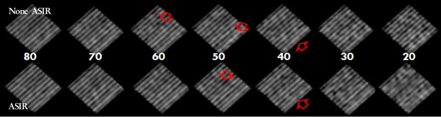
Fig. 3. Filtered back projection applied images which has no ASIR, and ASIR applied images where we had selection of 20 to 80%.
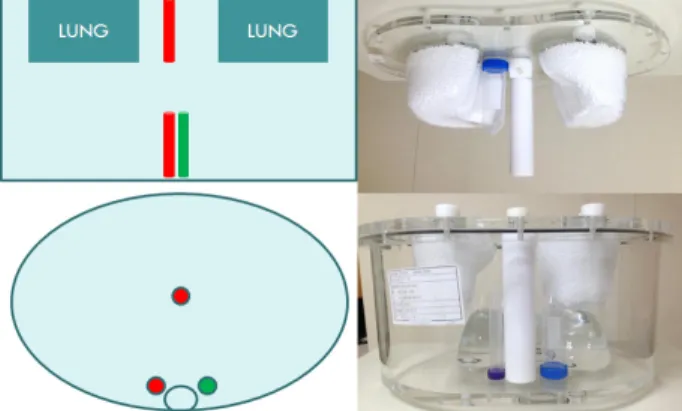
Fig. 4. One of tubes is in the middle of both lungs and the other two is located next to spine.
ative reconstruction (ASIR) 기법을 실제 bone SPECT/CT 검사에 적용하여 유용성을 알아보았다. AISR 기법은 CT image의 재구성 방법으로서 recon space의 projection data 영역에 statistical noise modeling을 적용시켜 영상의 noise를 감소시켜 결과적으로 영상의 질을 향상시킨다고 소개되고 있다.2)
ASIR의 적용방법은 검사 전 CT의 조건을 설정하는 화면 에서 간단히 설정할 수 있으며, 적용시킬 수 있는 ASIR의 level은 ASIR 10%부터 ASIR 100%까지 총 10단계가 있다.
ASIR를 적용시킴으로서 인해 줄어드는 noise index에 해당 하는 ASIR level을 선택하게 되면 결과적으로 환자의 dose 를 줄일 수 있다.3) 예를 들어 ASIR를 적용하지 않았을 때 100 mA에서의 noise index와 ASIR를 적용시킴으로 인하여 동일한 index가 60 mA image에 해당이 된다면 결과적으로 image의 질 저하 없이 40%의 dose reduction 효과를 볼 수 있다는 뜻이다(Fig. 1).
GE에서 제공하는 영상에서는 ASIR 기법을 이용하여 im- age의 질적 저하 없이 환자가 받는 방사선 피폭량을 줄일 수 있다고 소개하고 있다(Fig. 2). 본 논문은 bone SPECT/CT 에 ASIR 기법을 적용함으로서 감소하는 피폭량과 SPECT 와 CT image를 평가하고자 한다.
실험재료 및 방법
장비: Discovery NM/CT 670 scanner (GE, U.S.A)
Anthropomorphic Torso phantom, conical tube
ACR CT Accreditation Phantom, Gammex 464
Sectional Lower Torso Phantom, SK 250
1. ASIR 적용
모든 실험의 CT선량 조건은 120 kVp에 100 mA를 기준 으로 mA를 조금씩 변화시켜 실험을 하였다. 관전압은 일정 하게 유지시키고 관전류를 20, 30, 40, 50, 60, 70, 80, 100 mA로 변화시켜 영상을 획득하였다(Fig. 3). 변화하는 mA에 맞추어 ASIR level을 선택하여 reconstruction한 것과 ASIR 를 적용하지 않은, 즉 FBP 기법으로 reconstruction한 image 를 비교 평가하였다.
Fig. 5. Out of 4 modules, we used the second, third and fourth one.
Fig. 6. We used low torso phantom, SK250 and we particularly focused on spine images.
Fig. 7. All three tubes showed no significant difference regardless of ASIR.
2. Attenuation correction evaluation in SPECT image
ASIR level의 변화에 따른 SPECT image의 감쇠 보정 정 도를 측정하기 위하여 anthropomorphic torso phantom을 사용하였다. 길이 10 cm, 용적 50 cc, 15 cc인 3개의 conical tube를 팬텀내부의 구조물 중 양쪽 폐 중간에 한 개를 위치 시켰으며, 나머지 두 개는 spine 구조물 옆에 위치시켰다 (Fig. 4). Background에는 0.25 uCi/cc, 3개의 conical tube에 는 0.2 uCi/cc의 99mTcO4-을 넣어서 background와 hot lesion 의 activity가 1:8이 되도록 하였다.
3. Evaluation of CT image by changing the ASIR level
ASIR 적용 유무와 적용 level에 따른 CT image 평가를 위 해 ACR CT accreditation phantom, gammex 464를 사용하 였다(Fig. 5). 총 4개의 module 중 2번째 low contrast reso- lution 영역과 3번째 uniformity & noise, 4번째 high con- trast resolution 영역을 이용하여 획득한 image를 비교 평가 하였다.
4. Evaluation of spine image by changing the ASIR level
본원에서 주로 실시하는 bone SPECT/CT 검사 부위인 spine의 image가 ASIR 적용 유무와 적용 level의 변화에 따 라 어떤 변화를 보이는지 평가하고자 low torso phantom, SK250을 사용하였다(Fig. 6). Spine image를 검사하는데 있 어 어느 정도의 ASIR level을 수용할 수 있는지 평가하는 실 험이었고 blind test를 시행하였다.
결 과
1. Attenuation correction evaluation in SPECT image
팬텀 내부에 삽입시킨 3개의 conical tube에 대한 contrast ratio를 산출한 후 100 mA의 조건으로 획득한 영상을 100%
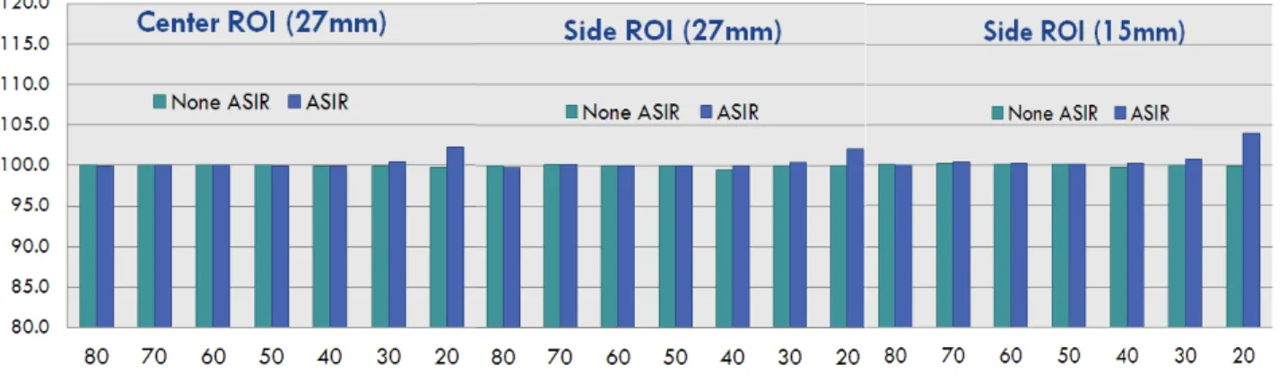
로 기준잡고, 나머지 영상들의 AC 정도를 그래프로 나타내 었다(Fig. 7).
/a % a
/C Q C
B H
B H
H 100
11 ×
−
= −
QH: Contrast ratio CH: Hot lesion count CB: Average of BKG aH: Hot lesion Activity aB: BKG lesion Activity
None ASIR
ASIR
Fig. 10. It showed that 12 line pairs can be distinguished from on 40 mA images regardless ASIR and 70 mA images without ASIR and 60 mA images with ASIR were evaluated as‘good’.
Fig. 8. When ASIR was used, the noise has been decreased from 17 up to 52%.
Fig. 9. When ASIR was used high CNRs were found from 80 to 40 mA which are higher than 0.81.
C % N SD
Av,B B
B = ×100
팬텀 중앙과 spine옆 3개의 tube 모두 ASIR의 적용 유무 에 무관하게 유의한 차이가 없음을 보여주었다. 5개의 slice 에 12개, 총 60개의 ROI를 그리고 percent background vari- ablity를 산출하였다. Percent background variabililty 또한 mA의 변화와는 무관함을 알 수 있었다.
2. Evaluation of CT image by changing the ASIR level
1) Uniformity & noise 영역
ASIR를 적용하지 않았을 때 noise index는 60 mA 이하의 관전류 조건에서 10 이상의 높은 수치를 보여준 반면, ASIR 를 적용했을 때 가장 낮은 20 mA에서도 8.1로 비교적 낮고 mA의 변화에 다소 균등한 noise를 보여주었다(Fig. 8). 전반 적으로 ASIR를 적용했을 때 noise는 최소 17%에서 52%까 지 줄어들었다.
2) Low contrast resolution 영역
ASIR를 적용하지 않았을 때 CNR은 40 mA에서 0.42를 보여준 반면, ASIR를 적용하였을 때 40 mA까지 0.8 이상의 높은 CNR을 유지함을 알 수 있었다(Fig. 9).
3) High contrast resolution 영역
8개의 각기 다른 line pair가 들어있는 high contrast reso- lution 영역에서 cm당 12개의 line pair 이미지를 비교 평가 하였다(Fig. 10). 5년 이상의 경력을 가진 방사선사 10명의 blind test 결과 ASIR 적용 유무에 관계없이 40 mA image부 터 12개의 line pair를 모두 구별할 수 있었다. 또한 ASIR를 적용하지 않았을 때 70 mA, 적용하였을 때 60 mA의 image 부터 영상이 ‘우수하다’라고 평가되었다.
3. Evaluation of spine image by changing the ASIR level
Torso phantom, SK250의 spine image는 ASIR 적용 유무와 적용 level의 변화에 따라 육안적 비교에서 큰 차이를 보이지
NB: Percent BKG variability SDB: Standard deviation CAv,B: Average of BKG
Fig. 11. As mA was decreased, small amount of noise appeared on the images but there was no significant difference on spine images.
Fig. 12. Even though we reduced 40 mA, the image quality was not affected by it.
Fig. 13. Small amount of noise has increased on the image with ASIR but there was no significant difference on spine images.
않았다. 관전류가 줄어들수록 noise가 증가하였다(Fig. 11).
4. Evaluation of patient images
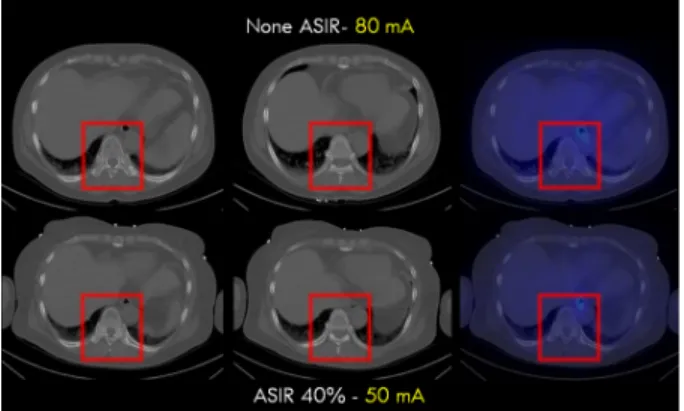
L-spine에 병변이 있는 환자로 follow up 환자에게 ASIR 를 적용하여 검사하였다. 이전에 AISR를 적용하지 않은 100 mA의 이미지와 ASIR 40%를 적용하여 60 mA로 획득한 이 미지를 비교하였다(Fig. 12). Image quality는 별다른 차이가 없지만 100 mA일 때 장비에서 제공하는 CTDIvol과 DLP 값은 5.44 mGy, 233.95 mGy인 반면, 60 mA일 때 3.26 mGy, 140.37 mGy로 약 40% 정도 줄어들었다.
다음은 131I whole body 환자로서 bone metastasis가 의심 되어 SPECT/CT를 시행한 환자의 image이다(Fig. 13). ASIR 40%를 적용한 영상에서 약간의 노이즈가 증가되었지만, spine의 image quality는 별 차이가 없음을 볼 수 있다.
결 론
본 논문의 결과는 SPECT/CT에서 ASIR 기법을 사용했을 때 SPECT와 CT image의 특별한 영상의 질 저하 없이 radi- ation dose를 줄일 수 있다는 것을 보여준다. SPECT image의 감쇠 보정 정도는 ASIR의 변화와는 무관하게 나타났으며, 오히려 ASIR를 적용했을 때, CT image의 CNR, noise, res- olution 모두 향상된 결과를 보여주었다. ASIR 40%를 적용 한 bone SPECT/CT 검사에서 영상의 질적 저하 없이 환자 의 radiation dose는 약 40%까지 낮출 수 있었다. 또한 관심 부위가 bone에 한정되어 있는 bone SPECT/CT 특성상 더 높은 ASIR level도 가능할 것으로 사료된다.
요 약
ASIR기법은 statistical noise modeling을 사용하여 CT im- age를 reconstruction하는 방법으로, mA를 낮춰도 이미지 질 을 보전하며 noise reduction 효과가 있다고 알려져 왔다. 본 논문은 본원에서 주로 하는 bone SPECT/CT에 ASIR 기법 을 적용하여 이미지를 평가하였다. GE의 Discorvery 670을 이용하여 120 kVp, 100 mA를 기준으로 mA를 변화시켜서 ASIR의 적용 전과 적용 후 영상을 비교하였다(ASIR level:
20-80%). Anthropomorphic phantom으로 ASIR (%)의 변화 에 따른 SPECT image의 감쇠 보정 정도를 측정하였다. 두 번째로 ACR phantom으로 CT image의 CNR, image noise, spatial resolution을 평가하였다. 세 번째로 lower torso phantom을 이용하여 spine에 최적화할 수 있는 ASIR level을 선택한 후 2명의 bone SPECT/CT follow up 환자에게 ASIR 를 적용하여 영상을 획득한 후 5년 이상의 경험이 있는 10명 의 방사선사에게 blind test를 시행하였다. SPECT의 영상의
감쇠 보정 정도는 ASIR의 변화와는 무관하게 모두 유의한 차이가 없었다(P>0.05). ASIR을 적용했을 때 CT image의 noise는 mA의 변화에 따라 최소 17%에서 최대 52%까지 감소하였다. ASIR를 적용하지 않았을 때 CNR은 40 mA에 서 0.42를 보여준 반면 ASIR를 적용한 40 mA (ASIR 60%) 에서도 0.8 이상을 유지하였다. High contrast 영역의 비교 에서는 ASIR 적용과 상관없이 40 mA까지 12 lp/cm 영역을 구별할 수 있었다. Lower torso phantom의 spine image에서 100 mA image와 육안적으로 비슷한 ASIR level은 60% (40 mA) 정도였고, bone SPECT/CT에 적용한 후 blind test에서 육안적으로 ASIR를 적용하지 않았을 때와 차이를 구별하지 못하였다. 본 논문의 결과는 SPECT/CT에서 ASIR 기법을 사용했을 때 SPECT와 CT image의 특별한 영상의 질 저하 없이 radiation dose를 줄일 수 있다는 것을 보여준다. 또한 관심부위가 bone에 한정되어 있는 bone SPECT/CT 특성상
더 높은 ASIR level도 가능할 것으로 사료된다.
REFERENCES
1. Hara AK, Paden RG, Silva AC, Kujak JL, Lawder HJ, Pavlicek W. Iterative reconstruction technique for reducing body radia- tion dose at CT: Feasibility study. AJR Am J Roentgenol 2009;193:764-771.
2. William P. Shuman. Adaptive iterative reconstruction in CT:
What does it do? How can I use it? Available at: http://www.i- magewisely.org/Imaging-Modalities/Com-puted- Tomography/Imaging-Physicians/Articles/Adaptive-Iterative -Reconstruction-in-CT. Accessed May 1, 2010.
3. Prakash P, Kalra MK, Kambadakone AK, Pien H, Hsieh J, Blake MA, et al. Reducing abdominal CT radiation dose with adaptive statistical iterative reconstruction technique. Invest Radiol 2010;45:202-210.