최 병 철 ^ 중앙대학교 약학대학
(Received September
8, 2008; Revised November 24, 2008)
E f f e c t o f C a f f e ic A c i d o n t h e P r o d u c t i o n o f R e a c t i v e O x y g e n S p e c i e s i n R a w 2 6 4 . 7 C e l l s
Byung-Chul Choi^
College of Pharmacy, Chung-Ang University
Abstract — To investigate effect of caffeic acid on the intracellular reactive oxygen species production, we used DHE for intracellular superoxide anion production, DCF for intracellular H
2O
2production and DHR for intracellular hydroperoxide production in Raw 264.7 cells. DPPH assay showed that antioxidant activity of caffeic acid with 39.5 |iM of IC
50values was similar to that of ascorbic acid with 41.3 |j.M of IC
5Q values. Caffeic acid dose-dependently inhibited silica-induced H
2O
2and hydroperoxide production but did not affect superoxide anion production in Raw 264.7 cells, which suggest that antioxidant effect of caffeic acid acts on the post-step of superoxide anion. On the other hand, caffeic acid showed a potent antioxidant effect in ICuSO^-induced lipid peroxidation. Furthermore, plasma superoxide dismutase activity (3.43±0.23 U/m/) in 10 mg/
kg caffeic acid-fed mice was significantly higher than that (2.32±0.24 U/m/) of control. From the above results, it is referred that caffeic acid appears to have potent anti-oxidant activity in both cell system and
in vivosystem.
Keywords □ caffeic acid, reactive oxygen species, nitric oxide, superoxide dismutase
세포에서 생성되는 reactive oxygen species(ROS)의 종류는 superoxide anion, hydrogen peroxide 와 hydroxyl radical 등이 있으며, 또한 nitric oxide는 superoxide anion파 결함하여 peroxynitirite롤 형성하여 세포에 비가역 손상을 일으킨다.^^ 이 러한 ROS를 제거하는데 있어서 항산화제는 중요한 역할을 담당 하고 있는데 식물체내에 존재하는 다양한 종류의 flavonoid나 phenylpropanoid 등은 강력한 항산화 기능을 갖고 있으므로써 이 들의 섬취는 인체 내에서 생성되는 ROS로부터 신체기능을 보호 해주고 있다
Caffeic acid는 phenylpropanoid 계열의 화합물로서 벤젠기에 2개 hydroxyl 기를 갖으며 항산화 작용을 비롯하여 다양?! : 약러 활성을 갖는 물질로 알려져 있다. 지금까지 알려진 caffeic acid의 작용을 살펴보면 흰쥐에서 caffeic acid는 지질괴산화물 생성을 억 제하고, ROS틀 제거하는데 관여하는 gluthathion peroxidase의 활 성을 증가시키는 기전을 통해 심근의 산화성 스트레스를 감소시 키며,^^ 열 손상에 의한 신장과 폐 조직의 산화성 스트레스를 억
제하였다.하 또한 caffeic acid 유도체돌은 Kaw 264.7 세포에서 lipopolysaccharide에 의한 NO 생성을 억제하였다.^^ 이러한 항산 화 작용이외에 항염증 작용도 보고 되고있다. Caffeic acid phenethyl ester가 사람 구강 상피세포에서 C0X-2의 발현을 억
제하며가 lipoxigenase룸 억 제한다는 보고가 있다.판 ^ 또한 흰쥐의 복강■비만세포에서 ATP에 의한 histamine 유리률 caffeic acid가 억제한다는 보고도 있다.^^
세포에서 생성되는 superoxide anion^ : superoxide dismutase 에 의해 로 선환되며, 세포내에 존재하는 catalase나 peroxidase에 의하여 무해한 산소와 물로 전환된다. 이 실험에서 는 세포내에서 생성되는 여러 가지 종류의 ROS를 측정하는 시 약들을 이용하여 caffeic acid가 세포내에서 생성되는 어떠한 유 해산소의 생성을 억제하며, 생체내에서도 항산화 기능을 나타내 는 지률 관찰하였다.
실험 방법
현 논문에 관한 문의는 저자에게로 (선화) 02-3426-5664 (팩스) 02-3426-2638 (E-mail) [email protected]
재료
Caffeic acid, l-diphenyl-2-picrylhydroazyl(DPPH), xanthine oxidaseCXO), NBT(nitroblue tetrazolium chloride), hypoxanthine,
441
442 최 병 철
2-thiobarbituric acid, malonyldialdehyde, hexadecyltrimethyl ammonium bromide(HTAB), 3,3',5,5'-tetramethyI benzidine (TMB), N,N-dimethylformamide(DMF)들은 Sigma사로부터 구 입하였으며, dihydrorhodamine(DHR), dihydroethidium, 2\T- dichlorofluorescin diacetate(DCF-DA)는 Molecular Probe Co.
에서 구입하였다.
세포 배양
Raw 264.7 세포는 10% fetal bovine serum 파 penicillin/
streptomysindOO RJ/50 |ig/m/)을 함유한 Dulbecco's modified Eagle's medium(DMEM) 용액으로 37X 로 유지되는 5% CO^
배양기에서 배양하여 superoxide 생성, 세포내 생성, 세포 내 hydroperoxide 생성 및 지질과산화를 죽정하였다.
DPPH 라디칼 소거 정량
96 well plate에 에탄을에 녹인 0.1 mM DPPH 용액 180 |o/와 각 농도별로 조제한 caffeic acid 20 j i 를 가하고 37°C에서 3Q분 배양한 푸 FL600 spectrofluorometer(Bio-Tek, Winooski, USA)를 이용하식 517nm에서 흡광도률 측정하였다.
세포내 superoxide anion 생성 촉정
Raw 264.7 세포률 10 m/의 Krebs buffer 용액 (mM: NaCl 137, KCl 2.7, Na2HP04 0.4, MgCl; 으 0.5, HEPES[pH 7.4] 10, CaCl2 1.8, glucose 5)에 분산시킨 후 10|iM dihydroethidium을 가하고 1 시간 빛을 차단한 곳에서 배양하였다. Dihydroethidium 이 없는 Krebs 용액으로 한번 세척한 후 10^cells/m/로 분주하 j l caffeic acid률 견처처 한 후 silica 1 mg/m/을 가하여 3Q분간 superoxide anion 생성을 유도하였다. 원심분리 루 cell pellet울 200 m/의 Krebs 용액에 분산시킨 후 형광(Ex: 480 nm: Em; 586 nm)을 측정하였다.
세포내 H2O2 생성 측점
Raw 264.7 세포률 10 m/의 Krebs buffer 용액에 suspend 시 킨 후 2(HiM DCF-DA를 가하고 1시간 빛을 차단한 곳에서 배 양하였다. DCF-DA가 없는 Krebs 용 액 로 한번 세척한 후 10'^
cells/m/로 분주하고 caffeic acid를 전처처 한 후 silica 1 mg/m/
을 가하여 30분간 H2O2 생성을 유도하였다. 원심분러 후 cell pellet을 200(0/의 Krebs 용액에 분산시킨 후 형광(Ex: 485 nm;
Em; 535 nm)을 측정하였다.
10^ cells/m/로 분주하고 여러 농도의 caffeic acid률 전처처 한 후 silica 1 mg/m/을 가하여 30분간 hydroperoxide 생성 을 유도하였다. 원심분리 후 cell pellet을 200 나의 Krebs 용 액에 분산 시킨 후 형광(Ex: 488 nm: Em : 515 nm)을 측정 하였다.
Nitrite의 측정
NO의 생성은 Nitrite률 측정함으로서 간접적으로 확인하였으 며, Griess reagent A(l% sulfanilamide in 5% phosphoric acid) 와 Griess reagent B(0.1% naphtylenediamine in 5%
phosphoric acid)를 사용하였다. RAW264.7 세포에 caffeic acid 와 lipopolysaccharide 1 iig/m/을 처 처하고 24시간 배양하였다.
배양액에 Griess reagent A와 B를 동량 섞은 용액을 가하고 5 분 간 37X에서 빈옹 시킨 후 ELISA reader틀 이용하여 550 nm 에서 흡광을 측정하였였다.""
지질과산화 측정
세포막의 지질과산화를 죽정하기 위하여 thiobarbituric acid reactive substances(TBARS)의 형광을 이용하였다. caffeic acid 와 100 나M C11SO4를 1시간 처러한 Raw 264.7 세포를 0.5 mM DTT률 함유?]: PBS buffer로 세척한 루 sonication하고 50%
trichloroacetic acid를 가하여 최종 5% 농노로 조절하였다, 동량 의 0.325% 2-thiobarbituric acid(in 50% acetic acid)!- 첨기하여 95T 에서 30 min 배양하였다. 원심분리 후 200 률 96 well plate에 옮기고 Ex: 485 nm, Em: 535 파고에서 형광 측정하였다.
표준물질로서 는 malcmylciialdehyde(l,l,33-tetraetlioxypropaiie, Sigma)를 사용하였다.조오스
혈장 superoxide dismutase 측정
체중이 2 0 ^ 5 g 정도 되는 숫컷 Balb-c 생쥐에 매일 10있간 10 mg/kg caffeic acid틀 경구투여 한 후 혈액을 채취하여 혈장내 superoxide dismutase의 활성을 아래의 방법으로 측정하였다. 0.6 mM hypoxanthine, 1 mM EDTA, 0.2 mM NBT률 힘■유하는 50 mM potassium phosphate buffer(pH 7.4) 400 나에 superoxide dismutase 및 혈장 10 틀 가하여 잘 흔합하였다. 반응은 xanthine oxidased U/m/) 100 pi룰 가하던서 시 작하였다. 반응 혼합액을 37T 에 서 5분간 배 양한 후 96 well plate에 200 |o/씩 소분하고 FL600 spectrofluorometer를 이용하여 645nm에서 몹 광도틀 측정하였다.노 크 ^
세포내 hydroperoxide 생성 측정
Raw 264.7 세포률 10 m/의 Krebs buffer 용액에 suspend 시킨 후 l(H iM DHR를 가하고 30분간 빛을 차단?t 장소에 서 배양하였다. DHR이 없는 Krebs 용액으로 한번 세척한 후
자료분석 및 통계적 검정
실험 결과는 평균±표준편차로 표기하였으며 , 실험 성적은 non- paired Student's t test와 ANOVA로 검정하였고 P 값이 5% 머 만일 때 통계적으로 유의하다고 간주하였다.
}. Pharm. Soc. Korea
해산소를 억제하는지 관찰하였다. 유해산소의 생성온 Raw 264.7 세포률 silica로 자극하였다. Silica는 다양한 세포에서 유해 산소 생성을 중가시킨다는 것이 이미 보 고 되 었 으 며 Table I에서 보 다시피 Raw 264.7 세포에서 silica는 농도 의존적^®_로 유해산소 생성을 증가시켰다.
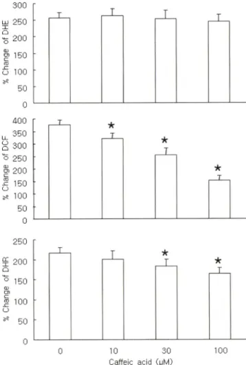
먼저 세포내에서 생성되는 superoxide anion 생성은 dihy- droethidium를 이용하여 측정하였다.비 Silica 1 mg/m/은 Raw 264.7 세포에서 superoxide anion 생성을 2.5배 증가시켰다. 그 러나 caffeic acid는 silica에 의해 생성되는 세포내 superoxide anion에는 벌 다른 영향을 주지 않았다(Fig. 2). 이 결과로 미루 어 볼 때 caffeic acid는 superoxide anion의 생성과 분해에 있어 서 직접적인 작용은 없는 것으로 사료된다.
세포내 유해산소의 일중인 H2O2는 DCF-DA률 이 용 하 측 정 하였다.^® Silica 1 mg/m/은 Raw 264.7 세포에서 H2O2 생성을
실험결과 및 고찰
Raw 264.7 세포내에서 caffeic acid의 항산화 작용
Caffeic acid는 phenylpropanoid 계열의 화합물로서 벤센기에 2개 hydroxyl기를 갖으며 항산화 작용을 비롯하여 다양한 약리 활성을 갖는 물질로 알려져 있다. 먼저 caffeic acid의 자체 항산 화 효과를 관찰하기 위하여 DPPH radical 소거률 관찰하였다.
Caffeic acid는 농도 의존적으로 DPPH radical을 소거하였다(Fig.
1). Caffeic acid의 ICjo 값은 39.5 나M로서 ascorbic add의 IC50 값인 41.3 nM과 유사하였다. 이러한 걸과는 caffeic acid 자체가 강력한 항산화 작용을 갖고 있옴을 제시하여 준다.
배양세포에서 생성되는 superoxide anion은 superoxide dismutase에 의해 H; P 2로 전환되며, 세포내에 존재하는 catalase 나 peroxidase에 의하여 무해한 산소와 물로 선환되지만 만약 유 해산소의 제거가 원활히 이루어지지 않는 경우 DNA, 단백질 및 지질의 산화가 일어나 세포 손상을 유래하게 된다. 이 실험에서 는 세포내에서 생성되는 여러 가지 종류의 ROS를 측정하는 시 약돌을 이용하식 caffeic acid가 세포내에서 생성되는 어떠한 유
Ascorbic acid Caffeic acid
10 30
Caffeic acid (gM)
100
Fig. 2 - Effect of caffeic acid on 1 silica-induced ROS produc
tion in Raw 264.7 cells. DHE was used for measurement of intracellular superoxide anion production, DCF for meas
urement of intracellular H
2O
2production and DHR for measurement of intracellular hydroperoxide production.
Results are means ±SD from 4 separate experiments.
* Significantly different from silica alone (p<0.05).
Concentrations (pM)
Fig.
1- DPPH radical scavenging activity of caffeic acid. A solution of 180
ixlof 100 i^M DPPH solution in ethanol was gently mixed with 20 |o/ of caffeic acid for 30 min and the absorbance was measured at 517 nm. Results are means ± SD from 4 separate experiments.
Table I - Dose-response of reactive oxygen species generation to silica in Raw 264.7 cells
Silica
{mg/ml)% Increase of Control
DHE DCF DHR
0 1 0 0 .0 1 0 0 .0 1 0 0 .0
0.5 164.2 ±15.2 225.2±21.2 173.2±11.4
1 .0
256.0±17.1 377.4±28.2 217.0±12.4
2 .0
432.2+26.3 622.2 ±33.9 410.2±24.8 4.0 553.2 ±33.2 751.7±42.3 520.2 ±52.8 DHE: used for measurement of intracellular superoxide anion production.
DCF: used for measurement of intracellular
H2O2production.
DHR: used for measurement of intracellular hydroperoxide production.
VoL 52, No. 6, 2008
★
'k -k
-k
★
m s
io
%
o o o o o o o o
0 5 0 5 0 5
0 5
4 3 3 2 2 1 1
joa
io
%yiHd
io
%
B
>l
u e ralj!6u이>e:JSle^jpe
j IHddQ
3.7배 증가시켰다. Caffeic acid는 silica에 의해 생성되는 세포내 H2O2를 농도 의존적으로 억제하였다(Fig. 2). 한편 세포내 peroxynitite률 포함한 hydroperoxide 생성을 DHR틀 이용하여 측정한 실험에서 ^* silica 1 mg/m/은 Raw 264.7 세포에서 2.1 배 증가시켰다(Fig. 2). 이러한 결과는 caffeic add의 세포내 항산화 효과가 H2O2 이후 단계에서 나타나고 있옴을 제시하식 준다.
Raw 264.7 세포에서 NO 생성에 미처는 영향
NO도 ROSSI 일종으로 혈관을 이완시키며 다양한 생리 작용 을 나타내고 있다. NO는 superoxide와 결합하여 peroxynitrite 률 형성하며 이는 단백질을 nitration 시키므로써 세포에 비가역 적 손상을 일으키는 유해한 물질로 알려져 있 다 . 앞 선 DHR을 이용한 실험에서 caffeic acicKr silica에 의한 hydroperoxide의 생 성을 유의하게 억제하였다. 이러한 결과를 확인하기 위하식 같 은 세포에서 lipopolysacharide에 의한 NO 생성을 족정한 결과 농도 의 존 적 억 제 하 였 다 (Fig. 3). 이 결과는 항산화 기능을 갖는 물질이 대식세포에서 NO 생성을 억제한다는 결과와 일치 하고 었다.1구 ' 이 러 한 결과는 caffeic add가 NO 생성을 억제하 며 세포내 ROS 생성도 억제하므로써 비가역적 손상을 일으키는 peroxynitrite의 생성을 억제하므로써 세포의 손상을 방지 할 것 으로 ^1•료된다.
Raw 264.7 세포에서 CuSO.i에 의한 지질과신화에 미치는 영향 세포에서 생성되는 reactive oxygen species들은 세포막이나 세포내 소기관의 막을 산화시켜 생성되는 지질파산$1"물을 생성 한다.페 CuSOt 100 나서은 Raw 264.7 세포에서 대조군에 비하여 2배 증가시켰다. caffeic add는 CuSC»4에 의한 지질과산화물 생 성도 유의하게 억제하였다(Fig. 4). 이 결과도 세포내에서 생성되 는 ROS틀 caffeic add가 유의하게 감소시킴으로써 나타나는 결 파일 것으로 사료된다.
Control 10 30
Caffeic acid (uM)
100
Fig. 4 - Effect of caffeic acid on 100 CuS
0 4-induced lipid peroxidation in Raw 264.7 cells. The cells were incubated with 100
나M C
11SO
4in the presence or absence of caffeic acid. Results are means ±SD from 4 separate experiments.
*SignificantIy different from CuSO. alone (p<0.05).
Control 0 1 3 10
Caffeic acid (uM)
30 100
Fig. 3 - Effect of caffeic acid on
1(Ag/m/ lipopolysaccharide-induced NO production in Raw 264,7 cells. The cells were in
cubated with
1fig/m/ lipopolysaccharide in the presence or absence of caffeic acid. Results are means ±SD from 4 separate experiments. ^Significantly different from lipopoly
saccharide alone (p<0.05).
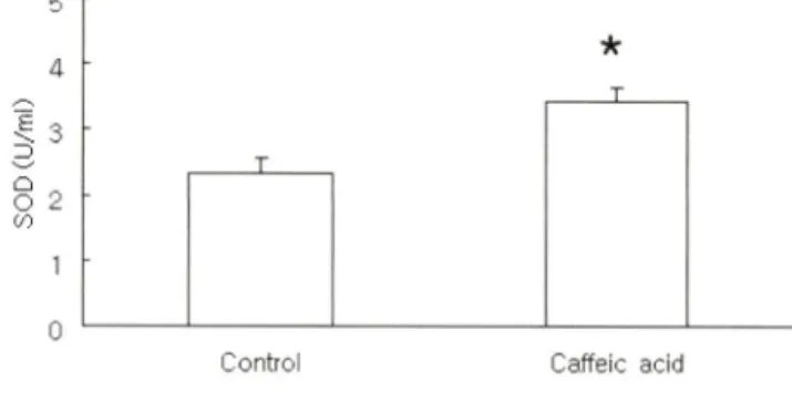
Control Caffeic acid
Fig. 5 - Effect of caffeic acid on plasma superoxide dismutase (SOD) in caffeic acid-fed mice. Results are means ±SD from
6separate experiments. *Significantly different from control (p<0.05).
Caffeic acid를 경구 투여한 생쥐 혈장에서 superoxide dismutase 활성 변화
이전 결과로 볼 때 caffeic acid는 세포내에서 생성되는 ROS 를 유의하게 억제하는 것으로 보인다. 이러한 caffeic acid의 항 산화 효과가 in 에서도 나타나는 지를 확인하기 위하지 caffeic acid를 생쥐에 10일간 경구 투여한 후 혈장내 SOD 활성을 측정 하였다. 대조군의 혈장내 SOD 활성은 2.32±024U/m/이였으며, caffeic acid를 류극한 시험군의 혈장내 SOD 휠성은 3.42±0.23U/
m/로 대조군에 비하여 유의하게 증가하였다(Fig. 5). 항산화제의 일종인 propofol온 혈관이 막힌 후 재관류시킨 사람에서 혈장내 SOD의 활성을 증가시킨다는 보고가 있다.^^^ 이 결과들을 종합 하늬 볼때 caffeic acid는 세포내에서 발생하는 ROS의 생성을 유 의하게 억제할 뿐만 아니라 생체내에서도 항산화 효과를 나타내 고 었움을 보여주고 있다.
문 헌
1) Kuo, W N., Kanadia, R. N. and Shanbhag, V R : Denitration of
J. Pharm. Soc. Korea
444 최 병 철
★ ★
* ★
★
★
cl/ro
os a
★
★
4
3
2
1
<
wf^ALPPJI'elpLlolelAI
peroxynitrite-treated proteins by "protein nitxatases" from dog prostate.
Biochem. Mol BioL Int.47, 1061 (1999).
2) Lee, J. Y., Yoon, J. W, Kim, C. X and Lim, S. T. : Antioxidant activity of phenylpropanoid esters isolated and identified from Platycodon grandiflorum A. DC.
Phytochemistry65, 3033 (2004).
3) Beyer, G. and Melzig, M. E : Effects of selected flavonoids and caffeic acid derivatives on hypoxanthine-xanthine oxidase- induced toxicity in cultivated human cells.
Planta. Med.69, 1125 (2003).
4) Okutan, H., Ozcelik, N., Ramazan Yilmaz, H. and Uz, E. : Effects of caffeic acid phenethyl ester on lipid peroxidation and antioxidant enzymes in diabetic rat heart.
Clin. Biochem.38,
191 (2005).
5) Gurel, A., Armutcu, E, Hosnuter, M., Unalacak, M., Kargi, E.
and Altinyazar, C. : Caffeic acid phenethyl ester improves oxidative organ damage in rat model of thermal trauma
Physiol Res.
53, 675 (2004).
6
) Da Cunha, E M., Duma, D., Assreuy, J., Buzzi, E C., Niero, R., Campos, ML M. and Calixto, J. B. : Caffeic acid derivatives:
in vitroand
in vivoanti-inflammatory properties.
Free. Radic. Res.38, 1241 (2004).
7) Michaluart, E, Masferrer, J. L, Carothers, A. M., Subbaramaiah, K., Zweifcl, B. S., Koboldt, C., Mestre, J. R., Grunberger, D., Sacks, R G., Tanabe, T. and Dannenberg, A. J. : Inhibitory effects of caffeic acid phenethyl ester on the activity and expression of cyclooxygenase
- 2in human oral epithelial cells and in a rat model of inflammation.
Cancer. Res.59, 2347 (1999).
8
) Mufti, N. A. and Shuler, M. L ,: Possible role of arachidonic acid in stress-induced cytochrome P450IA1 activity.
BiotechnoL Prog.12, 847 (1996).
9) Lee, Y. H., Lee, S. J., Seo, M. H., Kim, C. J. and Sim, S. S. : ATP-induced histamine release is in part related to phospholipase A2-mediated arachidonic acid metabolism in rat peritoneal mast cells.
Arch. Pharm. Res.24, 552 (2001).
10) Naoaki, M., Isao, S., Toru, M,, Naoto, U. and Eikai, K. : Aged garlic extract enhances production of nitric oxide.
Life Sciences71, 509 (2002).
1 1
) Toshifumi, T, Baier, L. D. and Morrson, A. R. : Antioxident inhibit interrukin
- 1-induced cyclooxygenase and nitrc-oxide synthase expression in rat mesangial cells./.
Biol Chem.271,
11689 (1996).
12) Waters, S., Fae, A., Gondalia, J., Holm, JL, Karlstrom, L., Nilsson, U. and Jonsson, 0. : Effects of pretreatment with a xanthine oxidase inhibitor on free radical levels during carotid endarterectomy.
Free. Radic. Res.38, 283 (2004).
13) Cho, Y. J., Seo, M. S., Kim, J. K., Lim, Y., Chae, G., Ha, K. S.
and Lee, K. H. : Silica-induced generation of reactive oxygen species in Rat2 fibroblast: role in activation of mitogen- activated protein kinase.
Biochem. Biophys. Res. Commun.262, 708 (1999).
14) Siegel, D., Gustafson, D. L., Dehn, D. L., Han, J. Y., Boonchoong, R, Berliner, L. J. and Ross, D. : NAD(P)H:
quinone oxidoreductase 1: role as a superoxide scavenger.
MoL Pharmacol.65, 1238 (2004).
15) Woo, C. H., Eom, Y. W, Yoo, M. H., You, H. J., Han, H. J., Song, W K., Yoo, Y, J., Chun, J. S. and Kim, J. H. : Tumor necrosis factor-alpha generates reactive oxygen species via a cytosolic phospholipase A
2-linked cascade.
J. BioL Chem.275, 32357 (2000).
16) Nomura, K., Imai, H., Koumura, T, Kobayashi, T. and Nakagawa, Y. : Mitochondrial phospholipid hydroperoxide glutathione peroxidase inhibits the release of cytochrome c from mitochondria by suppressing the peroxidation of cardiolipin in hypoglycaemia-induced apoptosis.
Biochem. J.351, 183 (2002).
17) Matsuda, H., Kageura, X, Oda, M., Morikawa, T, Sakamoto, Y. and Yoshikawa, M. : Effects of constituents from the bark of Magnolia obovata on nitric oxide production in lipopolysaccharide-activated macrophages.
Chem. Pharm. Bull (Tokyo)49, 716 (2001).
18) Arteel, G. E., Mostert, V, Oubrahim, H., Briviba, K., Abel, J.
and Sies, H. : Protection by selenoprotein P in human plasma against peroxynitrite-mediated oxidation and nitration.
BioL Chem.379, 1201 (1998).
19) Boland, A., Delapierre, EX, Mossay, D., Hans, E and Dresse, A. : Propofol protects cultured brain cells from iron ion-induced death: comparison with trolox.
Eur. J. Pharmacol.404, 21 (2000).
20) Turan, R., Yagmurdur, H., Kavutcu, M. and Dikmen, B. : Propofol and tourniquet induced ischaemia reperfusion injury in lower extremity operations.
Eur. J. Anaesthesiol.24, 185
(2007).
Vol. 52, No. 6, 2008